

4 курс / Лучевая диагностика / ПРИМЕНЕНИЕ_КОМПЛЕКСНОЙ_МАГНИТНО_РЕЗОНАНСНОЙ_ТОМОГРАФИИ_ПРИ_РАЗЛИЧНЫХ
.pdf191
CHAPTER 3. RESULTS OF THE STUDY
3.1 Results of comprehensive magnetic resonance imaging
3.1.1 Standard MRI examination
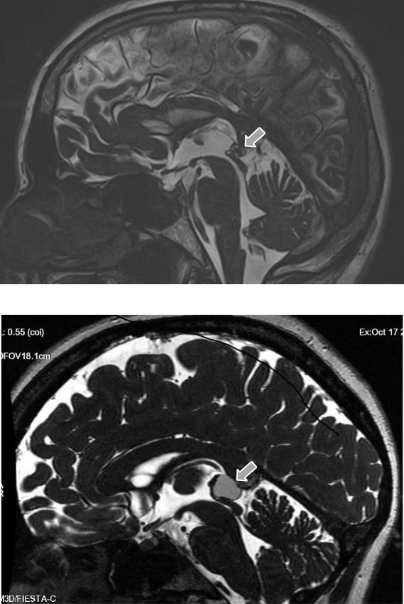
149 volunteers underwent a standard MRI stady. In 70 of them, the pineal gland had a normal structure (Figure 8). The mean thickness of the pineal gland was 4.13 0.23 mm.
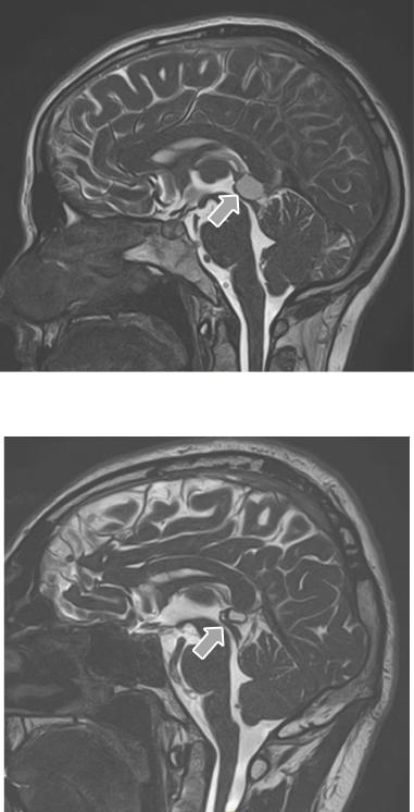
Figure 8 – MR image of patient K. (OC No. 32556, 2020), T2-PS, in the sagittal plane with normal pineal gland structure (indicated by arrow)
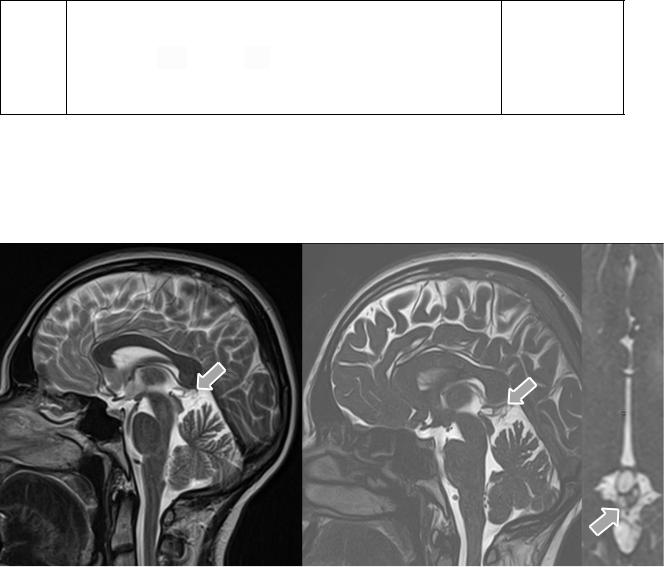
Cystic transformation of varying nature was detected in 79 (Figure 9). In the group with cysts less than 10 mm, the pineal gland thickness was 4.71 0.33 mm and in the group with large cysts it was 8.19±2.07 mm.
Рекомендовано к изучению сайтом МедУнивер - https://meduniver.com/
192
А
B
Figure 9 – MR images of patient D., (OC № 34557, 2021), SSFP-PS, in the sagittal plane with a cyst less than 10mm (A) and patient Sh., (OC № 34876, 2021), SSFPPS, in the sagittal plane with a cyst over 10mm (B) (indicated by arrows)
193
All patients were divided into 5 groups based on the structural changes of the pineal gland according to standard MR imaging based on a previously presented classification (Sirin S, 2016) (Table 4).
Table 4 — Distribution of patients according to structural changes in the pineal
gland
|
|
|
|
|
|
|
|
|
|
|
|
|
|
|
|
|
|
|
|
|
|
|
|
|
|
|
|
|
|
|
|
|
|
|
|
|
|
|
|
|
|
|
|
|
|
|
|
|
|
|
|
|
|
|
|
|
|
|
|
|
|
|
|
|
|
|
|
|
|
|
|
|
|
|
|
|
|
|
|
|
|
|
|
Structural changes |
|
|
|
|
|
|
a (≤5 |
|
|
|
|
|
|
b (6-9 |
|
|
|
|
|
c (≥10 |
|
|
|
Number of |
|
|
Distribution by |
|
|||||||||||||||||||||||||||||||||||||||||||||||||||
|
|
of the pineal gland |
|
|
|
|
|
|
|
m) |
|
|
|
|
|
|
|
|
mm) |
|
|
|
|
|
|
|
mm) |
|
|
|
|
|
|
patients |
|
|
|
|
|
gender, people |
|
|
|||||||||||||||||||||||||||||||||||||||
|
|
|
|
|
|
|
|
|
|
|
|
|
|
|
|
|
|
|
|
|
|
|
|
|
|
|
|
|
|
|
|
|
|
|
|
|
|
|
|
|
|
|
|
|
|
|
|
|
|
|
|
|
|
|
|
|
|
|
|
|
|
|
|
|
|
|
|
|
|
|
|
|
|
|
|
|
|
|
|
||
|
|
|
|
|
|
|
|
|
|
|
|
|
|
|
|
|
|
|
|
|
|
|
|
|
|
|
|
|
|
|
|
|
|
|
|
|
|
|
|
|
|
|
|
|
|
|
|
|
|
|
|
|
|
|
|
|
|
|
|
|
|
|
|
|
|
|
|
|
|
|
|
|
|
|
|
|
|
|
|
||
|
|
|
|
|
|
|
|
|
|
|
|
|
|
|
|
|
|
|
|
|
|
|
|
|
|
|
|
|
|
|
|
|
|
|
|
|
|
|
|
|
|
|
|
|
|
|
|
|
|
|
|
|
|
|
|
|
|
|
|
|
|
|
|
|
|
|
|
|
|
|
|
|
|
|
м - 33 |
||||||
|
|
|
|
|
|
|
|
No cysts - 0 |
|
|
|
|
|
|
|
|
|
|
|
|
|
|
|
|
|
- |
|
|
|
|
|
|
|
|
|
|
|
|
- |
|
|
|
|
|
|
|
|
|
|
|
- |
|
|
|
|
|
|
|
70 (47%) |
|
|
|
|
||||||||||||||||||
|
|
|
|
|
|
|
|
|
|
|
|
|
|
|
|
|
|
|
|
|
|
|
|
|
|
|
|
|
|
|
|
|
|
|
|
|
|
|
|
|
|
|
|
|
|
|
|
|
|
|
|
|
|
|
|
|
|
|
|
ж - 37 |
|
|
|
||||||||||||||||||
|
|
|
|
|
|
|
|
|
|
|
|
|
|
|
|
|
|
|
|
|
|
|
|
|
|
|
|
|
|
|
|
|
|
|
|
|
|
|
|
|
|
|
|
|
|
|
|
|
|
|
|
|
|
|
|
|
|
|
|
|
|
|
|
|
|
|
|
|
|
|
|
|
|
|
|
||||||
|
|
|
|
|
|
|
|
|
|
|
|
|
|
|
|
|
|
|
|
|
|
|
|
|
|
|
|
|
|
|
|
|
|
|
|
|
|
|
|
|
|
|
|
|
|
|
|
|
|
|
|
|
|
|
|
|
|
|
|
|
|
|
|
|
|
|
|
|
|
||||||||||||
|
|
|
|
|
|
|
|
|
|
|
|
|
|
|
|
|
|
|
|
|
|
|
|
|
|
|
|
|
|
|
|
|
|
|
|
|
|
|
|
|
|
|
|
|
|
|
|
|
|
|
|
|
|
|
|
|
|
|
|
|
|
|
|
|
|
|
|
|
|
|
|
|
|
||||||||
|
|
|
|
|
|
|
|
|
|
|
|
|
|
|
|
|
|
|
|
|
|
|
|
|
24 |
|
|
|
|
2 |
|
|
|
|
|
|
|
|
|
|
|
|
|
|
|
|
|
|
|
|
|
|
|
|
|
|
|
м - 7 |
|||||||||||||||||||||||
|
|
|
|
|
|
Single cyst - 1 |
|
|
|
|
|
|
|
|
|
|
|
|
|
|
|
|
|
|
|
|
0 |
|
|
|
|
|
26 (17,5%) |
|
|
|
|
||||||||||||||||||||||||||||||||||||||||||||
|
|
|
|
|
|
|
|
|
|
|
|
|
|
|
(16,1%) |
|
|
|
(1,4%) |
|
|
|
|
|
|
|
|
|
|
|
|
|
ж - 19 |
|
|
|
|||||||||||||||||||||||||||||||||||||||||||||
|
|
|
|
|
|
|
|
|
|
|
|
|
|
|
|
|
|
|
|
|
|
|
|
|
|
|
|
|
|
|
|
|
|
|
|
|
|
|
|
|
|
|
|
|
|
|
|
|
|
|
|
|
|
|
|||||||||||||||||||||||||||
|
|
|
|
|
|
|
|
|
|
|
|
|
|
|
|
|
|
|
|
|
|
|
|
|
|
|
|
|
|
|
|
|
|
|
|
|
|
|
|
|
|
|
|
|
|
|
|
|
|
|
|
|
|
|
|
|
|
|
|
|
|
|
|
|
|
|
|
|
|
|
|
|
|
|
|
|
|
||||
|
|
|
|
|
|
|
|
|
|
|
|
|
|
|
|
|
|
|
|
|
|
|
|
|
|
|
|
|
|
|
|
|
|
|
|
|
|
|
|
|
|
|
|
|
|
|
|
|
|
|
|
|
|
|
|
|
|
|
|
|
|
|
|
|
|
|
|
|
|
|
|
|
|
|
|
|
|
|
|
|
|
|
|
Multicystic pineal |
|
|
|
|
|
|
|
|
|
|
|
|
|
|
|
|
|
|
|
|
|
|
|
|
|
|
|
|
|
|
|
|
|
|
|
|
|
|
|
|
|
|
|
|
|
|
|
|
|
|
|
|
|
|
|
|
|
||||||||||||||||||||||
|
|
|
|
|
|
|
|
|
|
25 |
|
|
|
|
|
|
|
|
|
1 |
|
|
|
|
|
|
|
|
|
|
|
|
|
|
|
|
|
|
|
|
|
|
|
|
|
|
|
|
м - 12 |
|
|
|
|
||||||||||||||||||||||||||||
|
|
|
|
|
|
|
|
|
|
gland (no |
|
|
|
|
|
|
|
|
|
|
|
|
|
|
|
|
|
|
|
|
|
|
|
|
|
|
|
|
|
0 |
|
|
|
|
|
26 (17,5%) |
|
|
|
|
|
||||||||||||||||||||||||||||||
|
|
|
|
|
|
|
|
|
|
|
|
|
|
|
|
|
|
|
|
|
|
|
(16,7%) |
|
|
(0,68%) |
|
|
|
|
|
|
|
|
|
|
|
|
ж - 14 |
|
|
|
|||||||||||||||||||||||||||||||||||||||
|
|
magnification) |
- 2 |
|
|
|
|
|
|
|
|
|
|
|
|
|
|
|
|
|
|
|
|
|
|
|
|
|
|
|
|
|
|
|
|||||||||||||||||||||||||||||||||||||||||||||||
|
|
|
|
|
|
|
|
|
|
|
|
|
|
|
|
|
|
|
|
|
|
|
|
|
|
|
|
|
|
|
|
|
|
|
|
|
|
||||||||||||||||||||||||||||||||||||||||||||
|
|
|
|
|
|
|
|
|
|
|
|
|
|
|
|
|
|
|
|
|
|
|
|
|
|
|
|
|
|
|
|
|
|
|
|
|
|
|
|
|
|
|
|
|
|
|
|
|
|
|
|
|
|
|
|
|
|
|
|
|
|
|
|
|
|
|
|
|
|
|
|
|
|
||||||||
|
|
|
|
|
|
|
|
|
|
|
|
|
|
|
|
|
|
|
|
|
|
|
|
|
|
|
|
|
|
|
|
|
|
|
|
|
|
|
|
|
|
|
|
|
|
|
|
|
|
|
|
|
|
|
|
|
|
|
|
|
|
|
|
|
|
|
|
|
|
|
|
|
|
|
|
|
|
|
|
|
|
|
|
Multicystic pineal |
|
|
|
|
|
|
|
|
|
|
|
|
|
|
|
|
|
|
|
|
|
|
|
|
|
|
|
|
|
|
|
|
|
|
|
|
|
|
|
|
|
|
|
|
|
|
|
|
|
|
|
|
|
|
|
|
|
||||||||||||||||||||||
|
|
|
|
|
gland (enlarged |
|
|
|
|
|
|
|
2 |
|
|
|
|
|
|
|
|
|
|
|
|
|
|
|
|
|
|
|
|
4 |
|
|
|
|
|
|
|
|
|
|
|
|
|
|
|
|
|
м - 3 |
|
||||||||||||||||||||||||||||
|
|
|
|
|
|
|
|
|
|
|
|
|
|
|
|
|
|
|
|
|
|
|
|
|
3 (2%) |
|
|
|
|
|
|
|
|
|
9 (6%) |
|
|
|
|
|
|
|
|
||||||||||||||||||||||||||||||||||||||
|
|
|
|
|
|
|
without edge |
|
|
|
|
|
|
(1,3%) |
|
|
|
|
|
|
(2,68%) |
|
|
|
|
|
|
|
|
|
|
ж - 6 |
|
||||||||||||||||||||||||||||||||||||||||||||||||
|
|
|
|
|
|
|
|
|
|
|
|
|
|
|
|
|
|
|
|
|
|
|
|
|
|
|
|
|
|||||||||||||||||||||||||||||||||||||||||||||||||||||
|
|
|
|
|
displacement)-3 |
|
|
|
|
|
|
|
|
|
|
|
|
|
|
|
|
|
|
|
|
|
|
|
|
|
|
|
|
|
|
|
|
|
|
|
|
|
|
|
|
|
|
|
|
|
|
|
|
|
|
|
|
|
|
|
|
|
|
|
|
||||||||||||||||
|
|
|
|
|
|
|
|
|
|
|
|
|
|
|
|
|
|
|
|
|
|
|
|
|
|
|
|
|
|
|
|
|
|
|
|
|
|
|
|
|
|
|
|
|
|
|
|
|
|
|
|
|
|
|
|
|
|
|
|
|
|
|
|
|
|
|
|
||||||||||||||
|
|
|
|
|
|
|
|
|
|
|
|
|
|
|
|
|
|
|
|
|
|
|
|
|
|
|
|
|
|
|
|
|
|
|
|
|
|
|
|
|
|
|
|
|
|
|
|
|
|
|
|
|
|
|
|
|
|
|
|
|
|
|
|
|
|
|
|
|
|
|
|
|
|
|
|
|
|
|
|
|
|
|
|
Multicystic pineal |
|
|
|
|
|
|
|
|
|
|
|
|
|
|
|
|
|
|
|
|
|
|
|
|
|
|
|
|
|
|
|
|
|
|
|
|
|
|
|
|
|
|
|
|
|
|
|
|
|
|
|
|
|
|
|
|
|
||||||||||||||||||||||
|
|
|
|
|
|
|
|
|
gland (with |
|
|
|
|
|
|
|
|
|
|
|
|
|
|
|
|
|
|
|
|
|
|
|
|
|
|
|
|
|
|
|
|
|
|
|
|
|
|
|
|
|
|
|
|
|
|
|
|
|
|
|
|
|
|
|
|
|
|
|
|
|
|||||||||||
|
|
|
|
|
|
|
|
|
|
|
|
|
|
|
|
|
|
|
|
|
|
|
|
|
|
|
|
|
|
|
|
|
|
|
|
|
|
|
|
|
|
|
|
|
|
|
|
|
|
|
|
|
|
|
|
|
|
|
|
|
|
|
|
|
|
|
|
|
|
|
м - 6 |
|
|
|
|
|
|||||
|
|
|
enlargement and |
|
|
|
|
|
|
|
|
|
|
|
0 |
|
|
|
|
|
|
|
6 (4%) |
|
|
|
12 (8%) |
|
|
|
18 (12%) |
|
|
|
|
|
|
|
|||||||||||||||||||||||||||||||||||||||||||
|
|
|
|
|
|
|
|
|
|
|
|
|
|
|
|
|
|
|
|
|
|
|
|
|
|
|
|
|
|
|
|
ж - 12 |
|
|
|
||||||||||||||||||||||||||||||||||||||||||||||
|
edge displacement) |
|
|
|
|
|
|
|
|
|
|
|
|
|
|
|
|
|
|
|
|
|
|
|
|
|
|
|
|
|
|
|
|
|
|
|
|
|
|
|
|
|
|
|
|
|
|
|
|
|
|
|
|
||||||||||||||||||||||||||||
|
|
|
|
|
|
|
|
|
|
|
|
|
|
|
|
|
|
|
|
|
|
|
|
|
|
|
|
|
|
|
|
|
|
|
|
|
|
|
|
|
|
|
|
|
|
|
|
|
|
|
|
|
|
|
|
|
|
|
|||||||||||||||||||||||
|
|
- 4 |
|
|
|
|
|
|
|
|
|
|
|
|
|
|
|
|
|
|
|
|
|
|
|
|
|
|
|
|
|
|
|
|
|
|
|
|
|
|
|
|
|
|
|
|
|
|
|
|
|
|
|
|
|
|
|
|
|
|
|
|
|
|
|
|
|
|
|
|
|
|
|||||||||
|
|
|
|
|
|
|
|
|
|
|
|
|
|
|
|
|
|
|
|
|
|
|
|
|
|
|
|
|
|
|
|
|
|
|
|
|
|
|
|
|
|
|
|
|
|
|
|
|
|
|
|
|
|
|
|
|
|
|
|
|
|
|
|
|
|
|
|
|
|
|
|
|
|
|
|
|
|
|
|
|
|
Table 4 shows that all the volunteers examined were divided into 2 roughly equal groups based on MRI findings: 70 (47%) people without signs of morphological changes
Рекомендовано к изучению сайтом МедУнивер - https://meduniver.com/

194
in the pineal gland structure and 79 (53%) with the presence of different variants of cystic transformation, of which 26 (17.5%) had a single cyst in the pineal gland structure, the same number had a multicystic pineal gland without signs of its enlargement. 9 (6%) had multicystic pineal gland with signs of enlargement and no edge displacement and 18 (12%) had multicystic pineal gland with signs of enlargement and edge displacement.
In a two-factor analysis of variance, there was a statistically significant difference in mean age between individuals without a cyst (0) and multicystic pineal gland with signs of enlargement and edge displacement (4) (p=0.042). Individuals in the latter group were younger, with a mean age of 33±3.4 years, compared with 45±1.7 years in the former group. In the group with multicystic pineal gland with enlargement and without edge displacement (3), the mean age was also quite young, 33±5 years, but due to the small number of individuals in the group, no statistically significant difference was found with other groups (Table 5).
There was also no effect of gender on cyst type (p=0.840) and the combination of gender and cyst variant showed no effect on mean age (p=0.838).
Table 5 — Comparison of groups by average age
Type |
|
|
|
Mean age in the group, M±m |
p-value |
|||||||
of cyst |
|
|
|
|
|
|
|
|
|
|
|
|
|
|
|
|
|
|
|
|
|
|
|
|
|
0 |
|
|
|
|
|
|
|
|
|
|
|
p0,4 = 0.042 |
|
|
|
|
|
|
|
.1; |
|
|
|
.2; total45±1.7 |
|
|
|
|
m - 45±13 |
g - 45±16 |
In the |
|||||||
|
|
|
|
|
|
|
|
|
||||
|
|
|
|
|
|
|
|
|
|
|
|
remaining |
1 |
|
|
|
|
|
|
|
|
|
|
|
|
|
|
|
|
|
|
|
|
|
|
|
groups |
|
|
|
|
|
|
|
|
|
|
|
|
|
|
|
|
m - 33±11 |
.8; |
g - 37±14.3; total 35±3.2 |
||||||||
|
|
|
||||||||||
|
|
|
|
|
|
|
|
|
|
|
|
p>0,05 |
|
|
|
|
|
|
|
|
|
|
|
|
|
2 |
|
|
|
|
|
|
|
|
|
|
|
|
|
|
|
|
|
|
.3; |
|
|
.1; total42±2.8 |
|
||
|
|
|
m - 40±13 |
g - 43±16 |
|
|||||||
|
|
|
|
|
|
|
|
|
|
|
|
|
3 |
|
|
|
|
|
|
|
|
|
|
|
|
|
|
|
|
|
37±27; |
|
.3; total33±5 |
|
||||
|
|
|
|
m - |
g - 30±10 |
|
||||||
|
|
|
|
|
|
|
|
|
|
|
|
|
195
4
m - 32±12; g - 36±10; total33±3.4
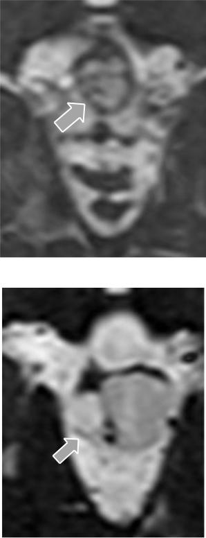
Additionally, individuals with PGC greater than 10 mm were examined using a protocol that also included an SSFP-PS in the sagittal plane in order to clarify the nature of structural changes in the pineal gland. The imaging benefits of the SSFP-PS are shown in Figure 10.
A B C
Figure 10 — MR images of the brain. Comparison of two-dimensional T2-PS in the sagittal plane (A) and three-dimensional SSFP-PS in the sagittal and axial planes
(B, C) in the same patient G., (OC №34748, 2021). In the image (A), the cyst appears to be unicameral, but in the 3D sequence (B, C), the PGC appears to be multicameral and large in size. The content of one chamber has a slightly reduced MR signal, suggesting increased protein content. This multicameral cyst can be considered atypical. The changes described are indicated by arrows
In 8 subjects, the original interpretation of 'solitary pineal gland cyst' changes was changed to multicystic changes. An additional was also assessed criterion for edge displacement in the case of a large cyst (Figure 11).
Рекомендовано к изучению сайтом МедУнивер - https://meduniver.com/
196
А
B
Figure 11 — MR images of the brain, SSFP-PS, axial plane. Variants of cystic transformation without edge displacement in patient L. (OC №32234, 2020) (A) and with edge displacement in patient A. (OC №32256, 2020) (B) (indicated by arrows)
197
Subsequently, based on the changes identified by the SSFP-PS, DWI-PS from the second phase of the dataset were taken data of 48 people to assess whether they had MR patterns of central venous hypertension and to assess the impact of the pineal gland structure on the surrounding structures.
Volunteers were conventionally divided into 3 groups of equal numbers based on cyst size: the first group without cysts, the second with small cysts (less than 10 mm) or small cystic degeneration and the third with a large cyst over 10 mm (Table 6).
In the group with large cysts, out of 16 people, 11 had category 4c, where 4 is a multicystic pineal gland with an increase in size and edge displacement.
5 people were assigned category 3c, where 3 is a multicystic pineal gland with an increase in size and no edge displacement.
In the group with small cysts, out of 16 people — 9 people were given category 1a, 6 people were given category 2a, 1 person was given category 4b.
Analysis of variance between the three groups revealed that the group of individuals with large cysts was significantly (p1,3 = 0.025) younger than the group without evidence of cysts, but not significantly different from those with small cysts. The mean age of patients with large cysts was the youngest at 30.81±10.8 years.
In addition, the mean thickness of the pineal gland in the group of individuals with a large cyst was 8.19±2.07, compared with individuals with a small cyst (4.55±1.03) and without a cyst (4.14±0.78), which was statistically significantly greater (p1,3 = 0.0004, p2,3 = 0.0004).
The same ratios were obtained when assessing the significance of the difference in the mean distance from the corpus callosum to the tectal plate in each group.
The mean value in the group with large cysts was 10.39±1.6, which was significantly greater (p1,3 =0.002, p2,3 =0.0004) compared with the other groups.
Рекомендовано к изучению сайтом МедУнивер - https://meduniver.com/
198
Table 6 - MRI-detected changes in patients with different PGC variants
Indicators |
Without |
With small |
With large |
F- |
p-value |
|
|
PGC |
PGC (less |
PGC (over |
criterion |
|
|
|
|
than |
10mm) |
|
|
|
|
|
10mm) |
|
|
|
|
|
|
|
|
|
|
|
Number of |
16 |
16 |
16 |
- |
|
- |
patients, |
|
|
|
|
|
|
people |
|
|
|
|
|
|
|
|
|
|
|
|
|
Average age, |
42,5±14,2 |
35,88±11, |
30,81±10,8 |
3,703 |
p1,3 |
= 0.025 |
years |
|
3 |
|
|
|
|
|
|
|
|
|
|
|
Average |
4,14±0,78 |
4,55±1,03 |
8,19±2,07 |
40,008 |
p1,3 = 0.0004 |
|
pineal gland |
|
|
|
|
p2,3 |
= 0.0004 |
thickness, |
|
|
|
|
|
|
mm |
|
|
|
|
|
|
|
|
|
|
|
|
|
Average |
8,4±1,4 |
7,96±1,7 |
10,39±1,6 |
11,130 |
p1,3 = 0.002 |
|
distance |
|
|
|
|
p2,3 |
= 0.0004 |
from the |
|
|
|
|
|
|
splenium of |
|
|
|
|
|
|
the corpus |
|
|
|
|
|
|
callosum to |
|
|
|
|
|
|
the tectal |
|
|
|
|
|
|
plate, mm |
|
|
|
|
|
|
|
|
|
|
|
|
|
Signs of |
0 |
0 |
8 |
|
p1,3 |
≤ 0.05; |
aqueduct |
|
|
|
|
p2,3 ≤ 0.05 |
|
stenosis, |
|
|
|
|
|
|
man |
|
|
|
|
|
|
|
|
|
|
|
|
|
Signs of |
0 |
0 |
1 |
|
p> 0,05 |
|
tectal plate |
|
|
|
|
|
|
compression, |
|
|
|
|
|
|
man |
|
|
|
|
|
|
|
|
|
|
|
|
|
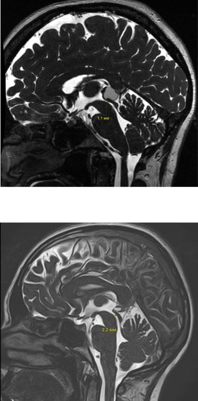
None of the patients showed evidence of occlusive hydrocephalus, but 8 had noncritical aqueduct stenosis (1.5 mm or less). In addition, patients with category 4 had the narrowest aqueduct lumen (1.1-1.2 mm) (Figure 12). One person showed signs of tectal plate compression (Figure 13).
199
А
B
Figure 12 - MR images of the brain, SSFP-PS, sagittal plane. Signs of aqueduct stenosis in the presence of a large cyst in patient A. (OC №32256, 2020) (A). Its width in the sagittal plane at its narrowest point is 1.1mm. For comparison, here is an image of patient B. (OC №33598, 2020) with PGC, who has a 2.2-mm wide ductus (B)
Рекомендовано к изучению сайтом МедУнивер - https://meduniver.com/
200
А
B
Figure 13 — MR images of the brain, SSFP-PS, sagittal plane. Signs of compression of the quadrigeminal plate in patient C. (OC №33768, 2020) (A). The lamina is thinned, flattened, and its upper contour is smoothed due to volumetric exposure. For comparison, there is an image of the PGC of patient L (OC №33786, 2020) without any signs of impact on the quadrigeminal plate. It has convex contours in the sagittal plane (B) (indicated by arrows)