


8THE CELL
Microtubules are also associated with proteins, known as microtubule-associated proteins (MAPs), which permit organelles, vesicles, and other components of the cytoskeleton to bind to microtubules.
•Most microtubules originate from the microtubuleorganizing center of the cell, located in the vicinity of the Golgi apparatus.
•These elements of the cytoskeleton are pathways for intracellular translocation of organelles and vesicles, and during cell division, chromosomes are moved into their proper locations.
•Two important MAPs, kinesin and dynein, are motor proteins that facilitate anterograde and retrograde intracellular vesicular and organelle movement, respectively.
•The axoneme of cilia and flagella, as well as a framework of centrioles, are formed mostly of microtubules.
Inclusions
Cytoplasmic inclusions, such as lipids, glycogen, secretory granules, and pigments, are also consistent constituents of the cytoplasm. Many of these inclusions are transitory in nature, although some pigments, for example, lipofuscin, are permanent residents of certain cells.
NUCLEUS
The nucleus is enclosed by the nuclear envelope, composed of an inner and an outer nuclear membrane with an intervening perinuclear cistern (see Graphic 1-2). The outer nuclear membrane is studded with ribosomes and is continuous, in places, with the rough endoplasmic reticulum. In areas the inner and outer membranes fuse with each other, forming circular profiles, known as
•nuclear pores that permit communication between the nucleoplasm and the cytoplasm.
•These perforations of the nuclear envelope are guarded by protein assemblies which, together with the perforations, are known as nuclear pore complexes, providing regulated passageways for the transport of materials in and out of the nucleus.
The nucleus houses chromosomes and is the location of
RNA synthesis.
•mRNA and tRNA, as well as microRNA, are transcribed in the nucleus,
•whereas rRNA is transcribed in the region of the nucleus known as the nucleolus.
The nucleolus is also the site of assembly of ribosomal proteins and rRNA into the small and large subunits of ribosomes.These ribosomal subunits enter the cytosol separately.
CELL CYCLE
The cell cycle is governed by the cell cycle control system that not only ensures the occurrence of the correct sequence of events in a timely fashion but also monitors and controls them. The cell cycle is subdivided into four phases, G1, S, G2, and M.
•During the presynthetic phase, G1, the cell increases its size and organelle content.
•During the S phase, DNA (plus histone and other chromosome-associated protein) synthesis and centriole replication occur.
•During G2,ATP is accumulated, centriole replication is completed, and tubulin is accumulated for spindle formation. G1, S, and G2 are also referred to as interphase.
•M represents mitosis, which is subdivided into prophase, prometaphase, metaphase, anaphase, and telophase (see Table 1-4). The result is the division of the cell and its genetic material into two identical daughter cells.
The sequence of events in the cell cycle is controlled by a number of trigger proteins, known as cyclin-dependent kinases and cyclins.

THE CELL |
9 |
TABLE 1-4 • Stages of Mitosis
Stage |
DNA Content |
Identifying Characteristics |
|
|
|
Prophase |
DNA content doubles in the |
|
S phase of interphase (4n); |
|
also, centrioles replicate. |
Nuclear envelope begins to disappear and the nucleolus disappears. Chromosomes have been replicated and each chromosome is
composed of two sister chromatids attached to each other at centromere.
Centrioles migrate to opposite poles where they act as microtubuleorganizing centers and give rise to spindle fibers and astral rays.
Prometaphase |
DNA complement is 4n. |
Nuclear envelope disappears. |
|
|
Kinetochores, additional microtubule-organizing centers, develop |
|
|
at centromeres and kinetochore microtubules form. |
|
|
|
Metaphase |
DNA complement is 4n. |
Chromosomes align at the equatorial plate of the mitotic spindle. |
|
|
|
Anaphase |
DNA complement is 4n. |
Sister chromatids separate at centromere and each chromatid |
|
|
migrates to an opposite pole of the cell along the microtubule, a |
|
|
process known as karyokinesis. |
|
|
In late anaphase, a cleavage furrow begins to form. |
|
|
|
Telophase |
Each new daughter |
Deepening of the cleavage furrow restricts the continuity between |
|
cell contains a single |
the two developing daughter cells forming the midbody. The |
|
complement of DNA (2n). |
two daughter cells separate from each other, a process known as |
|
|
cytokinesis. |
|
|
Nuclear envelope reforms, nucleoli reappear, and chromosomes |
|
|
disperse, forming new interphase nucleus in each daughter cell. |
|
|
|
CLINICAL CONSIDERATIONS
Lysosomal Storage Diseases
Certain individuals suffer from lysosomal storage diseases, which involve a hereditary deficiency in the ability of their lysosomes to degrade the contents of their endolysosomes. One of the best-characterized examples of these diseases is Tay-Sachs disease that occurs mostly in children whose parents are descendants of Northeast European Jews. Since the lysosomes of these children are unable to catabolize GM2 gangliosides, due to hexoaminidase deficiency, their neurons accumulate massive amounts of this ganglioside in endolysosomes of ever increasing diameters. As the endolysosomes increase in size, they obstruct neuronal function and the child dies by the third year of life.
Zellweger’s Disease
Zellweger’s disease is an inherited autosomal recessive disorder that interferes with normal peroxisomal biogenesis whose characteristics include renal cysts, hepatomegaly, jaundice, hypotonia of the muscular system, and cerebral demyelination resulting in psychomotor retardation.
Cancer
Recent studies have suggested that most cancers arise not from mutations in individual genes but from the
formation of aneuploidy. In fact, within the same tumor, the chromosomal configurations of individual cells vary greatly, and the DNA content of the cells may be 50% to 200% of the normal somatic cell. It is interesting to note that in the apparently chaotic reshuffling and recombination of chromosomes in cancer cells, there appears to be an order, as in Burkitt’s lymphoma, where chromosomes 3, 13, and 17 usually displayed translocations and chromosomes 7 and 20 were usually missing segments.
Hereditary Hemochromatosis
Excessive iron storage in hereditary hemochromatosis, untreated, can be a lethal disorder. The individual absorbs too much iron, which accumulates in the parenchymal cells of vital organs such as the liver, pancreas, and heart. Because it may affect organs in different sequence, the symptoms vary and diagnosis may be difficult. Testing the blood levels for high concentration of ferritin and transferrin can provide definitive diagnosis, which can be confirmed by genetic testing. Since this is a hereditary disorder, the close relatives of the positive individual should also undergo genetic testing.
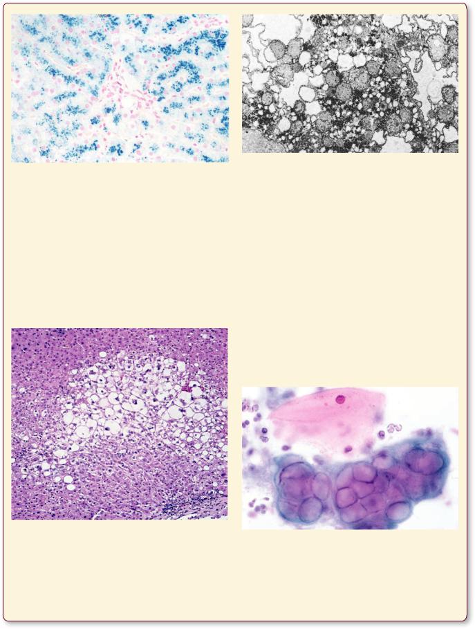
10 THE CELL
In the case of the liver, displayed in this photomicrograph of a Prussian blue-stained specimen, the lysosomes of hepatocytes are congested by large accumulations of iron (appearing as small, granular deposits). (Reprinted with permission from Rubin R, Strayer D, et al., eds. Rubin’s Pathology. Clinicopathologic Foundations of Medicine, 5th ed., Baltimore: Lippincott, Williams & Wilkins, 2008. p. 19.)
Hydropic Swelling
When cells become injured by coming into contact with toxins, are placed in areas of low or high temperature or low oxygen concentration, as well as being exposed to various inimical conditions, their cytoplasm swells and
This light photomicrograph of a liver of a patient with toxic hepatic injury displays hydropic swelling. Note that the affected cells are enlarged with accumulations of fluid, but the nuclei of most cells appear to be at their normal location. The cells at the periphery seem to be healthy. (Reprinted with permission from Rubin R, Strayer D, et al., eds. Rubin’s Pathology. Clinicopathologic Foundations of Medicine, 5th ed., Baltimore: Lippincott, Williams & Wilkins, 2008. p. 9.)
An electron micrograph of a liver with hydropic swelling displays enlarged cisternae of the endoplasmic reticulum that cause the liver cells to be swollen. (Reprinted with permission from Rubin R, Strayer D, et al., eds. Rubin’s Pathology. Clinicopathologic Foundations of Medicine, 5th ed., Baltimore: Lippincott, Williams & Wilkins, 2008. p. 9.)
takes on a pale appearance. This characteristic is usually reversible and is called hydropic swelling. Usually, the nuclei occupy their normal position, their organelle content remains unaltered, but the organelles are located farther away from each other, and viewed with the electron microscope, it is noted that the cisternae of their endoplasmic reticulum are dilated.
Genital Herpes Infection
One of the most common sexually transmitted diseases, herpes simplex virus (HSV-2, genital herpes) infection of the cervix (although HSV-1, usually associated with cold sores on the lips and, occasionally, the eyes, can also
Note the healthy epithelial cell with its pink cytoplasm with its healthy-appearing nucleus. The infected epithelial cells possess multiple nuclei with “ground glass” appearance and with peripherally located chromatin. (Reprinted with permission from Rubin R, Strayer, D, et al., eds. Rubin’s Pathology. Clinicopathologic Foundations of Medicine, 5th ed., Baltimore: Lippincott, Williams & Wilkins, 2008. p. 1268.)
THE CELL |
11 |
be a causative factor). Usually, infection by herpes simplex virus displays the presence of painful blisters that discharge a clear fluid, form a scab within a week or so, and disappear. During this episode, the genital area in females is painful and urination may be accompanied by a burning feeling. However, if the affected region is the cervix or the vagina, the pain may be much less severe.
When the blisters break, the fluid within them is filled with HSV and the individual is infectious. Subsequent to the outbreak of the blistering, the virus retreats, along nerve fibers, into the ganglion and remains there until the next episode. HSV infections cannot be cured, but the severity of the pain and the duration of the episode can be lessened by antiviral agents.
Cell The • 1-1 GRAPHIC
12 THE CELL
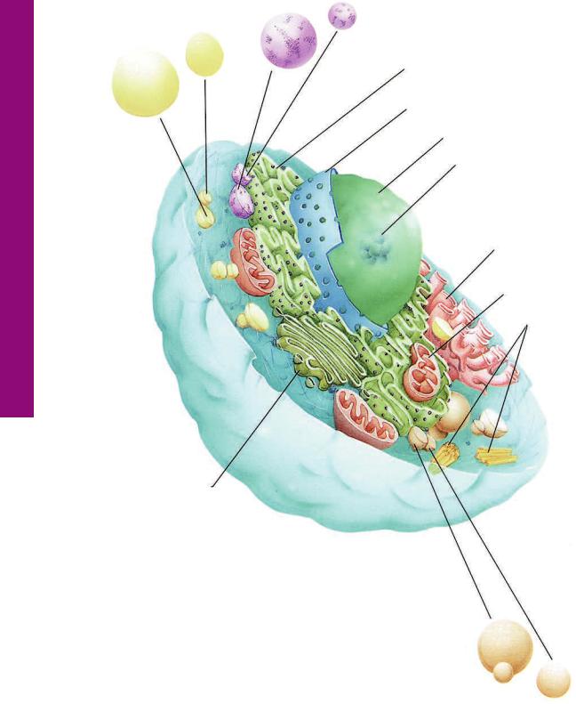
Lipid droplets
Golgi apparatus and trans-Golgi network
Lysosomes
Rough endoplasmic reticulum
Nuclear envelope
Nucleus
Nucleolus
Smooth endoplasmic reticulum
Mitochondrion
Centrioles
Secretory granules
Nuclear envelope is composed of inner and outer nuclear membranes
Rough endoplasmic reticulum is the site of synthesis of proteins that are to be packaged
Golgi-apparatus and the trans-Golgi network (TGN) function in posttranslational modification and packaging of proteins
THE CELL |
13 |
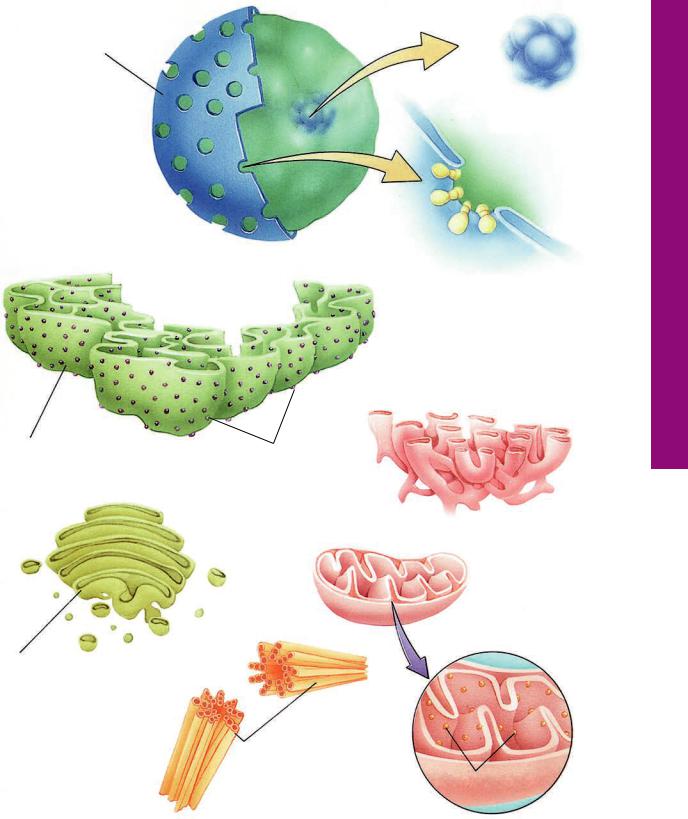
Nucleus
Nucleolus
(rRNA synthesis)
Nuclear pore complex
Smooth endoplasmic reticulum functions in synthesis of cholesterol-based lipids
Ribsomes
Mitochondria function in synthesis of ATP and certain lipids
Elementary particles couple oxidation to phosphorylation
Centrioles act as microtubule organizing centers
Elementary particles
Organelles The• 2-1 GRAPHIC
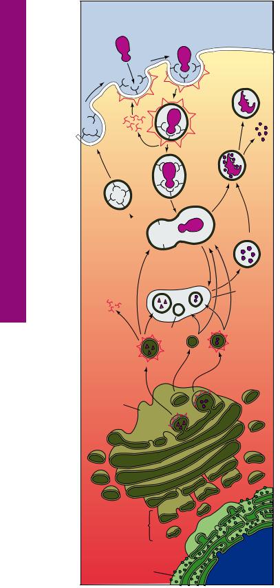
Trafficking Membrane and Membranes • 3-1 GRAPHIC
14 THE CELL
|
Ligand in |
|
Receptors |
solution |
|
|
|
|
for ligand |
|
|
|
|
Residual |
|
Coated |
body |
|
|
|
|
pit |
|
Clathrin |
|
|
|
Clathrin- |
|
Clathrin |
coated |
|
endocytoic |
|
|
triskelions |
|
|
vesicle |
|
|
recycle to plasma |
|
|
|
Degra- |
|
membrane |
|
|
|
|
dation |
|
Uncoated |
products |
|
|
|
|
endocytotic |
|
|
vesicle |
|
Recycling of 
 receptors to
  plasma membrane
Early endosome ph≈6.0
Late endosome ph≈5.5
Clathrin
Clathrin- |
|
coated |
Hydrolases |
vesicles |
|
containing |
|
lysosomal |
|
hydrolases or |
|
lysosomal |
|
membrane |
|
proteins |
|
trans-Golgi |
|
network |
|
(TGN) |
|
Golgi |
|
Vesiculartubular cluster (VTC)
Transitional Endoplasmic 
Reticulum element (TER)
Rough Endoplasmic
Reticulum (RER)
Lysosome
Lysosomal membrane
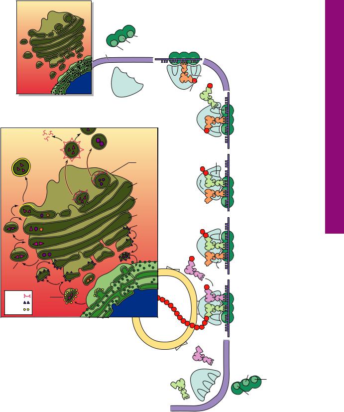
Signaling molecules bind to receptors (integral proteins) embedded in the cell membrane and initiate a specific sequence of responses. Receptors permit the endocytosis of a much greater concentration of ligands than would be otherwise possible. This process, receptor-mediated endocytosis, involves the formation of clathrin-coated endocytic vesicles. Once within the cell, the vesicle sheds its clathrin coat and fuses with an early endosome (pH 6) where the receptor is uncoupled from the ligand. The receptors are carried from the early endosome into a system of tubular vesicles, known as the recycling endosome, from which the receptors are returned to the cell membrane.
The ligand is transferred by the use of multivesicular bodies from the early endosome to another system
of vesicles, late endosomes, located deeper in the cytoplasm. Late endosomes are more acidic (pH 5.5) and it is here that the ligand begins to be degraded. Late endosomes receive lysosomal hydrolases and lysosomal membranes, and in that fashion late endosomes probably are transformed into lysosomes (pH 5.0). Hydrolytic enzymes of the lyosomes degrade the ligand, releasing the usable substances for utilization by the cell, whereas the indigestible remnants of the ligand may remain in vesicles, residual bodies, within the cytoplasm.
THE CELL |
15 |
Small |
As the mRNA enters the cytoplasm, it becomes associated |
||
with the small subunit of a ribosome. The small subunit has |
|||
ribosomal |
|||
a binding site for mRNA as well as three binding sites (A, P, |
|||
subunit |
|||
and E) for tRNAs. Once the initiation process is completed |
|||
|
|
||
|
|
and the start codon (AUG, for the amino acid methionine) is |
|
|
A site |
recognized, and the initiator tRNA (bearing methionine) is |
|
|
attached to the P site, the large subunit of the ribosome |
||
|
P site |
||
|
becomes attached, and protein synthesis may begin. |
||
mRNA |
E site |
|
|
|
|
tRNA |
|
|
|
Amino |
|
|
Large |
acid |
|
|
|
||
|
ribosomal |
The next codon is |
|
|
subunit |
||
|
recognized by the proper |
||
|
|
||
|
|
acylated tRNA, which then |
|
|
|
binds to the A site. |
|
|
Clathrin- |
Methionine is uncoupled |
|
|
coated |
from the initiator tRNA |
|
|
vesicle |
(at the P site), and a |
|
|
trans- |
peptide bond is formed |
|
|
Golgi |
between the two amino |
|
|
network |
acids, resulting in a |
|
|
trans |
dipeptide. |
|
|
face |
The initiator tRNA moves |
|
|
|
||
|
medial |
to the E site and the tRNA |
|
|
with the dipeptides moves |
||
|
face |
||
|
to the P site, leaving the A |
||
|
cis Golgi |
||
|
site empty. As the A site |
||
|
cis Golgi |
becomes occupied by a |
|
|
new amino acyl tRNA, the |
||
|
network |
||
|
initiator tRNA drops off the |
||
|
|
||
|
VTC |
E site and the mRNA |
|
|
move the distance of one |
||
|
|
||
|
TER |
codon (three nucleotides) |
|
|
and the new amino acyl |
||
|
|
tRNA’s amino acid forms a |
|
Non-clathrin- |
|
peptide bond with the |
|
|
dipeptide. The two tRNAs |
||
coated vesicle |
|
||
RER |
move to sites E and P, and |
||
(transport) |
|||
|
the cycle continues. |
||
|
|
||
Clathrin |
Newly |
After the signal |
|
recognition particle is |
|||
COP I |
synthesized |
||
bound to the completed |
|||
COP II |
protein |
||
signal protein, the entire |
|||
|
|
||
|
|
polysome docks on the |
|
The newly synthesized protein is modified in the |
RER membrane. A pore |
||
RER by glycosylation as well as by the formation of |
opens up in the RER |
||
disulfide bonds that transform the linear protein into |
membrane, so that the |
||
globular form. The proteins are transported to the |
forming protein chain can |
||
transitional ER (TER) elements from where they are |
enter the RER cisterna. |
||
delivered into the vesicular-tubular cluster (VTC) via COPII- |
Once protein synthesis is |
||
coated vesicles. The proteins are sent to the cis Golgi network |
|||
in COPI-coated vesicles for further processing. Phosphorylation |
completed, the two |
||
of proteins occurs within the cis face. Nonphosphorylated mannose |
P site |
||
groups are removed in the medial compartment. Final modification |
A site |
||
occurs in the trans face. Modified proteins are transported from the |
|
||
Golgi apparatus to the trans-Golgi network (TGN) for packaging |
E site |
||
and sorting. Lysosomal enzymes and regulated secretory proteins |
|||
leave the TGN in clathrin-coated vesicles. Membrane and |
ribosomal subunits fall off |
||
unregulated proteins are packaged in non-clathrin-coated vesicles. |
the RER and return to |
||
|
|
the cytosol. |
|
Exocytosis and Synthesis Protein• 4-1 GRAPHIC
Cell ypical • T1-1 PLATE
16 THE CELL
FIGURE 1. Cells. Monkey. Plastic section. ×1,323.
The typical cell is a membrane-bound structure that consists of a nucleus (N) and cytoplasm (C). Although the cell membrane is too thin to be visualized with the light microscope, the outline of the cell approximates the cell membrane (arrowheads). Observe that the outline of these particular cells more or less approximates a rectangle in shape. Viewed in three dimensions, these cells are said to be tall, cuboidal in shape, with a centrally placed nucleus. The nucleolus (n) is clearly evident, as are the chromatin granules (arrows) that are dispersed around the periphery as well as throughout the nucleoplasm.
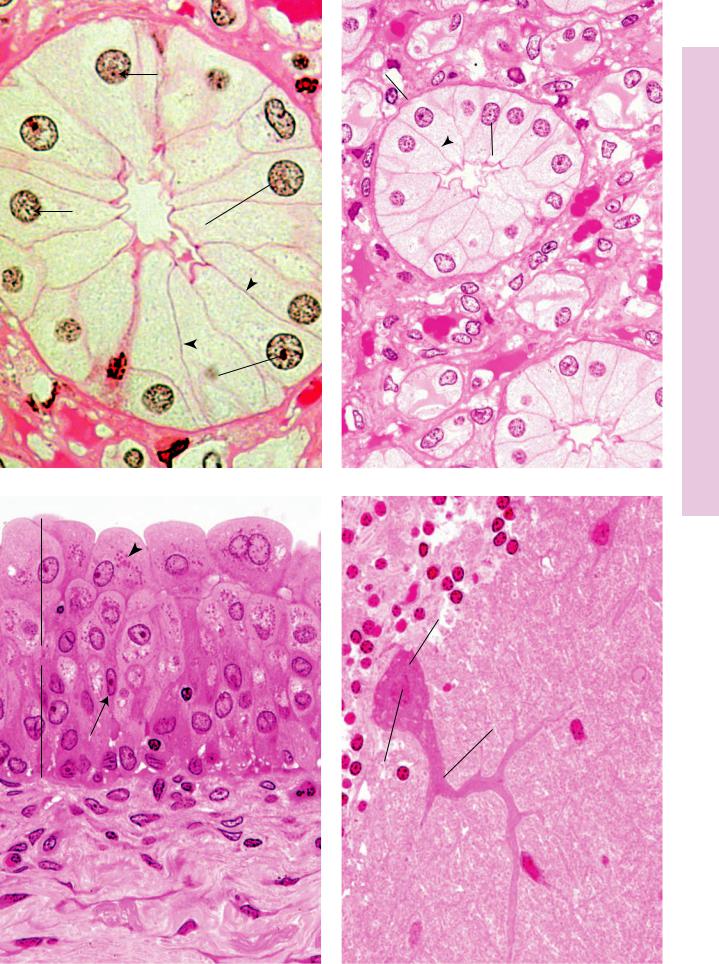
FIGURE 3. Cells. Monkey. Plastic section. ×540.
Cells come in a variety of sizes and shapes. Note that the epithelium (E) that lines the lumen of the bladder is composed of numerous layers. The surface-most layer consists of large, dome-shaped cells, some occasionally displaying two nuclei (N). The granules evident in the cytoplasm (arrowhead) are glycogen deposits. Cells deeper in the epithelium are elongated and narrow, and their nuclei (arrow) are located in their widest region
FIGURE 2. Cells. Monkey. Plastic section. ×540.
Cells may possess tall, thin morphologies, like those of a collecting duct of the kidney. Their nuclei (N) are located basally, and their lateral cell membranes (arrowheads) are outlined. Because these cells are epithelially derived, they are separated from connective tissue (CT) elements by a basal membrane (BM).
FIGURE 4. Cells. Monkey. Plastic section. ×540.
Some cells possess a rather unusual morphology, as exemplified by the Purkinje cell (PC) of the cerebellum. Note that the nucleus
(N) of the cell is housed in its widest portion, known as the soma (perikaryon). The cell possesses several cytoplasmic extensions, dendrites (De), and axon. This nerve cell integrates the numerous digits of information that it receives from other nerve cells that synapse on it.
Nucleus
Nucleolus
Cell
KEY
BM |
basal membrane |
De |
dendrite |
N |
nucleus |
C |
cytoplasm |
E |
epithelium |
n |
nucleolus |
CT |
connective tissue |
L |
lumen |
PC |
Purkinje cell |
BM
CT
C
N
N
CT
n
FIGURE 1 |
FIGURE 2 |
N
PC
E
De
N
Cell ypical • T1-1 PLATE
FIGURE 3 |
FIGURE 4 |
Inclusions and Organelles ell • C2-1 PLATE
18 THE CELL
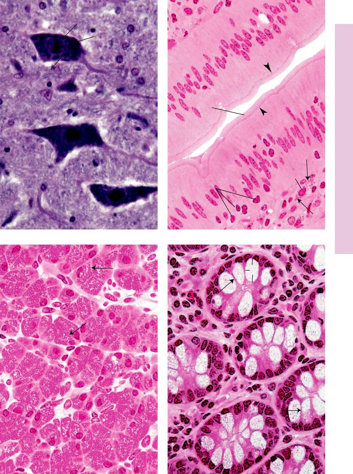
FIGURE 1. Nucleus and Nissl bodies. Spinal cord. Human. Paraffin section. ×540.
The motor neurons of the spinal cord are multipolar neurons because they possess numerous processes arising from an enlarged soma (S), which houses the nucleus (N) and various organelles. Observe that the nucleus displays a large, densely staining nucleolus (n). The cytoplasm also presents a series of densely staining structures known as Nissl bodies (NB), which have been demonstrated by electron microscopy to be RER. The staining intensity is due to the presence of ribonucleic acid of the ribosomes studding the surface of the RER.
FIGURE 3. Zymogen granules. Pancreas. Monkey. Plastic section. ×540.
The exocrine portion of the pancreas produces enzymes necessary for proper digestion of ingested food materials. These enzymes are stored by the pancreatic cells as zymogen granules (ZG) until their release is effected by hormonal activity. Note that the parenchymal cells are arranged in clusters known as acini (Ac), with a central lumen into which the secretory product is released. Observe that the zymogen granules are stored in the apical region of the cell, away from the basally located nucleus (N). Arrows indicate the lateral cell membranes of adjacent cells of an acinus.
FIGURE 2. Secretory products. Mast cell. Monkey. Plastic section. ×540.
The connective tissue (CT) subjacent to the epithelial lining of the small intestines is richly endowed with mast cells (MC). The granules (arrows) of mast cells are distributed throughout their cytoplasm and are released along the entire periphery of the cell. These small granules contain histamine and heparin, as well as additional substances. Note that the epithelial cells (EC) are tall and columnar in morphology and that leukocytes (Le) are migrating, via intercellular spaces, into the lumen (L) of the intestines. Arrowheads point to terminal bars, junctions between epithelial cells. The brush border (BB) has been demonstrated by electron microscopy to be microvilli.
FIGURE 4. Mucous secretory products. Goblet cells. Large intestines. Monkey. Plastic section. ×540.
The glands of the large intestine house goblet cells (GC), which manufacture a large amount of mucous material that acts as a lubricant for the movement of the compacted residue of digestion. Each goblet cell possesses an expanded apical portion, the theca (T), which contains the secretory product of the cell. The base of the cell is compressed and houses the nucleus (N) as well as the organelles necessary for the synthesis of the mucus— namely, the RER and the Golgi apparatus. Arrows indicate the lateral cell membranes of contiguous goblet cells.
Nucleus
Cell
KEY
AC |
acinus |
L |
lumen |
NB |
Nissl body |
BB |
brush border |
Le |
leukocyte |
S |
soma |
CT |
connective tissue |
MC |
mast cell |
T |
theca |
EC |
epithelial cell |
N |
nucleus |
ZG |
zymogen granule |
GC |
goblet cell |
n |
nucleolus |
|
|
S
n
NB
N
FIGURE 1
 ZG
ZG
Ac
N 
FIGURE 3
L
EC
BB 
EC
MC
Le
CT
FIGURE 2
T
 N
N
GC
FIGURE 4
Inclusions and Organelles ell • C2-1 PLATE
Modifications Surface ell • C3-1 PLATE
20 THE CELL
FIGURE 1. Brush border. Small intestines. Monkey. Plastic section. ×540.
The cells lining the lumen (L) of the small intestine are columnar cells, among which are numerous mucus-producing goblet cells (GC). The columnar cells’ function is absorbing digested food material along their free, apical surface. To increase their free surface area, the cells possess a brush border (BB), which has been demonstrated by electron microscopy to be microvilli—short, narrow, finger-like extensions of plasmalemma-covered cytoplasm. Each microvillus bears a glycocalyx cell coat, which also contains digestive enzymes. The core of the microvillus contains longitudinally arranged actin filaments as well as additional associated proteins.
FIGURE 3. Stereocilia. Epididymis. Monkey. Plastic section. ×540.
The lining of the epididymis is composed of tall, columnar principal cells (Pi) and short basal cells (BC). The principal cells bear long stereocilia (arrows) that protrude into the lumen. It was believed that stereocilia were long, nonmotile, cilia-like structures. However, studies with the electron microscope have shown that stereocilia are actually long microvilli that branch as well as clump with each other. The function, if any, of stereocilia within the epididymis is not known. The lumen is occupied by numerous spermatozoa, whose dark heads (asterisks) and pale flagella (arrowhead) are clearly discernible. Flagella are very long, cilia-like structures used by the cell for propulsion.
FIGURE 2. Cilia. Oviduct. Monkey. Plastic section. ×540.
The lining of the oviduct is composed of two types of epithelial cells: bleb-bearing peg cells (pc), which probably produce nutritional factors necessary for the survival of the gametes, and pale ciliated cells (CC). Cilia (arrows) are long, motile, finger-like extensions of the apical cell membrane and cytoplasm that transport material along the cell surface. The core of the cilium, as shown by electron microscopy, contains the axoneme, composed of microtubules arranged in a specific configuration of nine doublets surrounding a central pair of individual microtubules.
FIGURE 4. Intercellular bridges. Skin. Monkey. Plastic section. ×540.
The epidermis of thick skin is composed of several cell layers, one of which is the stratum spinosum shown in this photomicrograph. The cells of this layer possess short, stubby, finger-like extensions that interdigitate with those of contiguous cells. Before the advent of electron microscopy, these intercellular bridges (arrows) were believed to represent cytoplasmic continuities between neighboring cells; however, it is now known that these processes merely serve as regions of desmosome formation so that the cells may adhere to each other.
Cell
KEY
BB |
brush border |
GC |
goblet cell |
Pi |
principal cell |
BC |
basal cell |
L |
lumen |
|
|
CC |
ciliated cell |
pc |
peg cell |
|
|
L
GC
CC
pc
BB
L
FIGURE 1 |
FIGURE 2 |
Pi
BC
FIGURE 3 |
FIGURE 4 |
Modifications Surface ell • C3-1 PLATE

Microscopy Electron and Light osis, •Mit4-1 PLATE
22 THE CELL |
|
|
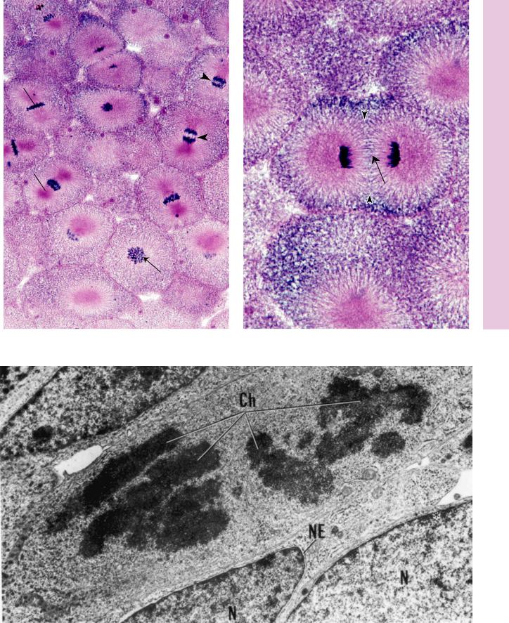
FIGURE 1. Mitosis. Whitefish blastula. Paraffin |
|
FIGURE 2. Mitosis. Whitefish blastula. Paraffin |
section. ×270. |
|
section. ×540. |
This photomicrograph of whitefish blastula shows different stages of mitosis. The first mitotic stage, prophase (P), displays the short, thread-like chromosomes (arrow) in the center of the cell. The nuclear membrane is no longer present. During metaphase (M), the chromosomes line up at the equatorial plane of the cell. The chromosomes begin to migrate toward the opposite poles of the cell in early anaphase (A) and proceed farther and farther apart as anaphase progresses (arrowheads). Note the dense regions, centrioles (c), toward which the chromosomes migrate.
FIGURE 3. Mitosis. Mouse. Electron microscopy. ×9423.
Neonatal tissue is characterized by mitotic activity, in which numerous cells are in the process of proliferation. Observe that the interphase nucleus (N) possesses a typical nuclear envelope (NE), perinuclear chromatin (asterisk), nucleolus, and nuclear pores. A cell that is undergoing the mitotic phase of the cell cycle loses its nuclear membrane and nucleolus, whereas its chromosomes (Ch) are quite visible. These chromosomes are no longer lined up at the equatorial plate but are migrating to opposite poles, indicating that this cell is in the early to mid-anaphase stage of mitosis. Observe the presence of cytoplasmic organelles, such as mitochondria, RER, and Golgi apparatus.
During the early telophase stage of mitotic division, the chromosomes (Ch) have reached the opposite poles of the cell. The cell membrane constricts to separate the cell into the two new daughter cells, forming a cleavage furrow (arrowheads). The spindle apparatus is visible as parallel, horizontal lines (arrow) that eventually form the midbody. As telophase progresses, the two new daughter cells will uncoil their chromosomes, and the nuclear membrane and nucleoli will become reestablished.
KEY
A |
anaphase |
M |
metaphase |
P |
prophase |
C |
centriole |
N |
nucleus |
|
|
Ch |
chromosome |
NE |
nuclear envelope |
|
|
THE CELL |
23 |
M
Ch Ch
c
c  A
A
P
Microscopy Electron and Light osis, •Mit4-1 PLATE
FIGURE 1 |
FIGURE 2 |
FIGURE 3
Microscopy Electron Cell, ypical • T5-1 PLATE
24 THE CELL
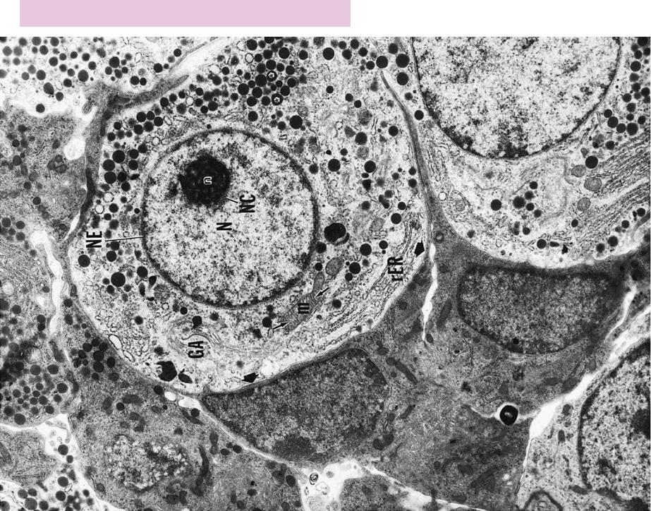
FIGURE 1. Typical cell. Pituitary. Rat. Electron microscopy. ×8936.
The gonadotrophs of the pituitary gland provide an excellent example of a typical cell because they house many of the cytoplasmic organelles possessed by most cells. The cytoplasm is limited by a cell membrane (arrowheads) that is clearly evident, especially where it approximates the plasmalemma of the adjacent electrondense cells. Mitochondria (m) are not numerous but are easily recognizable, especially in longitudinal sections, because their cristae (arrows) are arranged in a characteristic fashion. Because this cell actively manufactures a secretory product that must be packaged and delivered outside of the cell, it possesses a welldeveloped Golgi apparatus (GA), positioned near the nucleus
(N). Observe that the Golgi is formed by several stacks of flattened membranes. Additionally, this cell is well-endowed with rough endoplasmic reticulum, indicating active protein synthesis. The
cytoplasm also displays secretory products (asterisks), which are transitory inclusions.
The nucleus is bounded by the typical nuclear envelope (NE), consisting of a ribosome-studded outer nuclear membrane and an inner nuclear membrane. The peripheral chromatin and chromatin islands are clearly evident, as is the nucleolus-associated chromatin (NC). The clear area within the nucleus is the nucleoplasm representing the fluid component of the nucleus. The nucleolus (n) presents a sponge-like appearance composed of electron-lucent and electron-dense materials, suspended free in the nucleoplasm. The electron-dense region is composed of the pars granulosa and the pars fibrosa, whereas the electronlucent region is probably the nucleoplasm in which the nucleolus is suspended. (From Stokreef JC, Reifel CW, Shin SH. A possible phagocytic role for folliculo-stellate cells of anterior pituitary following estrogen withdrawal from primed male rats. Cell Tissue Res 1986;243:255–261.)
Nucleus
Nucleolus
Rough ER
Mitochondrion
Golgi apparatus
Cell
KEY
GA |
Golgi apparatus |
n |
nucleolus |
NE |
nuclear envelope |
m |
mitochondrion |
NC |
nucleolus-associated |
rER |
rough endoplasmic |
N |
nucleus |
|
chromatin |
|
reticulum |
•PLATE1-5 Typical Cell, Electron Microscopy
FIGURE 1
Microscopy Electron ytoplasm, C and Nucleus• 6-1 PLATE
26 THE CELL
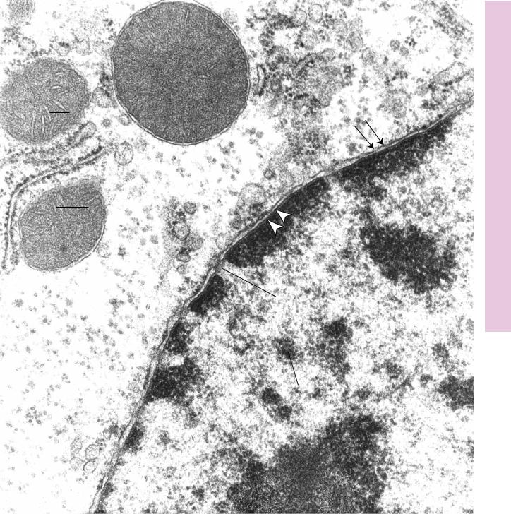
FIGURE 1. Nucleus and cytoplasm. Liver. Mouse. Electron microscopy. ×44,265.
The nucleus (N) displays its nucleoplasm and chromatin (c) to advantage in this electron micrograph. Note that the inner (arrowheads) and outer (double arrows) membranes of the nuclear
Rough endoplasmic reticulum
envelope fuse to form nuclear pores (NP). The RER is richly endowed by ribosomes (r). Note the presence of numerous mitochondria (m), whose double membrane and cristae (Cr) are quite evident.
Nuclear pore complex
THE CELL |
27 |
m
cr
cr
m
 r
r
NP
N
c
Microscopy Electron ytoplasm, C and Nucleus• 6-1 PLATE
FIGURE 1
Microscopy Electron ytoplasm, C and Nucleus• 7-1 PLATE
28 THE CELL
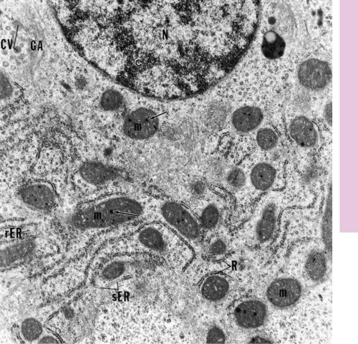
FIGURE 1. Nucleus and cytoplasm. Liver. Mouse. Electron microscopy. ×20,318.
This electron micrograph of a liver cell displays the nucleus (N), with its condensed chromatin (c), as well as many cytoplasmic organelles. Note that the mitochondria (m) possess
Golgi apparatus
electron-dense matrix granules (arrows) scattered in the matrix of the intercristal spaces. The perinuclear area presents the Golgi apparatus (GA), which is actively packaging material in condensing vesicles (CV). The rough endoplasmic reticulum is obvious due to its ribosomes (R), whereas the smooth endoplasmic reticulum is less obvious.
Mitochondrion
THE CELL |
29 |
Microscopy Electron ytoplasm, C and Nucleus• 7-1 PLATE
FIGURE 1

Microscopy Electron tus, Appara Golgi• 8-1 PLATE
30 THE CELL
FIGURE 1. Golgi apparatus. Mouse. Electron microscopy. ×28,588.
The extensive Golgi apparatus of this secretory cell presents several flattened membrane-bound cisternae (Ci), stacked one on top of the other. The convex face (cis face) (ff) receives transfer
Golgi apparatus
vesicles (TV) derived from the RER. The concave, trans-Golgi network (mf), releases condensing vesicles (CV), which house the secretory product. (From Gartner LP, Seibel W, Hiatt JL, et al. A fine-structural analysis of mouse molar odontoblast maturation. Acta Anat (Basel) 1979;103:16–33.)
Mitochondrion
THE CELL |
31 |
Microscopy Electron tus, Appara Golgi• 8-1 PLATE
FIGURE 1
Microscopy Electron ochondria, •Mit9-1 PLATE
32 THE CELL
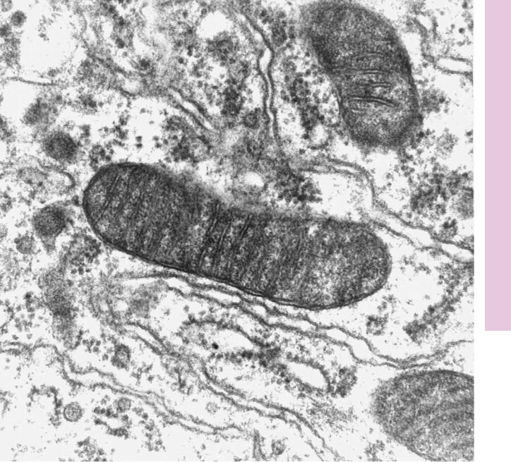
FIGURE 1. Mitochondria. Electron microscopy. ×69,500.
The basal aspect of this cell presents several mitochondria. The outer membrane of each mitochondrion is smooth, whereas its inner membrane is folded to form cristae (Cr) as is evident in the longitudinally sectioned mitochondrion.
Mitochondrion
THE CELL |
33 |
Microscopy Electron ochondria, •Mit9-1 PLATE
Cr
FIGURE 1