

compression and slowing down the flow of extracellular fluid, thus permitting more time for the exchange of materials by the cells and retarding the spread of invading microorganisms.
•Glycoproteins have also been localized in connective tissue proper. The best characterized are laminin, fibronectin, chondronectin, osteonectin, entactin, and tenascin.
Laminin and entactin are derived from epithelial cells, and tenascin is made by glial cells of the embryo, whereas the remainder are manufactured by cells of connective tissue.
Many cells possess integrins, transmembrane proteins, with receptor sites for one or more of these glycoproteins. Moreover, glycoproteins also bind to collagen, thus facilitating cell adherence to the extracellular matrix.
The basement membrane, interposed between epithelia and connective tissues, is described in Chapter 2, Epithelium and Glands.
Extracellular Fluid
Extracellular fluid (tissue fluid) is the fluid component of blood, similar to plasma, that percolates throughout the ground substance, carrying nutrients, oxygen, signaling molecules, and other blood-borne materials to and carbon dioxide and waste products from cells. Extracellular fluid leaves the vascular supply at the arterial end of the capillaries and returns into the circulatory system at the venous end of capillaries, the venules, and the excess fluid enters lymphatic capillaries.
CELLS
The following are cells of connective tissue proper—or more accurately, loose (areolar) connective tissue (see Graphic 3-2).
•Fibroblasts, the predominant cell type, are responsible for the synthesis of collagen, elastic and reticular fibers, and much, if not all, of the ground substance.
The morphology of these cells appears to be a function of their synthetic activities, and therefore, resting (or inactive fibroblasts) cells were often referred to as fibrocytes, a term that is rapidly disappearing from the literature.
•Macrophages (histiocytes) are derived from monocytes in bone marrow. They migrate to the connective tissue and function in ingesting (phagocytosing) foreign particulate matter. These cells also participate in enhancing the immunologic activities of lymphocytes.
C O N N E C T I V E T I S S U E 61
•Plasma cells are the major cell type present during chronic inflammation. These cells are derived from a subpopulation of lymphocytes and are responsible for the synthesis and release of humoral antibodies.
•Mast cells are usually observed in the vicinity of small blood vessels, although the relationship between them is not understood.
These cells house numerous metachromatic granules containing histamine, which is a smooth muscle contractant, and heparin, which is an anticoagulant.
Mast cells also release eosinophilic and neutrophilic chemotactic factors and leukotriene among their agents (see Table 3-2).
Because of the presence of immunoglobulins on the external surface of the mast cell plasmalemma, these cells, in sensitized individuals, may become degranulated (i.e., release their granules) and release membrane-derived factors, resulting in anaphylactic reactions or even in life-threatening anaphylactic shock.
•Pericytes are also associated with minute blood vessels, but much more closely than are mast cells, since they share the basal laminae of the endothelial cells.
Pericytes are believed to be contractile cells that assist in the regulation of blood flow through the capillaries.
Additionally, they may also be pluripotential cells, which assume the responsibilities of mesenchymal cells in adult connective tissue.
•Fat cells (adipocytes) may form small clusters or aggregates in loose connective tissue. They store lipids and form adipose tissue, which protects, insulates, and cushions organs of the body (see Adipose Tissue below).
•Leukocytes (white blood cells) leave the bloodstream and enter the connective tissue spaces. Here they assume various functions, which are discussed in Chapter 5.
CONNECTIVE TISSUE TYPES
•Mesenchymal and mucous connective tissues are limited to the embryo.
Mesenchymal connective tissue consists of mesenchymal cells and fine reticular fibers interspersed in a semifluid matrix of ground substance.
Mucous connective tissue is more viscous in consistency, contains collagen bundles and numerous fibroblasts, and is found deep to the fetal skin and in the umbilical cord (where it is known as Wharton’s jelly), surrounding the umbilical vessels.

62 C O N N E C T I V E T I S S U E
TABLE 3-2 • Mast Cells Factors and Functions
Substance |
Intracellular Source |
Action |
|
|
|
Primary mediators |
|
|
|
|
|
Histamine |
Granules |
Vasodilator; increases vascular permeability; causes |
|
|
contraction of bronchial smooth muscle; increases |
|
|
mucus production |
|
|
|
Heparin |
Granules |
Anticoagulant; inactivates histamine |
|
|
|
ECF |
Granules |
Attractant for eosinophils to site of inflammation |
|
|
|
NCF |
Granules |
Attractant for neutrophils to site of inflammation |
|
|
|
Aryl sulfate |
Granules |
Inactivates leukotriene C4, limiting inflammatory |
|
|
response |
|
|
|
Chondroitin sulfate |
Granules |
Binds and inactivates histamine |
|
|
|
Neutral proteases |
Granules |
Protein cleavage to activate complement; increases |
|
|
inflammatory response |
|
|
|
Secondary mediators |
|
|
|
|
|
Prostaglandin D2 |
Membrane lipid |
Causes contraction of bronchial smooth muscle; |
|
|
increases mucus secretion; vasoconstriction |
|
|
|
Leukotrienes C4, D4, E4 |
Membrane lipid |
Vasodilators; increases vascular permeability; |
|
|
contraction of bronchial smooth muscle |
|
|
|
Bradykinins |
Membrane lipid |
Causes vascular permeability; responsible for pain |
|
|
sensation |
|
|
|
Thromboxane A2 |
Membrane lipid |
Causes platelet aggregation; vasoconstriction |
Platelet-activating factor |
Activated by phospholipase A2 |
Attracts neutrophils and eosinophils; causes vascular |
|
|
permeability; contraction of bronchial smooth |
|
|
muscle |
ECF, eosinophil chemotactic factor; NCF, neutrophil chemotactic factor.
•Loose (areolar) connective tissue is distributed widely, since it constitutes much of the superficial fascia and invests neurovascular bundles. The cells and intercellular elements described above help form this more or less amorphous, watery tissue.
•Reticular connective tissue forms a network of thin reticular fi bers that constitute the structural framework of bone marrow and many lymphoid structures, as well as a framework enveloping certain cells.
•Adipose tissue is composed of fat cells, reticular fibers, and a rich vascular supply. There are two types of adipose tissue, white (unilocular) and brown (multilocular).
Unilocular adipose tissue is composed of fat cells, reticular fibers, and a rich vascular supply.
Cells of unilocular adipose tissue store triglycerides in a single, large fat droplet that occupies most of the cell. These cells make the enzyme lipoprotein lipase, which is transported to the luminal surface of the capillary endothelial cell membrane, where it hydrolyzes chylomicrons and very low density lipoproteins.
The fatty acids and monoglycerides are transported to the adipocytes, diffuse into their cytoplasm, and are reesterified into triglycerides.
Hormone-sensitive lipase, activated by cAMP, hydrolyzes the stored lipids into fatty acids and glycerol, which are released from the cell as the
C O N N E C T I V E T I S S U E 63
need arises, to enter the capillaries for distribution to the remainder of the body.
Unilocular adipose tissue acts as a depot for fat, a thermal insulator, and a shock absorber.
Multilocular adipose tissue is rare in the adult human. It is present in the neonate, as well as in animals that hibernate.
Cells of multilocular adipose tissue possess numerous droplets of lipid in their cytoplasm and a rich supply of mitochondria.
These mitochondria are capable of uncoupling oxidation from phosphorylation, and instead of producing adenosine triphosphate (ATP), they release heat, thus arousing the animal from hibernation.
•Dense irregular connective tissue consists of coarse, almost haphazardly arranged bundles of collagen fibers interlaced with few elastic and reticular fibers. The chief cellular constituents are fibroblasts, macrophages, and occasional mast cells. The dermis of the skin and capsules of some organs are composed of dense irregular connective tissue.
•Dense regular connective tissue may be composed either of thick, parallel arrays of collagenous fibers, as in tendons and ligaments, or of parallel bundles of elastic fibers, as in the ligamentum nuchae, the ligamentum flava, and the suspensory ligament of the penis. The cellular constituents of both dense regular collagenous and dense regular elastic connective tissues are almost strictly limited to fibroblasts.
64 C O N N E C T I V E T I S S U E
CLINICAL CONSIDERATIONS
Keloid Formation
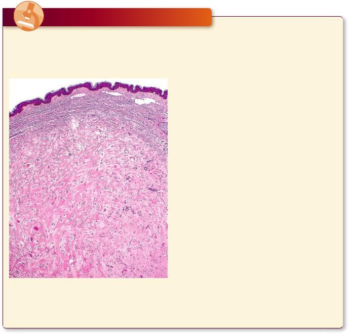
The body responds to wounds, including those caused by surgical intervention, by forming scars that repair the damage first with weak type III collagen that is later
Keloid formation at the site of injury is evidenced by the excessively thick layer of the dermis whose large, eosinophilic, type I collagen fibers are clearly apparent. (Reprinted with permission from Mills SE, Carter D, Greenson JK, Reuter VE, Stoler MH, eds. Sternberger’s Diagnostic Surgical Pathology, 5th ed., 2010. Figure 1.54. p. 29.)
replaced by type I collagen, which is much stronger. Some individuals, especially African Americans, form an overabundance of collagen in the healing process, thus developing elevated scars called keloids. The collagen fibers in keloids are much larger, more eosinophilic— said to have a “glassy” appearance—than the normal, fibrillar, collagen. Moreover, keloids are hypocellular, although they frequently display clusters of fibroblasts distributed among the large, glassy collagen fiber bundles.
Scurvy
Scurvy, a condition characterized by bleeding gums and loose teeth among other symptoms, results from a vitamin C deficiency. Vitamin C is necessary for hydroxylation of proline for proper tropocollagen formation giving rise to fibrils necessary for maintaining teeth in their bony sockets.
Marfan’s Syndrome
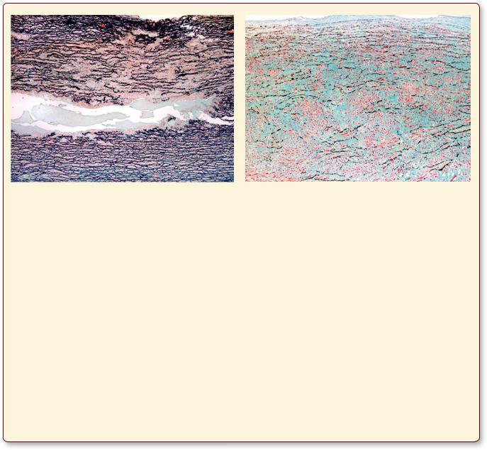
Patients with Marfan’s syndrome, a genetic defect in chromosome 15 that codes for fibrillin, possess undeveloped elastic fibers in their body and are predisposed to rupture of the aorta. Histologically, the aortas of a large portion of individuals with Marfan’s syndrome display cystic medial degeneration, a condition where the fenestrated membranes as well as the smooth muscles of the tunica media are reduced in quantity or are partially absent. In individuals with a less severe condition of cystic medial degeneration, the fenestrated membranes are less well organized, the smooth muscle cells are fewer in number, and the connective tissue is richer in ground substance than in normal aortas.
C O N N E C T I V E T I S S U E 65
A: Cystic medial degeneration, evident in the media of this aorta from a patient exhibiting Marfan’s syndrome, displays that the fenestrated membrane and smooth muscle cells have been replaced by amorphous ground substance. B: A less severe case of cystic medial degeneration is displayed in this patient. The tunica media evidences disorganized fenestrated membranes and smooth muscle fibers as well as an increase in the amorphous ground substance. (Reprinted with permission from Mills SE, Carter D, Greenson JK, Reuter VE, Stoler MH, eds. Sternberger’s Diagnostic Surgical Pathology, 5th ed. Philadelphia: Lippincott Williams & Wilkins, 2010. Figures 30.1A and B. P. 1228)
Edema |
Systemic Lupus Erythematosus |
The release of histamine and leukotrienes from mast cells during an inflammatory response elicits increased capillary permeability, resulting in an excess accumulation of extracellular fluid and, thus, gross swelling (edema).
Obesity
There are two types of obesity—hypertrophic obesity, which occurs when adipose cells increase in size from storing fat (adult onset), and hyperplastic obesity, which is characterized by an increase in the number of adipose cells resulting from overfeeding a new-born for a few weeks after birth. This type of obesity is usually lifelong.
Systemic lupus erythematosus is an autoimmune connective tissue disease that results in the inflammation in the connective tissue elements of certain organs as well as of tendons and joints. The symptoms depend on the type and number of antibodies present and can be anywhere from mild to severe and, due to the variety of symptoms, lupus may resemble other conditions such as growing pains, arthritis, epilepsy, and even psychologic diseases. The characteristic symptoms include facial and skin rash, sores in the oral cavity, joint pains and inflammation, kidney malfunction, neurologic conditions, anemia, thrombocytopenia, and fluid on the lungs.
66 C O N N E C T I V E T I S S U E
Collagen • 1-3 GRAPHIC
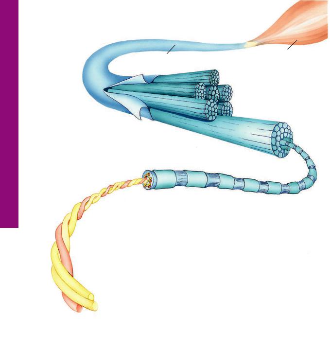
Muscle
Tendon
Bundle
Fiber
Fibril
Each collagen fiber bundle is composed of smaller fibrils, which in turn consist of aggregates of tropocollagen molecules. Tropocollagen molecules self-assemble in the extracellular environment in such a fashion that there is a gap between the tail of the one and the head of the succeeding molecule of a single row. As fibrils are formed, tails of tropocollagen molecules overlap the heads of tropocollagen molecules in adjacent rows. Additionally, the gaps and overlaps are arranged so that they are in register with those of neighboring (but not adjacent) rows of tropocollagen molecules. When stained with a heavy metal, such as osmium, the stain preferentially precipitates in the gap regions, resulting in the repeating light and dark banding of collagen.
C O N N E C T I V E T I S S U E 67
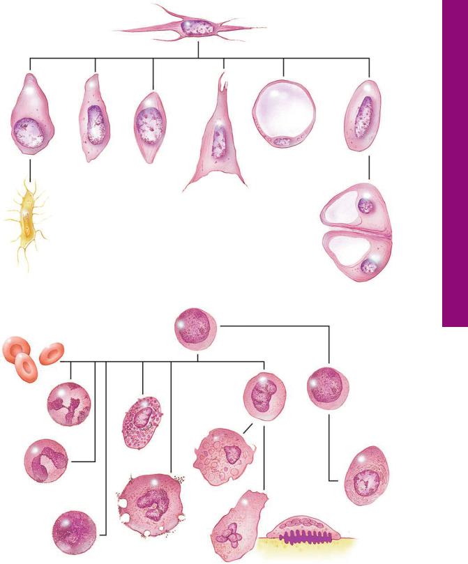
Undifferentiated mesenchymal cell
Osteoblast |
|
Adipocyte |
Chondroblast |
Endothelial |
|
|
|
|
Mesothelial |
|
|
|
cell |
|
|
|
cell |
|
|
|
|
|
|
|
|
Fibroblast |
|
Osteocyte |
|
Chondrocytes |
|
Hematopoietic stem cell
Red blood |
|
cells |
Monocyte |
B Lymphocyte
Neutrophil
Mast cell
Eosinophil |
Macrophage |
Plama cell
|
Megakaryocyte |
Basophil |
Osteoclast |
|
* The cells on this page are not represented in proportion to their actual diameters.
Cells Tissue Connective• 2-3 GRAPHIC
I Proper Tissue Connective and yonic Embr• 1-3 PLATE
68 C O N N E C T I V E T I S S U E
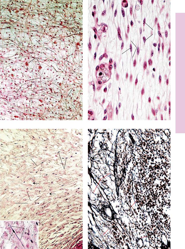
FIGURE 1. Loose (areolar) connective tissue. Paraffin section. ×132.
This photomicrograph depicts a whole mount of mesentery, through its entire thickness. The two large mast cells (MC) are easily identified, since they are the largest cells in the field and possess a granular cytoplasm. Although their cytoplasms are not visible, it is still possible to recognize two other cell types due to their nuclear morphology. Fibroblasts (F) possess oval nuclei that are paler and larger than the nuclei of macrophages
(M). The semifluid ground substance (GS) through which tissue fluid percolates is invisible, since it was extracted during the preparation of the tissues. However, two types of fibers, the thicker, wavy, ribbon-like, interlacing collagen fibers (CF) and the thin, straight, branching elastic fibers (EF), are well demonstrated.
FIGURE 3. Mucous connective tissue. Umbilical cord. Human. Paraffin section. ×132.
This example of mucous connective tissue (Wharton’s jelly) was derived from the umbilical cord of a fetus. Observe the obvious differences between the two embryonic tissues. The matrix of mesenchymal connective tissue (Fig. 2) contains no collagenous fibers, whereas this connective tissue displays a loose network of haphazardly arranged collagen fibers (CF). The cells are no longer mesenchymal cells; instead, they are fibroblasts (F), although morphologically they resemble each other. The emptylooking spaces (arrows) are areas where the ground substance was extracted during specimen preparation. Inset. Fibroblast. Umbilical cord. Human. Paraffin section. ×270. Note the centrally placed nucleus (N) and the fusiform shape of the cytoplasm
(c) of this fibroblast.
FIGURE 2. Mesenchymal connective tissue. Fetal pig. Paraffin section. ×540.
Mesenchymal connective tissue of the fetus is very immature and cellular. The mesenchymal cells (MeC) are stellate-shaped to fusiform cells, whose cytoplasm (c) can be distinguished from the surrounding matrix. The nuclei (N) are pale and centrally located. The ground substance is semifluid in consistency and contains slender reticular fibers. The vascularity of this tissue is evidenced by the presence of blood vessels (BV).
FIGURE 4. Reticular connective tissue. Silver stain. Paraffin section. ×270.
Silver stain, used in the preparation of this specimen, was deposited on the carbohydrate coating of the reticular fibers (RF). Note that these fibers are thin, long, branching structures that ramify throughout the field. Note that in this photomicrograph of a lymph node, the reticular fibers in the lower right-hand corner are oriented in a circular fashion. These form the structural framework of a cortical lymphatic nodule (LN). The small round cells are probably lymphoid cells (LC), whereas the larger cells, closely associated with the reticular fibers, may be reticular cells (RC), although definite identification is not possible with this stain. It should be noted that reticular connective tissue is characteristically associated with lymphatic tissue.
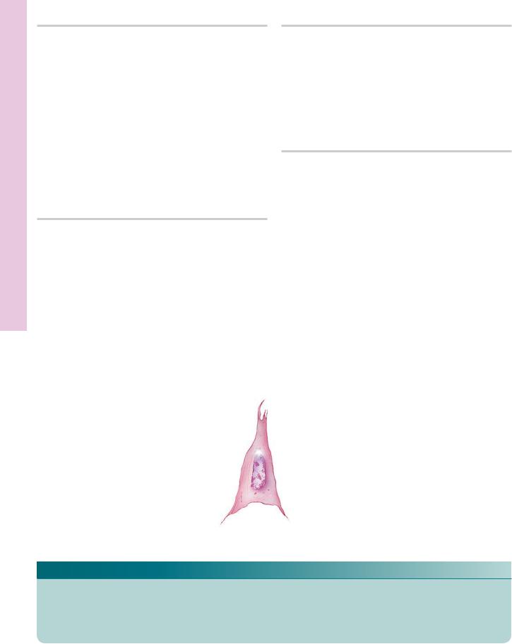
Fibroblast
KEY
BV |
blood vessel |
GS |
ground substance |
MeC |
mesenchymal cell |
C |
cytoplasm |
LC |
lymphoid cell |
N |
nucleus |
CF |
collagen fiber |
LN |
lymphatic nodule |
RC |
reticular cell |
EF |
elastic fiber |
M |
macrophage |
RF |
reticular fiber |
F |
fibroblast |
MC |
mast cell |
|
|
F
N
GS |
|
C |
MC |
F |
MeC |
|
||
|
|
|
M |
|
BV |
|
|
EF
CF
FIGURE 1 |
FIGURE 2 |
F
F
RF
CF
LN
C
LC |
RC |
N |
|
FIGURE 3 |
FIGURE 4 |
I Proper Tissue Connective and yonic Embr• 1-3 PLATE
II Proper Tissue onnective • C2-3 PLATE
70 C O N N E C T I V E T I S S U E
FIGURE 1. Adipose tissue. Hypodermis. Monkey. Plastic section. ×132.
This photomicrograph of adipose tissue is from monkey hypodermis. The adipocytes (A), or fat cells, appear empty due to tissue processing that dissolves fatty material. The cytoplasm (c) of these cells appears as a peripheral rim, and the nucleus (N) is also pressed to the side by the single, large fat droplet (FD) within the cytoplasm. Fat is subdivided into lobules by septa (S) of connective tissue conducting vascular elements (BV) to the adipocytes. Fibroblast nuclei (arrows) are clearly evident in the connective tissue septa. Note the presence of the secretory portions of a sweat gland (SG) in the upper aspect of this photomicrograph.
FIGURE 3. Dense regular collagenous connective tissue. l.s. Tendon. Monkey. Plastic section. ×270.
Tendons and ligaments present the most vivid examples of dense regular collagenous connective tissue. This connective tissue type is composed of regularly oriented parallel bundles of collagen fibers (CF), where individual bundles are demarcated by parallel rows of fibroblasts (F). Nuclei of these cells are clearly evident as thin, dark lines, whereas their cytoplasm (c) is only somewhat discernible. With hematoxylin and eosin, the collagen bundles stain a more or less light shade of pink with parallel rows of dark blue nuclei of fibroblasts interspersed among them.
FIGURE 2. Dense irregular collagenous connective tissue. Palmar skin. Monkey. Plastic section. ×132.
The dermis of the skin provides a good representation of dense irregular collagenous connective tissue. The thick, coarse, intertwined bundles of collagen fibers (CF) are arranged in a haphazard fashion. Although this tissue has numerous blood vessels (BV) and nerve fibers (NF) branching through it, it is not a very vascular tissue. Dense irregular connective tissue is only sparsely supplied with cells, mostly fibroblasts and macrophages, whose nuclei (N) appear as dark dots scattered throughout the field. At this magnification, it is not possible to identify the cell types with any degree of accuracy. The large epithelial structure in the upper center of the field is the duct (d) of a sweat gland. At higher magnification (Inset, ×540), the coarse bundles of collagen fibers are composed of a conglomeration of collagen fibrils (Cf) intertwined around each other. The three cells, whose nuclei (N) are clearly evident, cannot be identified with any degree of certainty, even though the cytoplasm (c) of the two on the left-hand side is visible. It is possible that they are macrophages, but without employing special staining techniques, the possibility of their being fibroblasts cannot be ruled out.
FIGURE 4. Dense regular collagenous connective tissue. x.s. Tendon. Paraffin section. ×270.
Transverse sections of tendon present a typical appearance. Tendon is organized into fascicles that are separated from each other by the peritendineum (P) surrounding each fascicle. Blood vessels (BV) may be observed in the peritendineum. Collagen bundles within the fascicles are regularly arranged; however, shrinkage due to preparation causes an artifactual layering (arrows), although in some preparations swelling of the tissue results in a homogenous appearance. The nuclei of fibroblasts (F) appear to be strewn about in a haphazard manner.
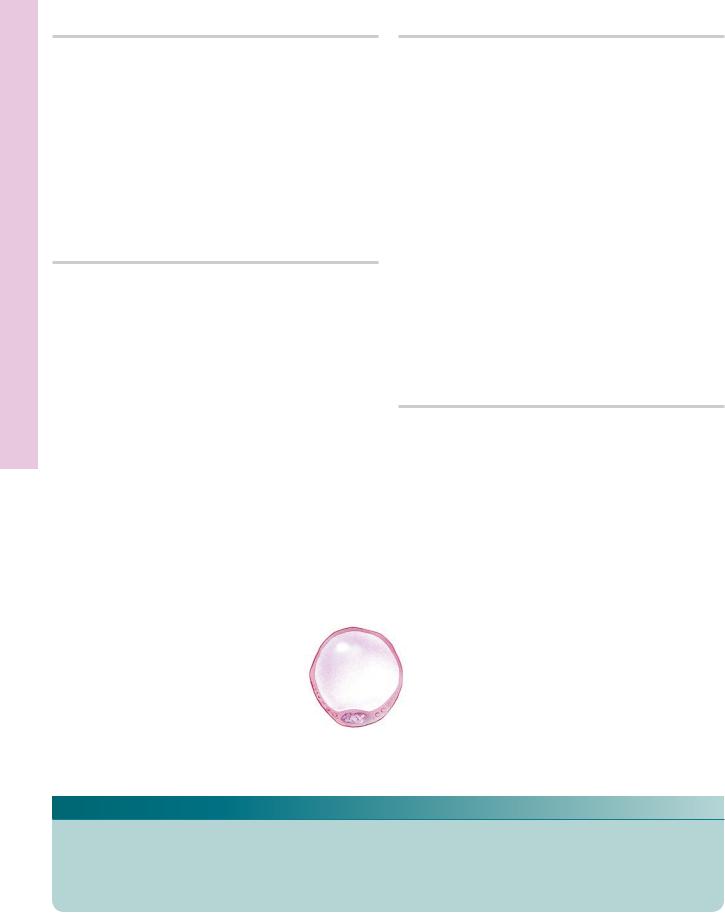
Adipocyte
KEY
A |
adipocyte |
D |
duct |
P |
peritendineum |
BV |
blood vessel |
F |
fibroblast |
S |
septum |
C |
cytoplasm |
FD |
fat droplet |
SG |
sweat gland |
CF |
collagen fibril |
N |
nucleus |
|
|
CF |
bundle of collagen fibers |
NF |
nerve fiber |
|
|
S N
c
FD
FD BV
S
A SG
FIGURE 1
C
F
 NF
NF
CF
 N
N
d
BV
C
N
Cf
FIGURE 2
 F
F
P
CF
BV
FIGURE 3 |
FIGURE 4 |
II Proper Tissue onnective • C2-3 PLATE
III Proper Tissue onnective • C3-3 PLATE
72 C O N N E C T I V E T I S S U E
FIGURE 1. Dense regular elastic connective tissue. l.s. Paraffin section. ×132.
This longitudinal section of dense regular elastic tissue demonstrates that the elastic fibers (EF) are arranged in parallel arrays. However, the fibers are short and are curled at their ends (arrows). The white spaces among the fibers represent the loose connective tissue elements that remain unstained. The cellular elements are composed of parallel rows of flattened fibroblasts. These cells are also unstained and cannot be distinguished in this preparation.
FIGURE 3. Elastic laminae (membranes). Aorta. Paraffin section. ×132.
The wall of the aorta is composed of thick, concentrically arranged elastic membranes (EM). Since these sheet-like membranes wrap around within the wall of the aorta, in transverse sections they present discontinuous, concentric circles, which in this photomicrograph are represented by more or less parallel, wavy, dark lines (arrows). The connective tissue material between membranes is composed of ground substance, collagen fibers (CF), and reticular fibers. Also present are fibroblasts and smooth muscle cells, whose nuclei may be discerned.
FIGURE 2. Dense regular elastic connective tissue. x.s. Paraffin section. ×132.
A transverse section of dense regular elastic connective tissue displays a characteristic appearance. In some areas the fibers present precise cross-sectional profiles as dark dots of various diameters (arrows). Other areas present oblique sections of these fibers, represented by short linear profiles (arrowhead). As in the previous figure, the white spaces represent the unstained loose connective tissue elements. The large clear area (middle left) is also composed of loose connective tissue surrounding blood vessels (BV).
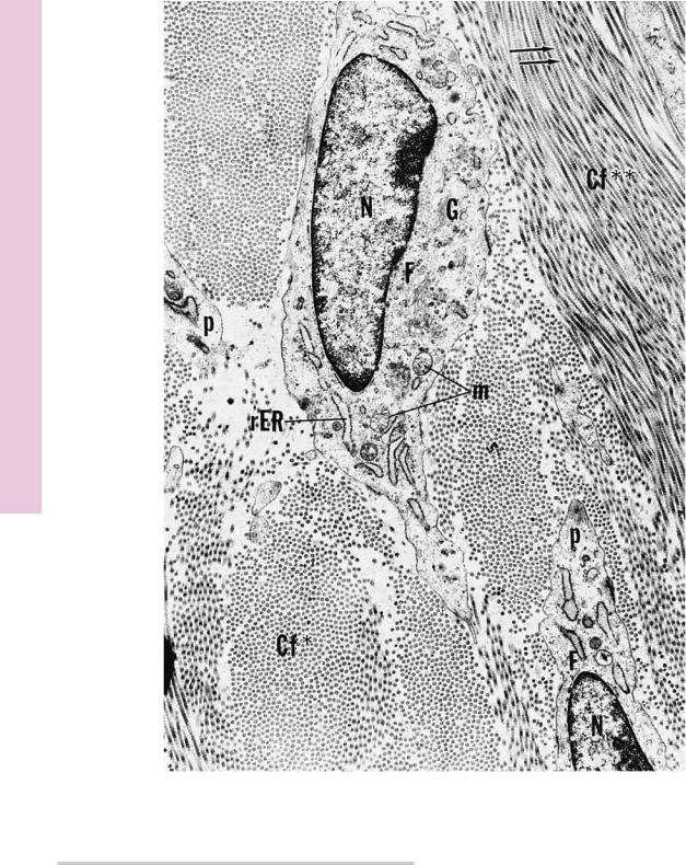
FIGURE 4. Mast cells, plasma cells, macrophages.
Mast cells (MC) are conspicuous components of connective tissue proper, Figure 4a (Tendon. Monkey. Plastic section. ×540), although they are only infrequently encountered. Note the round to oval nucleus and numerous small granules in the cytoplasm. Observe also, among the bundles of collagen fibers (CF), the nuclei of several fibroblasts. Mast cells are very common components of the subepithelial connective tissue (lamina propria) of the digestive tract, Figure 4b (Jejunum. Monkey. Plastic section. ×540). Note the basal membrane (BM) separating the connective tissue from the simple columnar epithelium (E), whose nuclei are oval in shape. The denser, more amorphous nuclei (arrows) belong to lymphoid cells, migrating from the connective tissue into the intestinal lumen. The lamina propria also houses numerous plasma cells (PC), as evidenced in Figure 4c (Jejunum. Monkey. Plastic section. ×540). Plasma cells are characterized by clock face (“cartwheel”) nuclei, as well as by a clear paranuclear Golgi zone (arrowhead). Figure 4d (Macrophage. Liver, injected. Paraffin section. ×270) is a photomicrograph of liver that was injected with india ink. This material is preferentially phagocytosed by macrophages of the liver, known as Kupffer cells (KC). These cells appear as dense, black structures in the liver sinusoids; vascular channels are represented by clear areas (arrow). An individual Kupffer cell (Inset. Paraffin section. ×540) displays the nucleus (N) as well as the granules of india ink (arrowhead) in its cytoplasm.
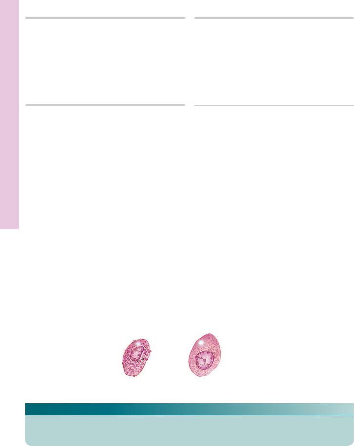
Mast cell |
Plasma cell |
|
KEY
BM |
basal membrane |
EF |
elastic fiber |
KC |
Kupffer cell |
BV |
blood vessel |
EM |
elastic membrane |
N |
nucleus |
CF |
collagen fiber |
MC |
mast cell |
PC |
plasma cell |
EF
BV
EF
FIGURE 1 |
FIGURE 2 |
b) |
E |
MC BM
BM
EM
CF
FIGURE 3 |
FIGURE 4 |
III Proper Tissue Connective• 3-3 PLATE
74 C O N N E C T I V E T I S S U E
Microscopy Electron Collagen, and ibroblasts • F4-3 PLATE
FIGURE 1
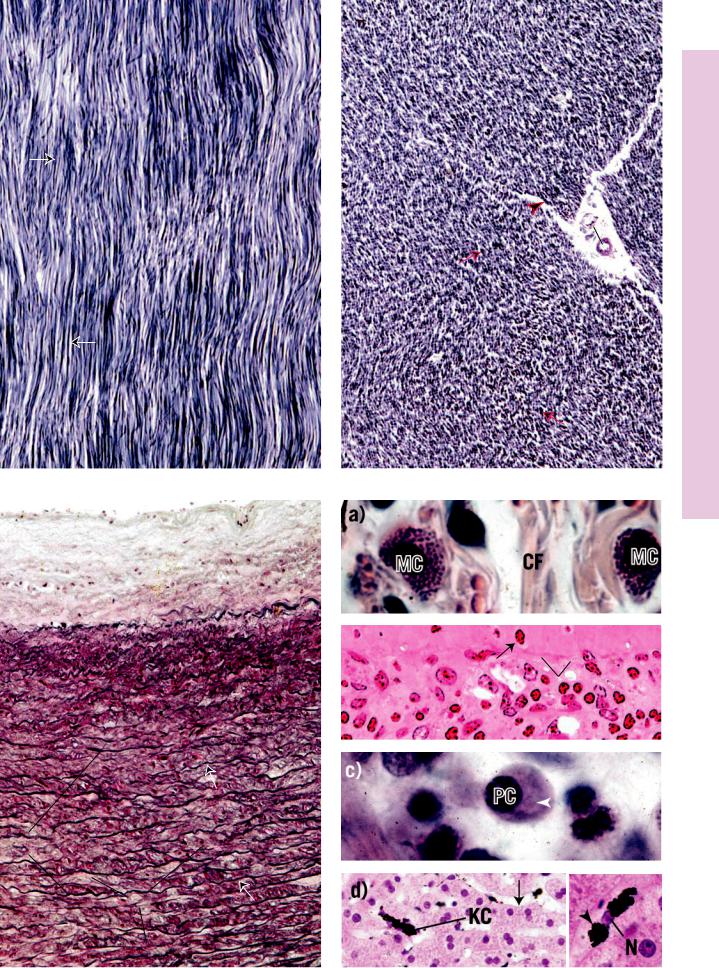
FIGURE 1. Fibroblast. Baboon. Electron microscopy. ×11,070.
This electron micrograph of fibroblasts (F) demonstrates that they are long, fusiform cells whose processes (p) extend into the surrounding area, between bundles of collagen fibrils. These cells manufacture collagen, reticular and elastic fibers, and the ground substance of connective tissue. Therefore, they are rich in organelles, such as Golgi apparatus (G), rough endoplasmic reticulum
(rER), and mitochondria (m); however, in the quiescent stage, as in
tendons, where they no longer actively synthesize the intercellular elements of connective tissue, the organelle population of fibroblasts is reduced in number, and the plump, euchromatic nucleus
(N) becomes flattened and heterochromatic. Note that the bundles of collagen fibrils (Cf) are sectioned both transversely (asterisk) and longitudinally (double asterisks). Individual fibrils display alternating transverse dark and light banding (arrows) along their length. The specific banding results from the ordered arrangement of the tropocollagen molecules constituting the collagen fibrils. (From Simpson D, Avery B. J Periodontol 1974;45:500–510.)
C O N N E C T I V E T I S S U E 75
Microscopy Electron ell, C Mast• 5-3 PLATE
FIGURE 1
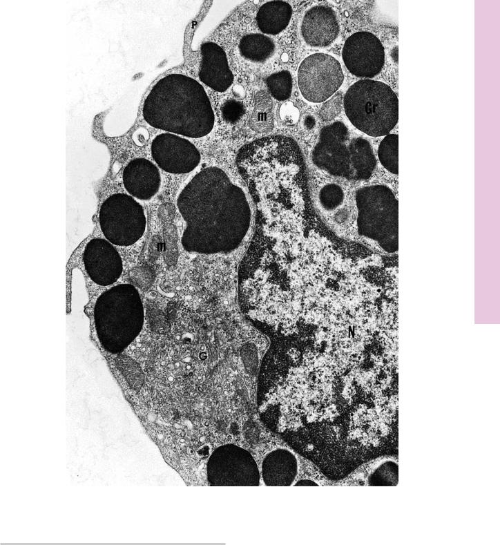
FIGURE 1. Mast cell. Rat. Electron microscopy. ×14,400.
This electron micrograph of a rat peritoneal mast cell displays characteristics of this cell. Note that the nucleus (N) is not lobulated, and the cell contains organelles, such as mitochondria
(m) and Golgi apparatus (G). Numerous processes (p) extend from the cell. Observe that the most characteristic component
of this cell is that it is filled with numerous membrane-bound granules (Gr) of more or less uniform density. These granules contain heparin, histamine, and serotonin (although human mast cells do not contain serotonin). Additionally, mast cells release a number of unstored substances that act in allergic reactions. (From Lagunoff D. Contributions of electron microscopy to the study of mast cells. J Invest Dermatol 1972;58:296–311.)
76 C O N N E C T I V E T I S S U E
Microscopy Electron Degranulation, ell C Mast• 6-3 PLATE
FIGURE 1
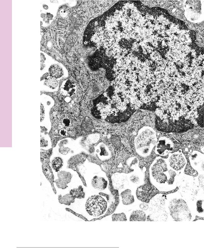
FIGURE 1. Mast cell degranulation. Rat. Electron microscopy. ×20,250.
Mast cells possess receptor molecules on their plasma membrane, which are specific for the constant region of IgE antibody molecules. These molecules attach to the mast cell surface and, as the cell comes in contact with those specific antigens to which it was sensitized, the antigen binds with the active regions of the IgE antibody. Such antibody-antigen binding on the mast cell surface causes degranulation, that is, the release of granules, as well as
the release of the unstored substances that act in allergic reactions. Degranulation occurs very quickly but requires both ATP and calcium. Granules at the periphery of the cell are released by fusion with the cell membrane, whereas granules deeper in the cytoplasm fuse with each other, forming convoluted intracellular canaliculi that connect to the extracellular space. Such a canaliculus may be noted in the bottom left-hand corner of this electron micrograph. (From Lagunoff D. Contributions of electron microscopy to the study of mast cells. J Invest Dermatol 1972;58:296–311.)
C O N N E C T I V E T I S S U E 77
Microscopy Electron Cell, Fat veloping • De7-3 PLATE
FIGURE 1
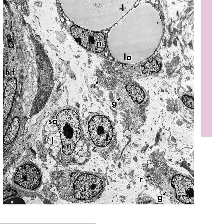
FIGURE 1. Developing fat cell. Rat. Electron microscopy. ×3,060.
This electron micrograph from the developing rat hypodermis displays a region of the developing hair follicle (hf). The peripheral aspect of the hair follicle presents a small adipocyte (sa) whose nucleus (n) and nucleolus are clearly visible. Although white adipose cells are unilocular, in that the cytoplasm of the cell contains a single, large droplet of lipid, during development lipid begins to accumulate as small droplets (l) in the cytoplasm of the small adipocyte. As the fat cell matures to become a large adipocyte (la), its nucleus (n) is displaced peripherally, and the lipid droplets (l)
fuse to form several large droplets, which will eventually coalesce to form a single, central fat deposit. The nucleus displays some alterations during the transformation from small to large adipocytes, in that the nucleolus becomes smaller and less prominent. Immature adipocytes are distinguishable, since they possess a well-developed Golgi apparatus (g) that is actively functioning in the biosynthesis of lipids. Moreover, the rough endoplasmic reticulum (r) presents dilated cisternae, indicative of protein synthetic activity. Note the capillary, whose lumen displays a red blood cell in the lower left-hand corner of this photomicrograph. (From Hausman G, Campion D, Richardson R, Martin R. Adipocyte development in the rat hypodermis. Am J Anat 1981;161:85–100.)