

4 курс / Акушерство и гинекология / Оптимизация_хирургического_метода_лечения_миомы_матки
.pdf131
INTRODUCTION
Relevance of reseach
Uterine fibroids are benign lesions of the myometrium and a most common disease of female reproductive system. About 20 to 50% of women develop uterine fibroids, with the incidence increases with age. According to histolopathology studies, uterine
fibroids are detected in 80% of women. Among women of reproductive age,
the incidence of uterine fibroids is 20 to 40%, which is presumably the underlying cause of female infertility in 5 to 10% of cases [38]. The negative impact of uterine fibroids on the female reproduction can manifestly challenge conception, gestation, or childbirth [14]. Most modern clinical recommendations report that conservative myomectomy can improve the chances to conceive, the course of gestation and childbirth, and should be performed within pregravity preparation efforts [17]. Often in such cases, it is pregnancy plans that are the main indication for myomectomy, rather than conventional factors such as: node size, submucous localization, rapid growth of fibroids [39]. However, opponents tend to question the expediency of myomectomy in nulliparous women, given that postoperative scarring of the myometrium can turn into a more critical complication of pregnancy and childbirth than the fibroid itself [119].
Since 2012 uterine fibroids are conservatively treated using selective progesterone receptor modulators, that directly affect leiomyomas, suppress their proliferation and induce apoptosis, leading to the decrease of nodes in size. These drugs can be prescribed for both pre-surgery preparation and as monotherapy [103]. Evidence shows that Ulipristal acetate allows to preserve fertility in women with fibroids. However, this evidence does not consider how Ulipristal acetate affects the reproductive ability [106]. The results of uterine fibroids surgical treatment evaluate the symptom-free rate, pregnancy incidence, uterine scar viability, and absence of relapses [51].
Existing studies of uterine scar consistency offer a comparative assessment of clinical evidence versus proliferative activity and apoptosis in myometrial biopsies, thus failing to provide a comprehensive overview of the intact myometrium morphology and architecture. The effects of such tissue factors as angiogenesis or structural integrity
132
(collagen type II) on scar formation remains unexplored. In addition, evidence-based
indicators of reparative activity allow us to suggest a mathematical model to assess
the chance of healthy childbirth in women having undergone myomectomy.
Current myomectomy techniques allow to apply various anti-adhesive barriers to avoid de-novo adhesion development [29]. Most often, methylcellulose-based drugs (gels or membranes) are utilized to effectively reduce postsurgical adhesions [27]. Most often, methylcellulose-based drugs (gels or membranes) are utilized to effectively reduce postsurgical adhesions. Moreover, anti–adhesion barriers containing hyaluronic acid and carbomethylcellulose, for example, Antiadhesin gel, are now available. By suppressing macrophage activity, as well as fibroblast and platelet adhesion, the implant inhibits fibrin production and, consequently, prevents damaged tissue from developing
protective barriers.
Therefore, today optimized treatment of uterine fibroids at the pre-pregnancy stage is nothing short of a solution [45]. Top priority objectives include individual planning of conservative, and surgical treatment, or a combination of both, for uterine fibroids, predictive evaluation of postoperative scar tissue, and identification of agents that stimulate post-op scar tissue proliferation. Once identified, morphological and
immunohistochemical factors that affect the ability of intact myometrium adjacent
to myomatous nodes to regenerate, allow to predict uterine scar viability and choose
the appropriate childbirth option, thus reducing the rate of unnecessary cesarean
sections and advancing surgical techniques, in particular biological membranes. These
developments are largely required due to surgery-associated traumatism, |
the choice |
of surgical energies (thermal, electrical, wave, laser), the reparative |
potential |
of myometrium surrounding the removed node [14;39]. |
|
Current research developments
Despite massive data on uterine myoma as a female reproductive pathology, this disease still inspires a lot of research. Due to the younger age of patients, on the one hand, and late pregnancy planning, on the other, the impact of uterine myoma on
Рекомендовано к изучению сайтом МедУнивер - https://meduniver.com/
133
pregnancy (challenged conception, pregnancy complications, limitations in the choice of childbirth options, childbirth and postpartum disorders) is gaining relevance [40].
While some authors associate uterine fibroids to 5 to 10% of female infertility [38;69], others believe that fibroid nodules, their number and size do not affect fertility, provided the uterine cavity is not deformed [11]. Currently, there is no reliable evidence to support recovery of fertility following myomectomy of subserosal and intramural nodules [55]. There is also an ongoing debate as to whether the presence of a uterine myoma or a post-myomectomy scar poses a greater risk to a healthy pregnancy in the future. Despite increased risk associated with intraoperative childbirth, the available research literature does consider uterine fibrosis a contraindication for natural childbirth [7]. Nowadays, indications for myomectomy and uterine extirpation during C-section are still a matter of debate [33;49].
This justifies the relevance of minimally invasive, informative, accessible and cost-effective methods for myometrial scar assessment following myomectomy. In most cases post-op myometrial scar assessment includes sonography, Doppler and MRI scanning. There is evidence that inflammatory process in the myometrium may be associated with uterine scar dehiscence manifest as a wall uterine defect at transvaginal imaging, i.e. an irregular triangular ‘niche’ with marked distal thinning. This sonographic pattern indicates partial uterine suture divergence, which is hardly ever possible to detect clinically [64].
There is evidence that myoma nodules differ from healthy myometrium by the ability to express specific biologically relevant molecules involved in vital signaling pathways. For example, the level of nuclear phosphoprotein p53 increases rapidly with cellular stress, while inactive p53 causes genomic instability [129]. Investigators disagree on the proliferating cell nuclear antigen (PCNA) expression dynamics at particular menstrual cycle phases. Some authors show that PCNA expression is higher throughout the proliferative phase of menstrual cycle than the secretory phase in both intact myometrium and myoma cells (by 4.6 and 3.7 times respectively) [92]. Collagen plays an important role in maintaining healthy uterine structure and physiology, while different collagen types can show significant fluctuation in expression levels due
134
to uterine pathology. Based on background reference data analysis, collagen expression can be considered as a potential indicator of leiomyoma; meanwhile, specific attention shall be paid to collagen type I expression, as its fluctuations have been the focus of most current studies. Although the link between leiomyoma pathogenesis and angiogenic disorders is no longer a matter of doubt, vascular endothelial growth factor (VEGF) expression in leiomyoma cells is still a controversy, with scarce reliable evidence.
Thus, relevant new data have both theoretical and practical importance.
The aim of the study is to develop a differentiated approach to the use of surgical techniques in pre-gravid women with uterine fibroids of different ages.
The research objectives are as follows:
1.to evaluate PCNA expression in women of different ages, indicative of cell proliferation potential in the scar area following myomectomy;
2.to study expression dynamics of angiogenesis molecules (VEGF, VEGFR),
markers of cellular aging and apoptosis (p53, p21, p16), type II collagen in
the scar area following myomectomy in women of different ages;
3.to identify potential prognostic markers of scar viability following myomectomy in women of older reproductive age;
4.to analyze the reproductive function parameters, the course of pregnancy and childbirth in women of different ages with uterine fibroids following laparoscopic myomectomy and conservative management;
5.to identify the criteria for C-section in patients of different ages following myomectomy.
Research novelty
Innovative evidence is presented to show that the expression area of p53, p21 and p16 aging and apoptosis markers in samples obtained from the uterine scar after
Рекомендовано к изучению сайтом МедУнивер - https://meduniver.com/
135
conservative myomectomy is 1.6, 2.1 and 6.7 times higher in women aged 36-46 years compared to younger women (aged 29-35 years). Innovative findings regarding PCNA expression show that the proliferative potential of uterine scar cells persists throughout reproductive age. Pioneering data demonstrate that the expression area of VEGF and
VEGFR angiogenesis factors in uterine scar tissue |
decreases 1.5 and 5.1 times |
respectively in women aged 36-46 years, compared |
to the younger age group |
(aged 29-35 years). At the same time, the average brightness of VEGF and VEGFR expression in post-op myometrial scar samples decreases with the age of patients by 2.8 and 4.3 times respectively. For the first time the paper shows that average type II
collagen expression brightness |
in post-op |
myometrial scar samples decreases |
by 1.4 times in women aged |
36-46 years, |
compared to younger patients (aged |
29-35 years), whereas type II collagen expression area in uterine tissue does not depend on age. These results provide pioneering molecular evidence that following myomectomy, post-op uterine scar tissue is less viable in women of older reproductive age compared to younger patients.
Theoretical and practical relevance of research
Though immunohistochemical studies report that post-op scar cells have preserved their proliferative potential in older women, there is evidence of decreased expression of key angiogenesis and collagen factors, as well as increased intensity of apoptotic processes and replicative aging of cells. The obtained results indicate that in women aged 36-46 years post-myomectomy scar shows less viability compared to women aged 25-35 years. Thus, in the case of pregnancy following laparoscopic myomectomy, C-section is preferable in older women in order to prevent complications associated with scar dehiscence. At the same time, the final decision regarding childbirth options should be made following a comprehensive assessment of the patient's condition, medical history, and uterine scar viability. Type II collagen, VEGF, VEGFR, p53, p21, p16 expression in the scar area allows to assess uterine scar viability in women of different ages.
136
Research methods and techniques
1 Clinical trial design
The study included 220 women with full-term pregnancy.
The study group included 160 patients with a history of laparoscopic
myomectomy performed in the Department of Operative Gynecology of the Research
Institute of Obstetrics, Gynecology and Reproductology named after D.O. Ott
from 2015 to 2016. From 2017 to 2018 all the patients included in the study underwent C-section (CS) at childbirth using the classical technique (intraperitoneal CS with crosssection in the lower segment of the uterus) and were managed in maternity wards of the D.O. Ott Research Institute.
Inclusion criteria:
1.primiparous women aged (29-47 years);
2.natural cycle or ART-assisted pregnancy;
3.full-term pregnancy (37-41 weeks);
4.uterine scar after laparoscopic myomectomy (for subserous or intramural uterine fibroids, 3 to 10 cm size);
5.informed consent signed to conduct the study.
Exclusion criteria:
1.history of surgical interventions for external genital endometriosis, uterine fibroids, reconstructive plastic surgery, prior childbirth by C-section.
2.adenomyosis;
3.intramural-submucous or submucous uterine fibroids;
4.severe somatic pathology, including type 1 and type 2 diabetes mellitus or blood clotting disorders.
In addition to general clinical examination, all patients underwent pelvic ultrasound examination prior to the CS operation to determine the number, size and localization of myomectomy scars.
Рекомендовано к изучению сайтом МедУнивер - https://meduniver.com/
137
The control group included 60 women having a full-term pregnancy and diagnosed uterine fibroids with no history of surgical or conservative treatment. Delivery was carried out by natural birth in maternity wards of the D.O. Ott Research Institute from 2017 to 2018. All women were divided into 3 subgroups.
Inclusion criteria:
1.primiparous women aged (29-46 years);
2.pregnancy occurred in a natural cycle or with the help of ART programs;
3.full-term pregnancy (37-41 weeks);
4.detected uterine fibroids (subserous or intramural, 3 to 10 cm size).
Exclusion criteria:
1.history of surgical interventions for external genital endometriosis, uterine fibroids, reconstructive plastic surgery, CS childbirth;
2.adenomyosis;
3.intramural-submucous or submucous uterine fibroids.
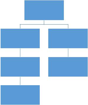
The figure below shows patient distribution across study groups (Figure 1).
Pregnant with uterine fibroids (n=220)
Pregnant with postlaparoscopic myomectomy uterine scar (n=60)
Childbirth by CS (n=60)
Trepan-biopsy
of intact myometrium in the myomectomy scar area (n=40)
Pregnant with surgically untreated uterine fibroids (n=30)
Natural childbirth (n=30)
Figure 1 – Clinical trial design
138
2 Laparoscopic myomectomy protocol (n=120)
In all patients, laparoscopic myomectomy was performed according to a specific standard technique. Laparoscopic myomectomy was performed using KarlStorz equipment (Germany) with an integrated SCB and HD endocamera. We applied endotracheal anesthesia. The Folley catheter was used to catheterize the bladder, and chromosalpingoscopy was performed through a uterine cannula, inserted in the cervix
(Cohen cervical hysterograph). If |
necessary, the cervical hysterograph allowed |
||
to manipulate the uterus in various |
directions, |
which improved intraopreational |
|
ergonomics. The laparoscopic myomectomy was performed |
using the following |
||
endoscopic instruments: laparoscope |
(0°), curved |
scissors – 1, |
forceps 5 mm – 1, |
forceps 10 mm – 1, bipolar Robi forceps – 1, Ultracision harmonic scalpel (ETHICON)
– 1, irrigator / aspirator – 1, needle holder – 1, thread pusher – 1, optical trocar (10-
12 mm) – 1, surgical trocars (5 mm) – 2, surgical trocar (10 mm) – 1, laparoscopic
hydrodissection needle (5 mm) – 1, cervical hysterograph – 1. To perform this surgical intervention, an advanced standardized surgical technique was used to minimize
surgical injury to intact myometrium and ensure formation of a solid post-surgery
uterine scar, which is critical for patients wishing to have children in the future. Abdominal access using standard technique was the option for patients with an uncomplicated medical history. Abdominal cavity and pelvis revision allowed to verify the scope of surgical intervention. The procedure was split into the following stages:
1. Intramyometrial injection at |
the incision |
area. |
Epinephrine |
and |
||
methylergobrevine |
solution |
(epinephrine |
hydrochloride 0.1 mg/mL 1 ml |
and |
||
methylergobrevine |
0.2 mg |
per saline 400mL,) was |
injected |
in the myometrium. |
||
Depending on the size of the myomatous node, 20-60 ml of solution was injected into
the myometrium using a 20 ml syringe and an endoscopic needle inserted into
the abdominal cavity through a 5 mm trocar. The main objective of the procedure was
to reduce intra-op |
blood loss, |
facilitate mechanical |
hydrodissection of intact |
||
myometrium from |
the myomatous |
node capsule, |
and improve further |
enucleation |
|
of fibroids. Meanwhile, practitioners shall avoid |
injecting |
the solution |
directly into |
||
Рекомендовано к изучению сайтом МедУнивер - https://meduniver.com/
139
the fibromyoma, as this will not trigger the desired effect, increasing the risk of tachycardia, elevated blood pressure or irregular heart rhythm, in case the solution enters nodular blood supply.
2. Myometrium incision. We should emphasize that the uterine incision was done to ensure that in the future it would be convenient for the surgeon to sew it up. We tried to avoid large incisions, although in intramural myomas the size of the incision was to ensure unconstrained fibroid enucleation. In order to minimize electrosurgical injury to the myometrium, firstly the incision was performed, followed by the node enucleation; a flap of serous membrane was excised with scissors in case of excess tissue; then precision bipolar hemostasis was performed prior to suturing. To incur minimal electrosurgical and thermal injury to the myometrium, ultrasonic energy was used to incise the uterus. We used Ultracision harmonic scalpel (ETHICON), as its ultrasonic energy has no thermal effect on intact myometrium. Another advantage of this tool is that it can be used both as a scalpel and for coagulation.
3. Myomatous node enucleation. At this stage 10 mm rigid forceps were inserted
into the central 10 mm port, allowing to reliably capture the myomatous node and
enucleate it using 5 mm forceps and a harmonic scalpel. Large vessels supplying blood
to the node were subject to bipolar coagulation and intersection. |
Notably, |
in |
case |
of a transmural deeply localized node extending into the uterine |
cavity, |
we |
tried |
to avoid open uterine cavity penetration. In all cases the myomatous node underwent intracapsular removal, while leiomyoma pseudocapsule was left in place to avoid open penetration into the uterine cavity in case of deep transmural myomatous nodes. At this stage we injected oxytocin 5 IU IV to reduce blood loss and let the uterus shrink to discharge and improve enucleation of fibroid remnants.
4. Myometrial defect suturing is a most important and technically difficult stage of the operation. Endostitch sutures with extracorporeal knotting technique were used for muscle closure permitting the knot to be tied outside by a thread pusher.
This ensured sustainability of nodes and smooth alignment of myometrial edges. Synthetic absorbable material (Vicryl+ 2-0 and/or Monocryl+ 2-0) was used. In case
140
of deep intramural fibroid localization, the myometrial defect was closed layer by layer applying several rows of endo-stitches in order to avoid hematomas in the myomatous bed area.
In a few cases precision point bipolar coagulation was used due to severe bleeding.
5. Myomatous node morcellation. The RotocutGl morcellator (KarlStorz, Germany) was inserted through the central 10 mm port instead of the trocar. The morcellator allowed to quickly, efficiently and safely remove the myomatous node from the abdominal cavity.
6. Chromohydrotubation was performed according to the standard protocol to assess fallopian tubal patency and exclude tubo-peritoneal infertility.
7. Abdominal cavity revision and sanation, elimination of small morcellated myomatous node fragments to prevent adhesions and morcellates. In most cases, no drainage is required after abdominal cavity and pelvis sanation due to high risk of adhesions.
8. Adhesion barriers. We used Interceed* (‘Interside’) absorbable adhesion barrier made of oxidized regenerated cellulose. It gelatinises within about 8 hours and is absorbed within 96 hours or 28 days if applied in layers or stacks. Interceed* would seal the injured area completely, provided hemostasis is accurate and irrigation fluid is promptly removed; otherwise the healing effect is lost.
3 Pelvic ultrasound examination techniques prior to C-section (n=40)
Determination of the myometrium thickness, structure and deformity areas, as well as uterine remodeling assessment after operative childbirth and timely detection of post-op scar insolvency was carried out using Voluson-730 expert transvaginal ultrasound device 3-6 months after the CS. The area under examination included the uterine wall at the site of the post-op scar. We looked at the following parameters:
Рекомендовано к изучению сайтом МедУнивер - https://meduniver.com/