

Lesson topic №25. Нарушения ритма (Cardiac arrhythmia in the outpatient setting)
.pdfSupraventricular tachycardia: orthodromic atrioventricular re-entrant tachycardia
Antidromic AVRT has the following ECG features:
1. a wide QRS complex (fully preexcited)
2. an RP interval that is difficult to assess as the retrograde P wave is usually inscribed within the ST−T segment.

Supraventricular tachycardia: atrioventricular re-entrant tachycardia
Synchronized cardioversion is recommended for haemodynamically unstable patients.
Vagal manoeuvres, preferably in the supine position with leg elevation, are recommended.
In orthodromic AVRT verapamil or diltiazem should be considered if vagal manoeuvres and adenosine fail.
In antidromic AVRT procainamide or profanenone or synchronized cardioversion should be considered if vagal manoeuvres fail.
In antidromic AVRT amiodarone may be considered in refractory cases.
Synchronized cardioversion is recommended when drug therapy fails to convert or control the tachycardia.

Supraventricular tachycardia: atrioventricular re-entrant tachycardia
The treatment of choice for patients with symptomatic and recurrent AVRT is catheter ablation.
If ablation is not desirable or feasible in patients with pre-excitation and symptomatic antidromic AVRT, and in whom structural or ischaemic heart disease has been excluded, class IC antiarrhythmic drugs act mainly on the accessory pathway and can be used in antidromic tachycardia.
Apart from class IC drugs, beta-blockers, diltiazem, or verapamil may also be considered in case of orthodromic tachycardias if no signs of pre-excitation are observed on the resting ECG.

The asymptomatic patient with preexcitation
Most patients with an asymptomatic WPW pattern will go through life without any clinical events related to their ventricular pre-excitation.
Approximately one in five patients will develop an arrhythmia related to their AP during follow-up. The most common arrhythmia in patients with WPW syndrome is AVRT (80%), followed by a 20–30% incidence of AF.
Performance of an electrophysiology study is recommended to risk stratify individuals with asymptomatic pre-excitation who have high-risk occupations/hobbies, and those who participate in competitive athletics.

Atrial fibrillation
Atrial fibrillation
Worldwide, AF is the most common sustained cardiac arrhythmia in adults.
A supraventricular tachyarrhythmia with uncoordinated atrial electrical activation and consequently ineffective atrial contraction.
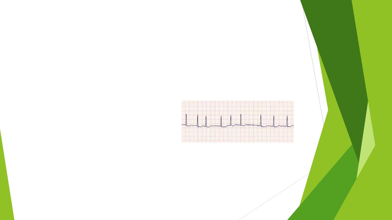
Electrocardiographic characteristics of AF include:
1.Irregularly irregular R-R intervals
2.Absence of distinct repeating P waves
3.Irregular atrial activations.
A standard 12-lead ECG recording or a single-lead ECG tracing of ≥ 30 s showing heart rhythm with no discernible repeating P waves and irregular RR intervals (when atrioventricular conduction is not impaired) is diagnostic of clinical AF

Atrial fibrillation: classification
First diagnosed |
AF not diagnosed before, irrespective of its |
|
duration or the presence/severity of AF-related |
|
symptoms. |
|
|
Paroxysmal |
AF that terminates spontaneously or with |
|
intervention within 7 days of onset. |
|
|
Persistent |
AF that is continuously sustained beyond 7 days, |
|
including episodes terminated by cardioversion |
|
(drugs or electrical cardioversion) after ≥7 days |
|
|
Long-standing persistent |
Continuous AF of >12 months’ duration when |
|
decided to adopt a rhythm control strategy. |
|
|
Permanent |
AF that is accepted by the patient and physician, |
|
and no further attempts to restore/maintain sinus |
|
rhythm will be undertaken. Permanent AF |
|
represents a therapeutic attitude of the patient |
|
and physician rather than a pathophysiological |
|
attribute of AF. |
|
|

Atrial fibrillation: evaluation
The ‘standard package’ for diagnostic evaluation of AF patients should include complete medical history and assessment of concomitant conditions, AF pattern, stroke risk, AF-related symptoms, thrombo-embolism, and LV dysfunction.
A 12-lead ECG is recommended in all AF patients, to establish the diagnosis of AF, assess ventricular rate during AF, and check for the presence of conduction defects, ischaemia, or signs of structural heart disease.
Laboratory tests (thyroid and kidney function, serum electrolytes, full blood count).
Transthoracic echocardiography (LV size and function, LA size, valvular disease, and right heart size and systolic function) are needed to guide treatment.

Atrial fibrillation: evaluation
As symptoms related to AF may range from none to disabling.
Symptom status should be characterized using the European Heart Rhythm Association (EHRA).
Palpitations, fatigue, dizziness, dyspnoea, chest pain, and anxiety during AF are evaluated with regard to how it affects the patient’s daily activity.
Score |
Symptoms |
Description |
|
|
|
1 |
None |
AF does not cause any symptoms |
|
|
|
2a |
Mild |
Normal daily activity not affected by symptoms |
|
|
related to AF |
|
|
|
2b |
Moderate |
Normal daily activity not affected by symptoms |
|
|
related to AF, but patient troubled by symptoms |
|
|
|
3 |
Severe |
Normal daily activity affected by symptoms |
|
|
related to AF |
|
|
|
4 |
Disabling |
Normal daily activity discontinued |
|
|
|

Atrial fibrillation: patient management
The simple Atrial fibrillation Better Care (ABC) holistic pathway ('A' Anticoagulation/Avoid stroke; ‘B’ Better symptom management; ‘C’ Cardiovascular and Comorbidity optimization) streamlines integrated care of AF patients.