Добавил:
Опубликованный материал нарушает ваши авторские права? Сообщите нам.
Вуз: Предмет: Файл:

Книги по МРТ КТ на английском языке / Advanced Imaging of the Abdomen - Jovitas Skucas

.pdf
Скачиваний:
8
Добавлен:
20.10.2023
Размер:
21.9 Mб
Скачать

629

KIDNEYS AND URETERS

A B

Figure 10.26. Wilms’ tumor. A: T2–weighted transverse MR image identifies a right renal tumor (arrows) with extension into the renal vein (arrowhead). B: Coronal T1-weighted image shows tumor extension into the vena cava (arrow). (Source: Burgener FA, Meyers SP, Tan RK, Zaunbauer W. Differential Diagnosis in Magnetic Resonance Imaging. Stuttgart: Thieme, 2002, with permission.)

A rare one even extends intraluminally in the ureter.

A rare Wilms’ tumor is multicystic and mimics a multilocular cystic nephroma or benign renal cystic disease. Likewise, differentiation from a neuroblastoma can be difficult, although a neuroblastoma tends to be ill-defined and more heterogeneous in appearance. Also, a neuroblastoma tends to metastasize early and extensively. Superficially, a Wilms’ tumor can be confused with xanthogranulomatous pyelonephritis.

Therapy and Outcome

Bilateral Wilms’ tumors present a therapeutic dilemma. Conservative surgical therapy with initial biopsy followed by chemotherapy and delayed tumor resection in children with bilateral Wilms’ tumors in the United Kingdom Children’s Cancer Study Group resulted in a mean preserved renal mass of 45%, but initial surgical resection followed by chemotherapy left a renal mass of 35% (74); children in both groups had similar survival and similar renal function.

Imaging after chemotherapy usually identifies necrosis, hemorrhage and calcifications. In one child CT after chemotherapy showed an increase in tumor size and extensive fatty infiltration (75); resection revealed no residual blastema.

Follow-up of the remaining kidney in children after unilateral nephrectomy for Wilms’ tumor reveals that only half develop renal hypertrophy.

Adults

A Wilms’ tumor is rare in adults. Histologically, these tumors are similar to the ones seen in children, and the therapy likewise is similar, but these tumors grow rapidly and prognosis is worse than in children, possibly due to their more advanced presentation. Even extrarenal Wilms’ tumors have developed in adults (76).

A typical Wilms’ tumor in an adult consists of a complex mass often containing a cystic component. These tend to be large, hypovascular and multilocular tumors containing a limited solid component. The differential includes a cystic carcinoma and a multilocular cystic nephroma.

Multilocular Cystic Nephroma/Cystic

Partially Differentiated Nephroblastoma

Clinical

These tumors consist of a mixture of epithelial and mesenchymal components. In the past, a multilocular cystic nephroma, also called multicystic nephroma, cystic lymphangioma, and

630

multilocular cyst, was considered a hamartoma, although the current thinking leans toward a neoplastic origin. Some authors classify multilocular cystic nephroma and cystic partially differentiated nephroblastoma in young children as one entity and believe that they represent cystic Wilms’ tumors having little potential for invasion or metastasis (77); in adults, however, these authors believe that multilocular cystic nephroma is a separate benign entity independent of Wilms’ tumor, although a rare one does undergo malignant transformation and develops into a carcinoma or sarcoma. In one adult, a cystic partially differentiated nephroblastoma exhibited cell maturation intermediate between a multilocular cystic nephroma and a Wilms’ tumor (78).

A similar condition is found in some infants with other gastrointestinal and skeletal abnormalities,including visceromegaly (Perlman syndrome), an autosomal-recessive condition.

In children (mostly young boys) a multilocular cystic nephroma is radiographically and grossly indistinguishable from a cystic partially differentiated nephroblastoma. Both are cystic thin-walled tumors containing multiple septations surrounded by a fibrous capsule. These tumors differ histologically in that a multilocular cystic nephroma is composed of mature differentiated tissue and does not contain blastemal elements, while septa in a cystic partially differentiated nephroblastoma do contain embryonal cells. Calcifications are uncommon in children.

Imaging

Imaging detects both of these cystic tumors but cannot differentiate between them. Their appearance is similar to other cystic tumors. They occur unilaterally. In children, the numerous small cysts can appear solid, and imaging findings resemble a Wilms’ tumor. In adults (mostly women), imaging and even aspiration biopsy findings mimic a cystic renal cell carcinoma, although these tumors are not related.

Computed tomography reveals multilocular cystic nephromas to be homogeneous, multicystic tumors containing thin septa without a solid components; for some reason a lower pole location predominates. The cystic component does not enhance with contrast. Their hallmark is that septa tend to be their only prominent

ADVANCED IMAGING OF THE ABDOMEN

solid component, and the presence of a solid component should suggest either malignant transformation or another diagnosis.

Magnetic resonance imaging of a multilocular cystic nephroma reveals a complex solitary renal cystic tumor often protruding into the renal collecting system and containing thin septa, with the cystic component varying from hypoto hyperintense on T1-weighted images. Any solid component and septa enhance postcontrast in all cases.

Nephrectomy in a patient with a multilocular cystic nephroma should be curative. With a cystic partially differentiated nephroblastoma, however, some tumors recur locally.

Metanephric (Embryonal) Adenoma

A curious tumor composed of small tubular, papillary, and glomeruloid structures and consisting of proliferating primitive epithelial cells mimicking an embryonal nephron was first described in 1980 and called a nephronogenic nephroma (79). Later, a similar benign mixed epithelial and mesenchymal renal tumor was termed an embryonal adenoma. Since then, a number of reports have used the term metanephric adenoma. Whether this tumor represents a benign counterpart of Wilms’ tumor is conjecture. Metanephric adenomas and metanephric adenofibromas (discussed below) are closely related neoplasms. In either case, a renal metanephric adenoma is a distinctive renal cortical neoplasm usually classified among nephroblastic tumors. Its histologic appearance is similar to that of metanephric hamartomas found in nephroblastomatosis. In spite of a morphologic similarity to Wilms’ tumor, genetics suggest a relationship to papillary adenomas and papillary renal cell carcinomas.

These tumors consist of differentiated cells containing few or absent mitoses; tubules and glomeruli are often identified. Its metanephric (embryonic) architectural and cytologic appearance is characteristic, although pathologic study of some tumors suggests a malignancy. Needle biopsy and aspiration cytology specimens have been misdiagnosed as Wilms’ tumor. The reverse is also true: a renal cell carcinoma and a metastasis from a poorly differentiated thyroid carcinoma can be misdiagnosed as metanephric adenomas. Most authors

631

KIDNEYS AND URETERS

consider these tumors to be benign, although a metanephric adenoma in a 7-year-old girl metastasized to lymph nodes (80).

There are two age peaks; half occur in children and the other half mostly in women in their 50s. Most tumors are discovered incidentally, with only a minority of patients having pain, hematuria, or a palpable tumor. Polycythemia and acquired von Willebrand’s disease develop in some patients. An association of a metanephric adenoma with embryonal hyperplasia of Bowman’s capsular epithelium was reported in a 9-year-old boy on peritoneal dialysis (81).

Metanephric adenomas are well-circum- scribed, unencapsulated tumors varying in size. Some contain fibrosis and calcifications. Computed tomography, US, and MRI usually reveal a solid tumor with occasional necrosis and cystic degeneration, suggestive of a hypovascular renal cell carcinoma. They are isoto hyperdense on CT. They range from hypoto hyperechoic on US.

Simple resection is curative in almost all patients.

Metanephric (Nephrogenic) Adenofibroma

The histology of metanephric adenofibromas (previously also called nephrogenic adenofibromas) reveals a proliferation of spindle cells, similar to what is found in congenital mesoblastic nephroma, encasing nodules of embryonal epithelium similar to the nephroblastomatosis associated with Wilms’ tumor; it is thus not surprising that these tumors have been misdiagnosed as Wilms’ tumors and mesoblastic nephromas.

These are childhood tumors. A not uncommon presentation is hematuria, hypertension, or polycythemia.

Metanephric Stromal Tumor

A metanephric stromal tumor is a renal medullary spindle cell (fibrous) tumor containing smooth-walled cysts that entrap native kidney tissue. Although such a description suggests that it should be classified under mesenchymal tumors, it is histologically identical to the stromal component of metanephric adenofibroma and is discussed with the latter entity (see above).

Histology reveals onion-skin cuffing around entrapped renal tubules, heterologous glial or cartilage differentiation, and vascular changes consisting of angiodysplasia of entrapped arterioles and juxtaglomerular cell hyperplasia of entrapped glomeruli (82).

The mean age of 31 patients with a metanephric stromal tumors was 2 years; most tumors presented as an abdominal tumor (82); they did not metastasize. The differential diagnosis includes renal clear cell sarcoma.

Mesoblastic Nephroma (Bolande’s Tumor)

Infants

Congenital mesoblastic nephroma, at times called congenital Wilms’ tumor, comprises about half of renal tumors encountered during the neonatal period. Most occur within the first month of life, and their prevalence decreases during childhood. They present as a large abdominal tumor. The prognosis depends on the infant’s age, being best for tumors discovered early in life.

Earlier publications considered mesoblastic nephromas to be a benign neoplasm, possibly even hamartomatous in origin. In fact, the favorable prognosis of Wilms’ tumors in neonates reported previously is suspect because many of these were probably mesoblastic nephromas rather than Wilms’ tumors.

The morphologic and cytogenetic evidence suggests a relationship between congenital mesoblastic nephromas and infantile fibrosarcomas. Not only are these tumors similar histologically, but also both are associated with similar chromosome polysomies and they may represent a single entity. Congenital mesoblastic nephromas and Wilms’ tumors also appear to share common genetic pathways and abnormalities.

Mesoblastic nephromas are mostly solid tumors which usually follow a benign course but tend to invade adjacent structures. Some authors subdivide these tumors into congenital and atypical, with the latter including aggressive and potentially malignant variants. The aggressive variety tends to recur or metastasize. A rare aggressive mesoblastic nephroma ruptures spontaneously.

Imaging reveals a solid, homogeneous, usually large tumor. Hemorrhage and necrosis

632

develop in the aggressive and malignant variants, although cystic degeneration is not common. Their imaging appearance mimics a Wilms’ tumor.

Some mesoblastic nephromas show uptake of Tc-99m-DMSA.

Adults

The rare similar adult renal tumor consisting of epithelial and stromal tissue is probably a different entity than the pediatric variety. Immunohistochemistry and electron microscopy aid in identifying this tumor. Some mesoblastic nephromas in adults may represent cystic hamartomas. An adult mesoblastic nephroma can grow either intraor predominantly extrarenal, at times even mimicking an adrenal tumor.

Lymphoma/Leukemia

Clinical

True lymphatics are not present in the kidneys and primary renal lymphoma is rare. Secondary involvement is either via contiguous spread from perirenal disease or from hematogenous dissemination. Involvement with nonHodgkin’s lymphoma is considerably more common than Hodgkin’s lymphoma. Kidneys of immune-deficient patients are more prone to involvement by lymphoma. Infiltration is usually bilateral.

Lymphoma involvement in other locations tends to dominate the clinical findings. Most renal involvement does not lead to symptoms until late in the course. A rare presentation is acute renal failure.

A granulocytic sarcoma (also called a chloroma) develops in a minority of patients after therapy for some leukemias. These solid renal tumors are hypointense on T1and hyperintense on T2-weighted images. They show little contrast enhancement.

Imaging

Imaging detects only a minority of lymphomatous renal involvement. No typical pattern predominates, and renal lymphoma ranges from

ADVANCED IMAGING OF THE ABDOMEN

solitary to multiple tumors, diffuse infiltration, or invasion from adjacent structures. Even perirenal involvement varies considerably from focal tumors to plaque-like infiltration. Retroperitoneal lymphoma generally invades the kidneys through renal sinuses, and thus the renal medulla is more often involved than the cortex.

Necrosis is uncommon in renal lymphomas, even when large. Likewise, lymphoma rarely results in a vascular tumor thrombus.

Computed tomography findings in the rare primary non-Hodgkin’s renal lymphoma range from a single nodule, multiple nodules, and diffuse infiltration, to a lymphomatous mass encasing the kidney. Lymphomatous foci are homogeneous and mostly slightly hyperdense to isodense on precontrast CT and do not enhance as much as normal renal parenchyma postcontrast. Focal tumors maintain a discrete margin. Diffuse infiltration results in an overall renal structure disorganization. A not unusual appearance is renal invasion by retroperitoneal lymphoma, but the lack of adjacent adenopathy is unreliable in excluding lymphoma. While CT allows evaluation of the site and size of involvement, it cannot establish a diagnosis; similar findings are seen with a renal cell carcinoma, transitional cell carcinoma, metastatic disease, or even some chronic inflammatory conditions.

Two sonographic patterns are evident: either a bilateral nodular infiltrate or diffuse renal enlargement with loss of differentiation between renal sinus echoes and parenchyma. The typical infiltration is hypoechoic, but at times homogeneous involvement results in an anechoic pattern mimicking a cyst.

Magnetic resonance imaging shows isoor slight hypointense tumors on T1and hypointense tumors on T2-weighted images. Postcontrast lymphomas show minimal tumor enhancement, in distinction to renal cell carcinomas where early enhancement is common. Whether CT or MR is superior in identifying and defining lymphomatous involvement is not clear. Ureteral involvement consists either of encasement or displacement by an adjacent lymphomatous focus.

Leukemic involvement results in symmetrically enlarged kidneys and compressed collecting systems.

633

KIDNEYS AND URETERS

Multiple Myeloma

Renal histology in patients with myeloma nephropathy revealed 14% with tubulointerstitial nephritis, 11% with amyloidosis, 7% with acute tubular necrosis, and 4% each with nodular glomerulosclerosis and plasma cell infiltration (83); at times renal biopsy pro-vides the first clue to a diagnosis of myeloma by identifying myeloma cast nephropathy.

Tubular precipitation of Bence-Jones proteins


is a cause of renal failure in multiple myeloma


patients. Haphazard IV contrast injection is a


cause of such precipitation, but with preplan-

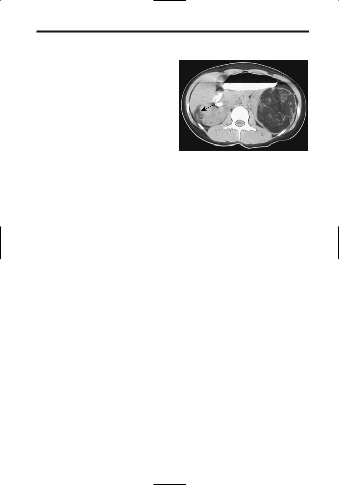
Figure 10.27. Renal angiomyolipomas in a 17-year-old with

ning and adequate hydration a number of these

tuberous sclerosis. The left fat-containing tumor is obvious. A

patients have undergone contrast studies

smaller angiomyolipoma is also present in the right kidney

without complication.

(arrow). (Courtesy of Luann Teschmacher, M.D., University of

Resorption of calcium from bone and the

Rochester.)

resultant hypercalcemia lead to nephrocalci-


nosis. The kidneys enlarge, and the collecting


systems are compressed. These patients are also


prone to developing uric acid calculi.

oleiomyomatosis, and tumors at other sites, but

Unexplained renal failure in a patient even

in general extrarenal angiomyolipomas are rare.

with absent skeletal lesions can still be due to

These tumors develop in the renal capsule,

multiple myeloma.

cortex, and medulla. Histologically they are


composed of haphazardly arranged blood

Mesenchymal Neoplasms

vessels, disorganized smooth muscle fibers, and

Angiomyolipoma

varying amounts of fat. Some contain sheets of

epithelioid cells,suggesting a malignancy. Those

Clinical

without fat mimic a leiomyoma. Histologically,

no sarcomatous transformation should be


Classification of angiomyolipomas has evolved

evident in either the leiomyomatous or

from their being considered hamartomas to

lipomatous components.

benign neoplasms, although a rare one does

Serial CT and US provide a measure of renal

undergo sarcomatous transformation and a

angiomyolipoma growth rates. The mean

propensity to metastasize. Even the usually

growth rate of isolated tumors is about 5% per

benign variety tends to be locally aggressive and

year, being considerably more in those with

produces considerable mischief. In some studies

tuberous sclerosis. No correlation exists

20% to 50% are detected in tuberous sclerosis

between the amount of fat in a tumor and its

patients, where they tend to be multiple and

growth rate. Growth tends to be unpredictable.

bilateral. Angiomyolipomas are associated to a

Some tumors are stable for years and then

lesser degree with von Recklinghausen’s disease,

undergo a fast increase in size, followed by

von Hippel-Lindau disease, and adult polycystic

another period of quiescence. Rapid growth

disease, and are found in about half of patients

occasionally occurs in pregnancy, suggesting

with pulmonary lymphangiomyomatosis. In

that hormones play a role in their growth. An

general, a finding of multiple angiomyolipomas

occasional angiomyolipoma is huge at initial

should suggest tuberous sclerosis (Fig. 10.27).

presentation.

In tuberous sclerosis, angiomyolipomas occur

Spontaneous, solitary tumors are most

earlier in life than sporadic ones, and most

common in middle-aged women and are rare in

are discovered incidentally. An occasional

children. Pain, a palpable tumor, or hematuria is

tuberous sclerosis patient develops bilateral

a common presentation, although many of these

renal angiomyolipomas,pulmonary lymphangi-

tumors are asymptomatic.

634

Pathologists describe a rare epithelioid angiomyolipoma as a separate entity. It tends to mimic renal cell carcinoma; it contains regions of hemorrhage and necrosis. Fatty tissue is not identified.

The rare liposarcoma is indistinguishable with imaging from an angiomyolipoma. This problem is especially pertinent when an incidental renal angiomyolipoma is detected in the work-up of another carcinoma and a metastatic liposarcoma is suspected.

Imaging

Angiomyolipomas are diagnosed with CT, US, or MR (Figs. 10.28 and 10.29); most contain sufficient focal fat to be identified with CT, appear as homogeneous and hyperechoic tumors containing smooth margins with US, and have hyperintense signals on T1and hypointense signals on T2-weighted MRI.

Focal fat is also found in lipomas and some liposarcomas, but these tumors are rare in the kidneys. Clear cell renal cell carcinomas contain intracellular lipid and glycogen, which is detectable with chemical shift MR, but this fat is diffuse. Focal regions of fat are rare in renal cell carcinomas.

Calcifications are not a feature of angiomyolipomas except secondary to hemorrhage.

ADVANCED IMAGING OF THE ABDOMEN

Even when associated with fat, calcifications should suggest a carcinoma.

Diagnostic difficulty occurs when a tumor is small or contains very little fat. Conventional CT may not detect fat in tumors smaller than several centimeters in size. Multislice helical CT is more sensitive and is preferred for small lesions. These low-fat tumors are homogeneously hyperdense on unenhanced CT, show homogeneous postcontrast CT enhancement, and are isoechoic on US. Recent hemorrhage tends to mask any fatty component. Occasionally in such tumors a small amount of fat is detected with chemical shift MRI but not CT; in these, a renal cell carcinoma is in the differential diagnosis.

The diagnosis appears more difficult in children. Some of these tumors in children contain little fat and imaging simply identifies a large renal tumor. Even a fine needle biopsy may be misleading and a Wilms’ tumor initially suspected. The reverse is also true; US detection of a hyperechoic tumor in a child with von HippelLindau disease suggests an angiomyolipoma, yet an occasional such tumor is a renal cell carcinoma.

A CT diagnosis is straightforward with most angiomyolipomas. Fat intermixed with other tissue detected on thin-section nonenhanced CT is strong presumptive evidence for an

A


Figure 10.28. Right renal angiomyolipoma. A: CT identifies a fat-


containing tumor in the superior renal pole (arrow). B: Selective


angiography shows a poorly vascular tumor that was sub-


sequently embolized. (Courtesy of David Waldman, M.D.,


University of Rochester.)

B

635

KIDNEYS AND URETERS

A B

C D

Figure 10.29. Angiomyolipoma with a retroperitoneal bleed. A: CT reveals a fat-containing left renal tumor (arrows). Arterial-phase

(B) and delayed-phase (C) arteriography also identify the tumor but did not detect a site of bleeding. D: An inferior vena cavagram study reveals caval compression from the left side by the extensive retroperitoneal bleeding (arrow). (Courtesy of Oscar Gutierrez, M.D., University of Chile, Santiago, Chile.)

angiomyolipoma. Precontrast CT should be used; postcontrast enhancement may increase the density to the level of water. Angiomyolipomas tend to be well marginated but do not have a capsule. Cystic regions are not common. Larger tumors often extend outside the kidney.

With an atypical CT appearance aspiration biopsy or cytology should provide a definitive diagnosis.

A typical sonographic appearance of an angiomyolipoma is that of a well-circum- scribed, homogeneous, hyperechoic tumor.

636

Shadowing is detected in some, a finding unusual for a carcinoma. Because many small renal cell carcinomas are also hyperechoic, it is necessary to confirm US findings with other imaging. In some instances, however, a homogeneously hyperechoic lesion is accepted without confirmation as a probable angiomyolipoma, especially in patients under the age of 50 and with a tumor smaller than 15mm or possibly 10mm in diameter. In some patients with tuberous sclerosis and a confluence of multiple angiomyolipomas, US simply reveals diffusely increased echogenicity in markedly enlarged kidneys.

Ultrasonography frequency-dependent attenuation is potentially useful in differentiating renal cell carcinomas from angiomyolipomas. Frequency-dependent attenuation values of renal cell carcinomas are significantly lower than those of angiomyolipomas. Some overlap does exist, and the practical application of this technique remains to be established.

The fat component even in small tumors is identified by fat-suppressed T1-weighted MRI. Fat in these tumors tends to be scattered throughout the lesion. Opposed phase MRI alone, being sensitive even to intracytoplasmic lipid found in some renal cell carcinomas, is inappropriate to evaluate the gross fat in angiomyolipomas and differentiate them from carcinomas. Magnetic resonance imaging can reliably identify small (<1.5cm) angiomyolipo-

ADVANCED IMAGING OF THE ABDOMEN

mas using a combination of sequences. Thus in-phase sequences show them to be minimally or moderately hyperintense relative to the renal cortex, and fat-suppressed sequences reveal a loss of signal intensity (84). Some tumors are difficult to identify due to limited signal differences or breathing artifacts.

Complications

Complications include compression of adjacent calyces and intratumoral, subcapsular, perirenal, or pararenal bleeding and hematoma (Fig. 10.30). These tumors undergo cystic degeneration. Spontaneous rupture is not uncommon and an angiomyolipoma should be high in the differential for the patient presenting with spontaneous rupture and bleeding of a renal tumor.

Extrarenal extension into the perirenal fat and lymph nodes is common, and these enlarged pararenal lymph nodes cause additional diagnostic and therapeutic dilemmas. Computed tomography shows these enlarged lymph nodes to be of soft tissue density, although an occasional one contains fat.

Extension into a renal vein and inferior vena cava as a tumor thrombus represents an additional complication. Computed tomography can occasionally detect fat within a thrombus. Renal vein involvement is more common on the right and does not necessarily imply malig-

A B

Figure 10.30. Bleeding left renal angiomyolipoma. Noncontrast CT reveals a fat-containing left renal tumor (A, arrow), surrounded by a well-marginated infiltrate having a CT density similar to aorta (B). (Courtesy of Algidas Basevicius, M.D., Kaunas Medical University, Kaunas, Lithuania.)

637

KIDNEYS AND URETERS

nant transformation. Complicating matters, an occasional one is associated with a renal cell carcinoma.

Therapy

Many of these tumors are still treated with a nephrectomy, although a trend is toward a more conservative approach—partial nephrectomy or enucleation is feasible in some patients. Incomplete tumor excision, however, leads to recurrence, often rapidly.

In patients with tuberous sclerosis, embolization is effective in long-term management of hemorrhage; most of these patients require several embolization sessions. If needed, a partial nephrectomy is then performed.

Symptomatic renal angiomyolipomas have been embolized with a mixture of iodized oil and absolute ethanol (85); some tumors decrease in size and others do not change, but most patients remain asymptomatic (except with incomplete embolization). Follow-up after embolization reveals that it is the angiomyogenic components that disappear, while fatty tissue undergoes liquefactive necrosis.

Lymphangiomyoma

Lymphangiomyomas, considered to be hamartomas rather than neoplasms by some authors, are discussed in more detail in Chapter 14. Lymphangiomyomas are related to angiomyolipomas and are occasionally associated with an angiomyolipoma; they are also suspected of being a manifestation of tuberous sclerosis.

One cause of chyluria is renal or retroperitoneal lymphangioleiomyomatosis.

These are cystic, hypervascular lesions, readily confused with renal cell carcinomas.

calcifications. Computed tomography and US reveal a heterogeneous and hypoechoic tumor.

Imaging shows most sarcomas as large, invasive, necrotic tumors. They tend to mimic a large renal cell carcinoma. Poorly defined calcifications are seen in a minority. Metastases at initial presentation are common.

Angiomatous Neoplasms

A primary renal angiosarcoma is very rare, with only case reports described. They are also known as a malignant hemangioendothelioma, hemangioendothelial sarcoma, and vascular sarcoma and appear as single or multiple tumors. They arise from blood vessel endothelial cells. Hemorrhage is a common presentation. Some contain fat cells suggesting an angiomyolipoma.

An epithelioid angiosarcoma is a very aggressive tumor mimicking a primary epithelial tumor.

Clear Cell Sarcoma

Clear cell sarcomas are rare tumors found in younger children. Initially thought to be a variant of Wilms’ tumor, this tumor has distinct immunohistochemical and flow cytometric findings differing from Wilms’ tumors. Its relationship to mesoblastic nephroma is not clear. Clear cell sarcomas exhibit p53 protein nuclear immunopositivity, suggesting alterations in the p53 tumor-suppressor gene.

The imaging findings of clear cell sarcomas are similar to those of a Wilms’ tumor, with differentiation made by histology.

This is an aggressive tumor having a poor prognosis and a predilection for bone metastasis.

Leiomyomatous Neoplasms

Leiomyosarcomas account for half or more of the rare renal sarcomas. Even after resection, differentiation between a leiomyoma and its malignant counterpart is difficult. These tumors originate mostly from blood vessels, although some of the reported renal leiomyomas probably represent atypical angiomyolipomas.

Most benign leiomyomatous tumors are solid and well marginated, but degeneration leads to a cystic appearance. An occasional one contains

Ossifying Renal Tumor of Infancy

The few reported ossifying renal tumors of infancy occurred in infants under the age of 14 months. They appear to be benign. These tumors contain varying proportions of osteoid, osteoblastic cells, and spindle cells. Whether the osteoid component represents a form of urothelial transformation is speculation. The proportion of osteoid and the degree of osseous maturation increases with age.

638

Gross hematuria is the most common presentation. An occasional infant presents with a palpable abdominal mass.

Typically these tumors are attached to a renal papilla and present as a pelvicalyceal system tumor. Some resemble a staghorn calculus.

Osteogenic Sarcoma

A primary renal osteogenic sarcoma is very rare and should be distinguished from a sarcomatoid variant of renal cell carcinoma with osteoid formation. Whether these tumors represent a sarcomatous version of an angiomyolipoma is speculation.

Patients with a primary renal osteogenic sarcoma have elevated serum alkaline phosphatase levels but no underlying bone disease.

These tumors contain calcifications. A “sunburst” type of calcification, if present, should suggest the diagnosis. The differential diagnosis for a calcified renal tumor includes a calcified inflammatory tumor, renal cell carcinoma, and a metastatic or invading osteogenic sarcoma.

ADVANCED IMAGING OF THE ABDOMEN

tension, which can be severe, often associated with hypokalemia and hyperaldosteronism. An occasional incidentally discovered reninoma is nonfunctioning. As its name implies, these tumors originate from juxtaglomerular cells. Whether they are hamartomas or neoplasms is not established. They are benign. Histologically, some of these tumors mimic a hemangiopericytoma, with the latter tumor occasionally also resulting in hypertension.

These are solid, hypovascular tumors on CT and hyperechoic on US. Most reninomas are smaller than 2 to 3cm in diameter. Even if a reninoma is too small to be detected with imaging, with strong clinical suspicion renal vein catheterization and renin-level sampling should suggest a tumor site. It should be kept in mind, however, that some renal cell carcinomas and other renal tumors on occasion also produce renin.

Resection leads to a marked drop in blood pressure in most but not all patients, with the latter presumably having a residual angiopathy.

Histiocytoma

A majority of renal histiocytomas are malignant. A renal capsule origin is common. A review of 44 malignant renal fibrous histiocytomas found an average patient age of 58 years, no sex predilection, and two thirds of tumors involving the left kidney (86). Symptoms tend to be few, and a late presentation is common. Sudden flank pain suggests spontaneous tumor rupture.

Imaging reveals a solid tumor tending to be hypodense on CT, hypoechoic on US, and hypovascular on angiography (86). A correct preoperative diagnosis is not common, with renal cell carcinoma or oncocytoma often being suspected.

These tumors have a poor prognosis. Metastases and local recurrence after resection are common.

Reninoma (Juxtaglomerular Tumor)

These rare renin-secreting tumors, known as reninomas and juxtaglomerular cell tumors, occur both in children and adults and are more common in females. Their primary importance is their renin production and resultant hyper-

Hemangiopericytoma

Hemangiopericytomas are rare in the kidneys. An occasional one leads to hypertension. These are well-marginated, solid, hypovascular renal sinus tumors involving the renal hilum to the point of suggesting a renal pelvis transitional cell carcinoma. Some are large at the initial presentation, compressing the renal pelvis and causing hydronephrosis and parenchymal atrophy.

Needle aspiration cytology may suggest the diagnosis, although the appearance is similar to that seen with other spindle cell tumors. Immunohistochemistry is helpful.

Renal Capsule Neoplasms

Neoplasms originating from the renal capsule are not common. They represent a spectrum of mesenchymal origin, with some earlier authors simply referring to them as capsulomas. They include a histiocytoma, solitary fibrous tumor, leiomyoma, lipoma, and even an angiomyolipoma, the latter a rare site for this tumor. Differentiating capsular from renal and extraperitoneal origin tumors is often difficult, especially with larger ones. Identifying a capsular artery by angiography as a primary tumor