

5 Musculoskeletal imaging
Contents
Basics of bones and joints 347 |
Basics of trauma 399 |
|
Arthritis 347 |
Basics of MR 400 |
|
Puttingittogether:evaluationofthe |
Foot and ankle 401 |
|
handsforarthritis:ABCDEs 362 |
Knee and knee MRI 411 |
|
Bonetumorsandtumor-likelesions: |
||
Hip and hip MRI 425 |
||
approach 365 |
||
Lumbar/thoracic spine 434 |
||
Bone lesions organized by cell of |
||
Cervical spine 435 |
||
origin 367 |
||
Musculoskeletal infection 385 |
Shoulder and shoulder MRI 438 |
|
Diffuse bone disease 390 |
Elbow and forearm 455 |
|
Differential diagnosis of common |
Wrist and hand 457 |
|
bone lesions 398 |
|
|
|
|
346
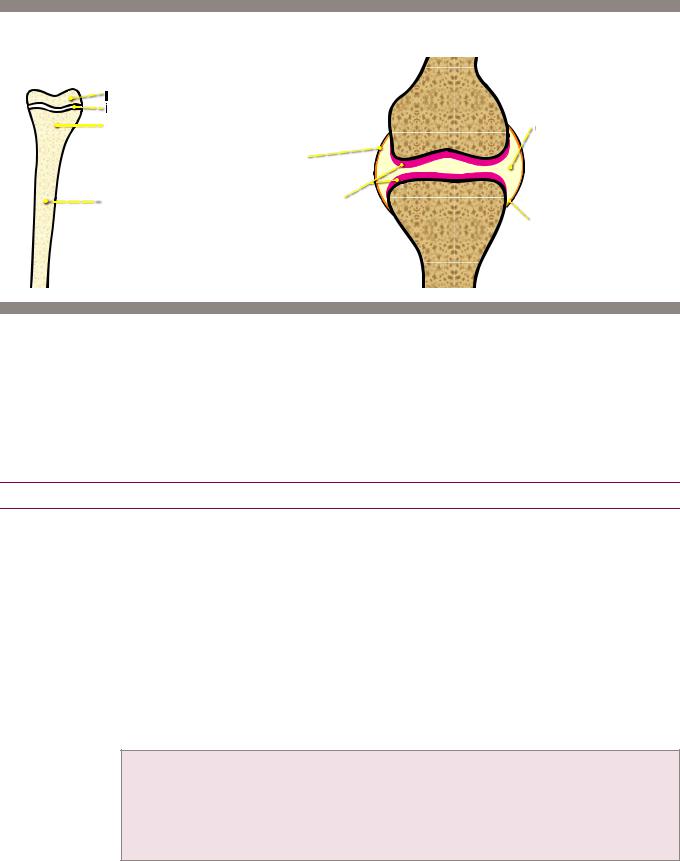
Basics of bones and joints
anatomy of a long bone |
anatomy of a synovial joint |
epiphysis |
|
physis (growth plate) |
|
metaphysis |
joint space lled with |
|
synovial uid |
|
synovium |
|
|
|
diaphysis |
articular cartilage |
|
|
|
||
|
|
|
|
bare area just inside |
|
|
|
|
synovium is site of |
|
|
|
|
rst erosions in rheumatoid |
Arthritis
The hallmark of arthritis is cartilage destruction, which may be evident on radiographs
as cartilage space narrowing.
n broad categories, arthritis can be divided into degenerative (osteoarthritis), inflammatory (rheumatoid arthritis, spondyloarthropathies, and juvenile idiopathic arthritis), crystal deposition (gout, calcium pyrophosphate dihydrate, and hydroxyapatite), hematologic (hemophilia), and metabolic categories.
Osteoarthritis (OA)
Overview of osteoarthritis
Also called osteoarthrosis degenerative joint disease, osteoarthritis (OA) is the result of articular cartilage breakdown from altered local mechanical factors in a susceptible individual. In addition to cartilage, OA is thought to involve the entire joint including bone, ligaments, menisci, joint capsule, synovium, and musculature.
OAisthemostcommoncauseofcartilagelossinthemiddle-agedandolderpopulation.
OA typically occurs in weight-bearing joints and the hands in a specific distribution.
When radiographic findings of OA are seen in younger patients or in unusual locations, such as the shoulder, elbow, or ankle, then there is usually a predisposing prior trauma or other underlying arthritis.
The radiographic hallmarks of OA, regardless of location in the body, include:
Asymmetrical joint space narrowing
Sclerosis of subchondral bone, stimulated by loss of hyaline cartilage and reactive remodeling.
Osteophytosis
Subchondral cystic change, due to herniation of joint fluid into bone through a cartilage defect.
Lack of periarticular osteopenia
Although joint space narrowing is present in all arthritides, osteoarthritis can be diagnosed with confidence when subchondral sclerosis, osteophytosis, and subchondral cystic changes are present and inflammatory erosions are absent.
When extensive subchondral cystic changes are present, calcium pyrophosphate dihydrate crystal
deposition disease (CPPD) should be considered as well.
347
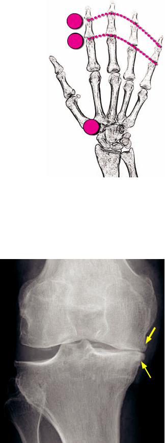
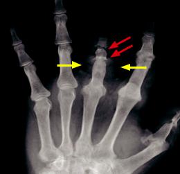
Osteoarthritis in the hand
Similar to osteoarthritis of other joints, the radiographic hallmarks of OA in the hand include cartilage space narrowing, subchondral sclerosis, and osteophytosis. Erosions are absent.
n order of decreasing involvement, typical sites of OA in the hand include the distal interphalangeal joints (DIPs), the base of the thumb at the first carpometacarpal joint (CMC), and the proximal interphalangeal joints (PIPs).
The most common site of osteoarthritis in the
hands is the second DIP.
Unlike rheumatoid arthritis, the metacarpophalangeal joints (MCPs) are less commonly affected.
Large osteophytes cause characteristic soft-tissue swelling surrounding the finger joints.
A Heberden node is soft-tissue swelling around the DIP.
A Bouchard node is soft-tissue swelling around the PIP.
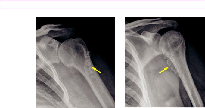
Osteoarthritis in the shoulder
DIPs
PIPs
CMC
TheGrasheyview(obtainedposteriorlyin40-degreeobliquedexternalrotation)shows
theglenohumeraljointinprofileandbestdemonstratescartilagespacenarrowing.
Osteoarthritis in the foot
ThemostcommonjointaffectedbyOAinthefootisthemetatarsophalangealjoint(MTP)
ofthegreattoe,whichmayleadtohalluxrigidus(astiffbigtoe)fromdorsalosteophytes.
Osteoarthritis also affects the talonavicular joint and is a cause of dorsal beaking.
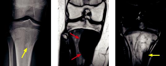
Osteoarthritis in the knee
Therearethreejointcompartments intheknee:Themedialandlateral tibiofemoralcompartmentsandthe patellofemoralcompartment.The typicalpatternforOAoftheknee isasymmetricalinvolvementofthe medialtibiofemoralcompartment.
Severeosteoarthritiscaninvolveall threecompartments.
Thefollowingruleofthumbapplies toOAingeneral,butespeciallyin theknee:Osteophytesdetermine whetherOAispresent.The degreeofjointspacenarrowing determinestheseverityofOA.
Thedegreeoftibiofemoralcartilage spacenarrowingisbestdetermined onstandingweight-bearingviews, oftenonstandingfilmsinflexion.
Bilateral involvement of the knees is typical.
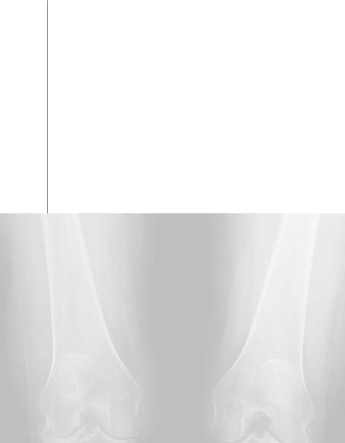
Osteoarthritis: Standing frontal radiograph of the knee shows severe cartilage space narrowing, sclerosis, and osteophytosis of the medial tibiofemoral compartment (arrows).
348
Osteoarthritis in the hip
Similar to the knee, the involvement of hip osteoarthritis tends to be bilateral.
n addition to the typical features of
OA including joint space narrowing, osteophytosis, subchondral cystic change, and sclerosis, hip OA also features characteristic migration of the femoral head in a superolateral direction. Less commonly, medial migration can be seen in hip OA.
n contrast, axial migration is seen more commonly in inflammatory arthritis.
Osteoarthritis: Hip radiograph shows severe cartilage space narrowing, sclerosis, and osteophytosis of the right hip with superolateral migration (arrow) of the femoral head.
superolateral migration
(most common in osteoarthritis)
axial migration
(most common in rheumatoid arthritis)
medial migration
(less common in osteoarthritis)
osteoarthritis |
|
|
|
rheumatoid arthritis |
|||
|
|
|
|
|
|
|
|
superolateral |
medial migration: |
axial migration: |
severe axial migration: |
||||
migration: superior-lateral |
inferomedial cartilage |
concentric cartilage |
protrusio deformity |
||||
cartilage space narrowing |
space narrowing |
space narrowing |
|
|
|||
Degenerative change in the spine
The vertebral body–disc articulations are cartilaginous joints. Vertebral body endplates are covered by hyaline cartilage that is analogous to articular cartilage in other joints. The intervertebral disk is composed of three components: The annulus fibrosus, nucleus pulposus, and the cartilaginous endplates.
Osteoarthritis only affects synovial joints. Therefore, in the spine, osteoarthritis can occur at the facet (zygapophyseal), atlantoaxial, uncovertebral joints (in the cervical spine at C3–C7), costovertebral, and sacroiliac joints.
The spectrum of intervertebral disk degeneration is best described as degenerative disk disease (DDD) which is characterized by dessication of the intervertebral discs, endplate sclerosis, and osteophytosis.
Gas in the intervertebral disc, also called vacuum phenomenon is commonly seen and is pathognomonic for degenerative disease.
t is important not to confuse vacuum phenomenon (gas in intervertebral disc) with Kümmel disease, which is gas in a vertebral body compression fracture representing osteonecrosis.
349
•Complications of DDD include spinal stenosis, neural foraminal stenosis, and degenerative spondylolisthesis.
•Diffuse idiopathic skeletal hyperostosis (DISH) is a distinct entity from degenerative disc disease, but appears similar due to exuberant osteophytosis. DISH is defined as flowing bridging anterior osteophytes spanning at least four vertebral levels, with normal disk spaces and sacroiliac joints. The etiology of DISH is unknown. It is usually asymptomatic but may be a cause of dysphagia when it affects the cervical spine. DISH occurs in elderly patients.
DISH is associated with ossification of the posterior longitudinal ligament (OPLL), which may be a cause of spinal stenosis. OPLL may be difficult to identify on MRI and is best seen on CT.
Osteoarthritis in the sacroiliac (SI) joint
•Only the inferior portion of the sacroiliac joint is a synovial (diarthrodial) joint. The superior portion is a syndesmotic joint.
•The typical changes of OA are only seen in the inferior (synovial) portion of the SI joint.
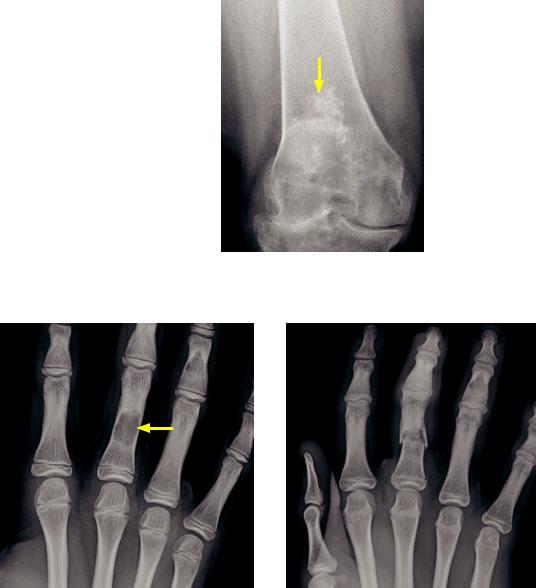
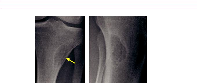
Erosive osteoarthritis
Overview of erosive osteoarthritis
•Erosive osteoarthritis combines the clinical findings of rheumatoid arthritis (e.g., swelling) with imaging findings and distribution that are more similar to osteoarthritis.
•Erosive OA typically affects elderly females.
Erosive osteoarthritis of the hands
•The distribution of erosive osteoarthritis is limited to the hands, where the distribution is the same as degenerative OA (DIPs, CMC of the thumb, and PIPs).
•Erosive OA features a characteristic gull-wing appearance of the DIP joint due to central erosion and marginal osteophytes.
Erosive osteoarthritis: Magnified radiograph of a digit demonstrates characteristic gullwing appearance of the DIP (yellow arrows).
Rheumatoid arthritis (RA)
Overview of rheumatoid arthritis
•Rheumatoidarthritis(RA)isanautoimmunedisorderwherethesynoviumisthetargetofa waxingandwaningimmuneresponse.Rheumatoidfactor(RF)istypicallypositive,although itisnotspecific.RFisanantibodydirectedagainstIgG,whichactivatesthecomplement cascade.RAclinicallypresentswithsymmetrical jointpain,swelling,andmorningstiffness.
•RA first affects the small joints in the hands and wrists. Foot involvement may occur early, so foot radiographs are routinely obtained in suspected cases of RA. In more advanced cases, RA affects the cervical spine, knees, shoulders, and hips.
•The radiographic hallmarks of RA include:
Marginal erosions,whichfirstoccurattheintracapsulararticularmarginsinthe“barearea.”Thebare areaisaregionofexposedbonejustwithinthejointcapsulethatisnotcoveredbythickcartilage.
Soft-tissue swelling.
Diffuse, symmetric joint space narrowing. Periarticular osteopenia.
Joint subluxations.
350
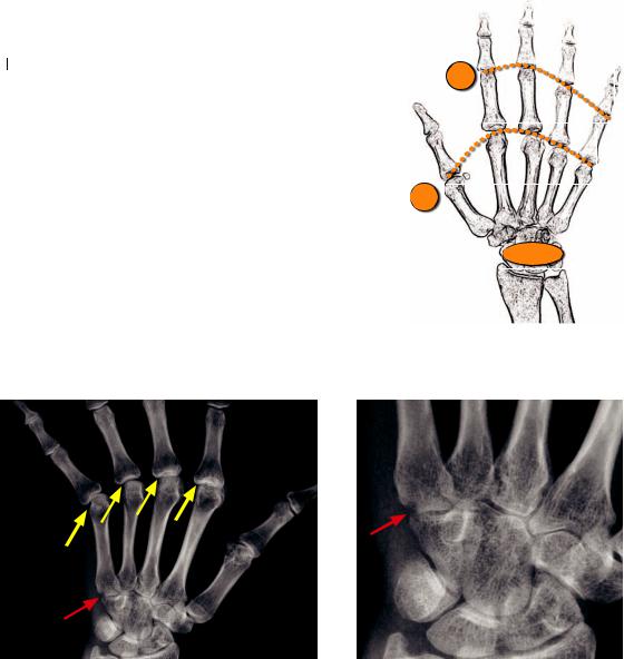
Rheumatoid arthritis in the hand and wrist
The hands are commonly affected in patients with RA.
Typical joints involved are the MCPs, PIPs, and the
carpal articulations. The DIPs are usually spared.
The earliest radiographic changes of RA are softtissue swelling and periarticular osteopenia, reflecting synovitis and hyperemia
Erosions occur early in disease, typically of the radial aspects of the second and third metacarpal heads, the radial and ulnar aspects of the bases of the proximal phalanges, and the ulnar styloid.
Joint subluxations are present in more advanced disease, which typically are not reducible and lead to several common deformities, including:
Boutonnière deformity (PIP flexion and DIP hyperextension).
Swan neck deformity (PIP hyperextension and DIP flexion).
Ulnar subluxation of the fingers at the MCPs.
PIPs
MCPs
Carpals
RA: PA hand radiograph shows ulnar deviation of the
2nd through 5th fingers at the MCPs (arrows). There is periarticular osteopenia. A 5th carpometacarpal joint erosion (red arrow) is better seen on the magnified image to the right.
Close up of the same image better shows the erosion across the 5th carpometacarpal joint (red arrow).
Late-stage rheumatoid arthritis may uncommonly cause ankylosis (fibro-osseous joint fusion occurring after complete cartilage loss) of the wrist. Juvenile idiopathic arthritis (discussed later), in contrast, has a higher propensity for carpal ankylosis.
Rheumatoid arthritis in the feet
The feet are commonly involved in RA. Typically, the metatarsophalangeal (MTP) joints in the forefoot and the talocalcaneonavicular joint in the midfoot are involved. Up to 20% of patients have the MTP joint as the first site of involvement.
Rheumatoid arthritis in the hip
RA causes concentric acetabular cartilage loss, leading to axial migration of the femoral head. In contrast, osteoarthritis more commonly causes superior acetabular cartilage space narrowing and superolateral femoral head migration.
nseverecases,RAmaycausea protrusio deformity,whichisdefinedas>3mmmedial deviationofthefemoralheadbeyondtheilioischiallineinmalesand>6mminfemales.
351
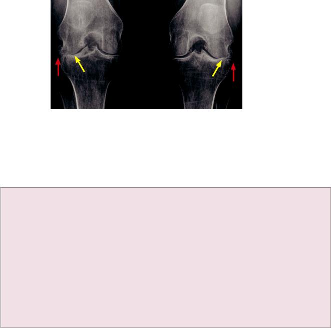
Rheumatoid arthritis in the knee
•All three joint spaces (medial and lateral tibiofemoral and patellofemoral) may be affected by RA in the knee. In contrast, OA tends to first affect the medial tibiofemoral articulation. If osteophytes and symmetrical cartilage space narrowing are present, then secondary osteoarthritis should be considered.
•Unlike the smaller joints affected by rheumatoid arthritis, erosions are not a prominent manifestation of rheumatoid arthritis of the knee.
Rheumatoid arthritis with secondary osteoarthritis:
Frontal PA weight-bearing view of the knees shows cartilage space narrowing of medial and lateral tibiofemoral articulations
bilaterally. The lateral tibiofemoral cartilage spaces are more markedly narrowed (yellow arrows). Prominent osteophytes laterally (red arrows) signify secondary osteoarthritis.
Rheumatoid arthritis in the spine
Atlantoaxial subluxation in |
rheumatoid arthritis |
•The cervical spine is involved in up to 70% of patients. Involvement is increased with more severe and long-standing disease. The thoracic and lumbar spine are almost never involved.
•The general pattern of rheumatoid arthritis in the cervical spine includes subluxation at multiple levels, osteopenia, and erosions of the odontoid, facet joints, vertebral endplates, and spinous processes. Unlike osteoarthritis, there is no bone production.
•A characteristic finding of rheumatoid arthritis is atlantoaxial (C1–C2) subluxation. Atlantoaxial
subluxation may occur in multiple directions, including anterior (most common), posterior, vertical (atlantoaxial impaction), rotatory, and lateral.
•Anterior atlantoaxial subluxation is caused by inflammation of adjacent bursa and resultant laxity of the transverse ligament. Anterior atlantoaxial subluxation may not be apparent if flexion radiographs are not obtained.
Anterioratlantoaxialsubluxationispresentiftheatlanto-dentalinterval(ADI)is>2.5mm(>5mmin children).Theatlanto-dentalintervalisthedistancebetweentheanterioraspectofthedensandthe posterioraspectoftheanteriorringofC1,asmeasuredattheinferioraspectoftheC1–C2articulation.
•Vertical atlantoaxial subluxation (also called atlantoaxial impaction) results from C1–C2 facet erosion and collapse, leading to protrusion of the odontoid through the foramen magnum. This may compress the midbrain.
Directvisualizationoftheodontoidisusuallynotpossibleonalateralradiograph,butimpactionmay causetheanteriorarchofC1(normallyinlinewiththeodontoid)tosinktothelevelofthebodyofC2.
•In the setting of RA, posterior atlantoaxial subluxation is usually due to odontoid erosion. It may also be caused by odontoid fracture.
Rheumatoid arthritis in the shoulder
•Rheumatoid arthritis causes chronic rotator cuff tears leading to the classic high riding humerus.
•Erosions tend to occur in the lateral aspect of the humeral heads. At the acromioclavicular (AC) joints, erosion may lead to “penciling” of the distal clavicle.
Rheumatoid arthritis in the elbow
•Rheumatoid arthritis involves the elbow in approximately one third of patients.
352
Seronegative spondyloarthropathies
Overview of seronegative spondyloarthropathies
•The seronegative spondyloarthropathies are a group of four inflammatory arthropathies, which by definition have negative rheumatoid factor. Patients are usually HLA-B27 positive.
•The four seronegative spondyloarthropathies are ankylosing spondylitis, psoriatic arthritis, reactive arthritis (previously called Reiters arthropathy), and inflammatory bowel disease (IBD) associated arthropathy.
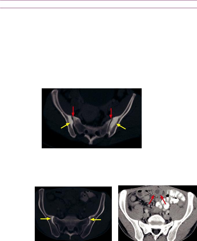
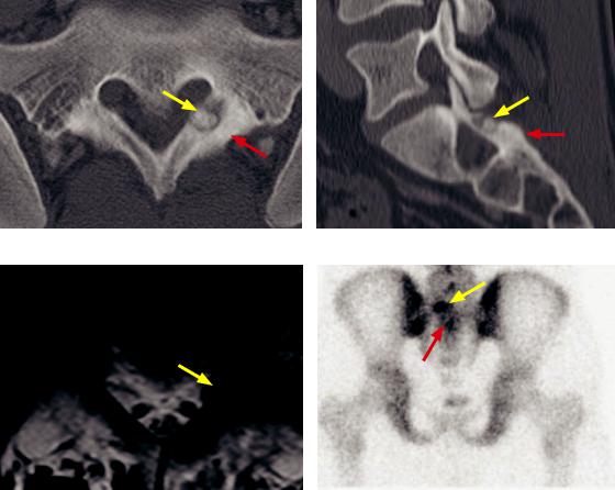
Sacroiliitis is a hallmark of the spondyloarthropathies
•Similar to inolvement in OA, only the inferior aspect of the sacroiliac (SI) joint is affected in seronegative spondyloarthropathies because only the inferior portion is a synovial (diarthrodial) joint. Erosions first involve the iliac aspect of the SI joint.
•Symmetric sacroiliitis is caused by IBD and ankylosing spondylitis (mnemonic: both start with vowels).
Symmetric sacroiliitis:
Axial CT through the pelvis at the level of the sacroiliac joints shows symmetric sclerosis (yellow arrows) and erosions (red arrows) at the iliac aspect of the sacroiliac joints bilaterally.
•Asymmetric sacroiliitis is caused by psoriatic arthritis and reactive arthropathy (mnemonic: both start with consonants).
•An important cause of unilateral sacroiliitis is septic arthritis, especially in an immunocompromised patient or with intravenous drug abuse. Septic arthritis usually presents with erosive changes in a patient with fever and SI joint pain.
Inflammatory bowel disease
Crohn sacroiliitis: Axial CT through the pelvis in bone window (left image) shows symmetric sclerosis (yellow arrows) and erosions of the iliac aspect of the sacroiliac joints bilaterally. Soft-tissue-window CT (right image) shows bowel wall thickening, mural stratification, and hyperenhancement (red arrow), consistent with Crohn enteritis.
•Sacroiliitis associated with inflammatory bowel disease can be seen in patients with ulcerative colitis, Crohn disease, Whipple disease, and status post gastric bypass.
•IBD-associated sacroiliitis is typically symmetrical.
353
Ankylosing spondylitis
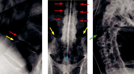
Three patients with ankylosing spondylitis:
Romanus and shiny corner lesions: Lateral radiograph of the upper lumbar spine (left image) shows an erosion of the anterior superior margin of a vertebral body at the discovertebral junction, representing a Romanus lesion (yellow arrow). The superiorly adjacent vertebral body demonstrates sclerosis of its anterior inferior margin, representing the shiny corner sign (red arrow). The shiny corner sign signifies evolution of a prior Romanus lesion.
Bamboo spine: Frontal radiograph of the sacroiliac joints and lumbar spine (middle image) demonstrates symmetrical sacroiliac joint ankylosis (yellow arrows). There are diffuse syndesmophytes, creating an undulating contour of the spinal column, representing the bamboo spine (red arrows). There is fusion of the spinous processes, creating the dagger sign (blue arrow).
Cervical fusion: Lateral radiograph of the cervical spine (right image) shows complete ankylosis of the cervical spine with fusion of the vertebral bodies and facet joints and a pseudarthrosis at C2–C3 (green arrow).
Cases courtesy Stacy Smith, MD, Brigham and Women’s Hospital.
•Ankylosing spondylitis (AS) is predominantly seen in young men with HLA-B27 and presents with back pain and stiffness. AS can be associated with pulmonary fibrosis (upper lobe predominant), aortitis, and cardiac conduction defects.
•The earliest radiographic signs of AS are symmetric erosions, widening, and sclerosis of the sacroiliac joints.
•Subsequently, the spine invariably becomes involved, with radiographic findings following a specific sequence, which ascends from the lumbar to the cervical spine.
Romanus lesions are erosions of the anterior superior or inferior edges of the vertebral body endplates caused by enthesitis (inflammation at a ligament or tendon insertion site) at attachment of the annulus fibrosus to the vertebral body.
Shiny corners represent sclerosis of prior Romanus lesions at the corners of the vertebral bodies. Squaring of the vertebral body disc margins develops due to erosions and bone loss.
Delicate syndesmophytes represents bony bridging connecting adjacent vertebral margins, which create the classic bamboo spine (spinal ankylosis) in late-stage disease.
•In advanced disease, the fully ankylosed spine is at a very high risk of fracture with even minor trauma. CT is necessary for evaluation of even minimal trauma in a patient with advanced AS and pain after trauma.
•An Andersson lesion is a pseudarthrosis occurring in a completely ankylosed spine.
354
Psoriatic arthritis
Psoriatic arthritis (arthritis mutilans form):
PA radiograph of the hand demonstrates swelling of the third digit. There are pencil-in-cup erosions of the DIP and PIP joints (red arrows). Multiple joint subluxations produce telescoping of the digits with a main-en-lorgnette (operaglass hand) deformity.
Case courtesy Barbara Weissman, MD, Brigham and Women’s Hospital.
•Psoriatic arthritis clinically presents as arthropathy in a patient with skin psoriasis.
Psoriatic arthritis most commonly affects the hands. In contrast to RA, mineralization is preserved. Sacroiliitis, when present, is usually asymmetric.
•There are several patterns of psoriatic arthritis, including oligoarthritis, polyarthritis, spondyloarthropathy (producing bulky asymmetric bridging), and arthritis mutilans (a severe form usually affecting the hands, less commonly the feet).
•In the hands, the radiographic hallmark of psoriatic arthritis is diffuse soft-tissue swelling of an entire digit, producing the sausage digit. Pencil-in-cup erosions are also characteristic, most commonly affecting the DIPs. Although hand findings are usually bilateral, involvement tends to be asymmetric. The severe arthritis mutilans variant can cause marked deformity and telescoping digits, also known as the main-en- lorgnette (opera-glass hand) deformity.
Additional findings in the hands include fluffy periostitis and ill-defined erosions of the joint margins.
•In the foot, the great toe IP and MTP joints are most commonly affected. An ivory phalanx represents osteosclerosis and is relatively specific for psoriatic arthritis. Psoriatic arthritis produces a plantar calcaneal spur with periosteal reaction. In contrast, a degenerative calcaneal spur will not feature reactive new bone.
•Inthespine,psoriaticarthritiscausesformationofcoarsebonybridging(bulkylateralbony outgrowths),sometimesindistinguishablefromreactivearthropathy(discussedbelow).
Reactive arthropathy (previously called Reiter disease)
•Reactivearthropathyisaninflammatoryarthritisthoughttobeasequelaofinfectious diarrhea,urethritis,orcervicitis.Sacroiliitisisusuallyasymmetric,asinpsoriaticarthritis.
•Reactive arthropathy predominantly affects the feet, where it has a similar appearance to psoriatic arthritis. Initial radiographic findings include diffuse soft-tissue swelling, joint space loss, aggressive marginal erosions, and juxta-articular osteopenia. Bony mineralization is preserved in the later stage of disease.
•In particular, the calcaneus is a common site of involvement with bony proliferative changes including erosions, enthesophytes, and fluffy periosteal reaction. The posterior-superior aspect of the calcaneus is a frequent site of erosion due to adjacent bursitis. There is often secondary Achilles tendinitis and thickening of the soft tissues.
•In the hands, reactive arthropathy affects the interphalangeal joints and MTPs with erosions and diaphyseal periostitis.
•Reactive arthropathy may affect the spine with formation of coarse bony bridging, which may be difficult to distinguish from psoriatic arthritis.
355

Connective tissue disorders affecting the joints
Systemic lupus erythematosus (SLE)
•Jointabnormalitiesareseenin~90%ofpatientswithsystemiclupuserythematosus(SLE).
•ThekeyradiographicfindingofSLEisreduciblesubluxationsoftheMCPsandPIPs. AlignmentmayappearnormalonaPAviewwhenthehandsarecompressedagainstthe radiographicplate.SubluxationsbecomeapparentintheNorgaard(“ballcatcher’s”or “you’reingoodhandswithAllstate”)orobliqueviewswhenthehandisnotconstrained.
Jaccoud arthropathy
•Jaccoud arthropathy was historically described as being secondary to recurrent rheumatic fever, but some authors feel that SLE and Jaccoud arthropathy are the same disease. Both entities share the same type III hypersensitivity mechanism and feature identical radiographic findings of reducible subluxations in the hand.
Scleroderma
•Sclerodermaisasystemiccollagenvasculardiseasecausedbycollagendepositioninthe skinandsoft-tissues.Thefingertipsareaffectedfirst,withatrophyofthedistalsofttissues.
•Acroosteolysis(resorptionofthedistalportionofthedistalphalanges)ischaracteristic, especiallyifthereisaccompanyingcalcification.Thedifferentialforacroosteolysisincludes:
Collagen vascular disease, including scleroderma.
Neuropathy.
Polyvinyl chloride exposure.
Thermal injury (burn or frostbite). In frostbite the thumb is usually spared because it is clenched in a fist.
Hyperparathyroidism, seen in conjunction with subperiosteal resorption.
Hajdu–Cheney, a rare autosomal dominant syndrome characterized by short stature, craniofacial changes, and progressive acroosteolysis.
•Dystrophic soft tissue and periarticular calcifications are common in scleroderma, which causes tightening and fibrosis of the skin and often leads to joint contractures.
Polymyositis and dermatomyositis
•Polymyositisanddermatomyositisareidiopathicconditionscharacterizedbyinflammation ofmuscle(polymyositis)ormuscleandskin(dermatomyositis).Jointabnormalitiesare rare,althoughperiarticularosteopeniamaybepresentintheseconditions.
•The imaging hallmark of polymyositis and dermatomyositis is soft-tissue calcification. Intramuscular calcifications are most common, although subcutaneous calcifications may also be seen, similar to scleroderma.
Crystal deposition arthropathies
Calcium hydroxyapatite deposition disease (HADD)
•Also called calcific tendinitis, calcium hydroxyapatite deposition disease (HADD) causes crystals to be deposited in the periarticular tissues. Hydroxyapatite crystals typically do not deposit directly within the joints (the articular cartilage and synovium are spared), but instead amorphous deposition of calcium forms within tendons.
•Thesupraspinatustendonintherotatorcuffismostcommonlyaffectedintheshoulder.
•Calcifictendinitisoftheprevertebrallonguscolimusclemaycauseneckpain,odynophagia, fever,andprevertebraleffusion,andmayclinicallymimicaprevertebralabscess.
•An intra-articular variant seen in the shoulder, called Milwaukee shoulder, leads to rapid destruction of the rotator cuff and the glenohumeral joint.
356
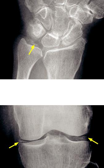
Calcium pyrophosphate dihydrate deposition disease (CPPD)
•Calcium pyrophosphate dihydate deposition disease (CPPD) is an inflammatory arthropathy caused by intra-articular deposition of calcium pyrophosphate dihydrate crystals. Microscopically, the rhomboid crystals of CPPD are positively birefringent.
•CPPD has been called the “great mimicker” of other arthropathies. Clinical manifestations of CPPD disease include pseudoosteoarthritis (common), pseudogout (10–20%), pseudorheumatoid (2–6%), pseudoneuropathic (<2%), and asymptomatic (10–20%). CPPD is rare in a patient under 50. Crystal deposition may be idiopathic or associated with hemochromatosis, hyperparathyroidism, and hypophosphatasia.
•The hallmark radiographic finding of CPPD disease is chondrocalcinosis, which is calcification of hyaline (articular) or fibro- (meniscal) cartilage. Radiographs of the knees, wrist, and pelvis (pubic symphysis) are nearly 100% sensitive for the detection of chondrocalcinosis.
•In the wrist, chondrocalcinosis tends to affect the triangular fibrocartilage complex (TFCC). Advanced disease may lead to scapholunate advanced collapse (SLAC) wrist. SLAC wrist is proximal migration of the capitate between the dissociated scaphoid and lunate, and may also be seen in RA or trauma.
Frontal radiograph of the wrist demonstrates chondrocalcinosis of the triangular fibrocartilage complex (TFCC, arrow), a common site of chondrocalcinosis.
•In the knee, the patellofemoral compartment is affected first, but all three compartments may become involved. Isolated degenerative changes of the patellofemoral joint are highly suggestive of CPPD. Prominent subchondral cysts are also especially suggestive of CPPD.
Frontal radiograph of the knee demonstrates chondrocalcinosis of the
menisci in the same patient.
•In the hands, involvement of the second and third MCP joints is typical, producing characteristic hook-like or drooping osteophytes from the radial aspect of the metacarpal heads. A similar appearance can be seen in hemochromatosis, which typically features more extensive involvement of the MCPs.
357
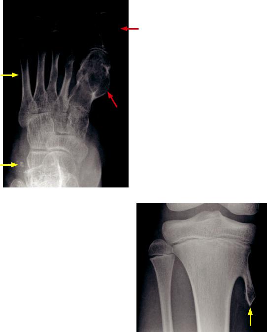
Gout
•Goutisacrystal-inducedinflammatory arthropathycausedbysodiumurate depositioninthejoints.Excessuricacid maybesecondarytounder-excretion (morecommon,typicallycausedbyrenal insufficiency)oroverproduction(muchmore rare,typicallyseeninyoungerpatients).
It takes about 10–20 years of hyperuricemia before the clinical syndrome of gout develops.
Microscopically,goutcrystalsarenegatively birefringentneedle-likecrystalswithinneutrophils.
•The great toe is most commonly involved, but gout can occur in any joint.
•Radiographic hallmarks are sharply marginated erosions with overhanging margins, associated with soft-tissue gouty tophi.
•Joint spaces are typically well preserved until late in the disease. Bony mineralization is preserved.
AP radiograph of the foot shows soft-tissue swelling surrounding the second MTP. An erosion of the head of the second metacarpal features a characteristic overhanging margin (arrow).
Metabolic, hematologic, and miscellaneous arthropathies
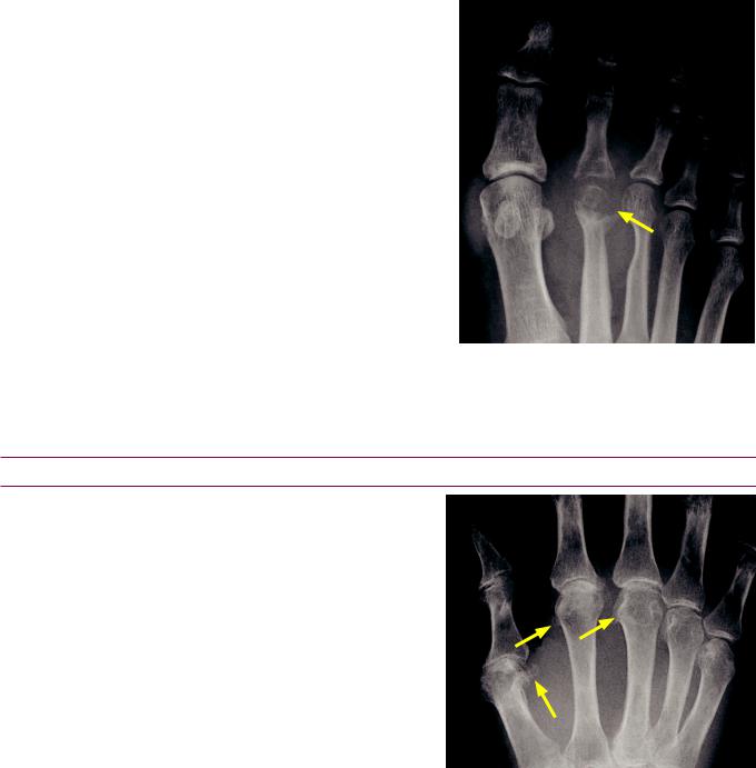
Hemochromatosis
•Hemochromatosis arthropathy affects 50% of those with hemochromatosis, an autosomal recessive disease of altered iron metabolism. The arthropathy is caused by deposition of iron and calcium pyrophosphate dihydrate crystals.
Hemochromatosis clinically presents with bronze pigmentation, diabetes, cirrhosis, congestive heart failure, and arthropathy.
•Inthehand,thetypicallocationof hemochromatosisarthropathyisthe MCPjoints,producingcharacteristic hook-likeosteophytesatthemetacarpal heads.Calciumpyrophosphatedihydrate depositiondisease(CPPD)canappear identicalwheninvolvementisisolatedto the2ndand3rdMCPs.IncontrasttoCPPD, hemochromatosismayinvolveallMCPs.
Hemochromatosis: Frontal radiograph of the hand shows cartilage loss and beak-like osteophytes of the 1st through 3rd MCPs (arrows).
Case courtesy Stacy Smith, MD, Brigham and Women’s Hospital.
Acromegaly
•Acromegaly (excess growth hormone) causes arthropathy due to enlargement of the articular cartilage and subsequent degeneration. In contrast to all other arthropathies, joint spaces are widened in early disease due to cartilage hypertrophy. Later in disease, secondary osteoarthritis occurs with cartilage space narrowing.
•In the hand, beak-like osteophytes of the metacarpal heads and spade-like enlargement of the terminal tufts are characteristic.
358
Amyloid arthropathy
•Amyloidarthropathyisararenoninflammatoryarthropathyduetoinfiltrationofbones, jointsandsofttissuesbybeta-pleatedsheetsofaminoacids.Primarysystemicamyloidosis isassociatedwithmonoclonalplasmacelldyscrasia.Secondaryamyloidosisisassociated withchronicunderlyinginflammationorinfection.Anotherformofamyloidosisiscaused byβ2-microglobulinaccumulationinpatientsonchronichemodialysis.
•Acharacteristicclinicalfindingofamyloidosisisbulkysoft-tissuenodulesintheshoulder superimposeduponatrophicshouldermuscles,producingtheshoulder-pad sign.
•Imaging findings of amyloid arthropathy are nonspecific but may resemble RA. Intra-articular deposits cause articular cartilage destruction. Soft-tissue nodules and erosions may be present.
Ochronosis (alkaptonuria)
•Ochronosis is the connective tissue manifestation of alkaptonuria. Alkaptonuria is caused by a defect in homogentisic acid oxidase, causing homogentisic acid polymers to accumulate in the visceral organs, intervertebral discs, and joints. Clinically, homogentisic acid in the urine turns black when exposed to air.
•A specific finding of ochronosis is intervertebral disc calcifications at every level with accompanying disk space narrowing.
Multicentric reticulohistiocytosis
•Multicentric reticulohistiocytosis is a rare disease where lipid-laden macrophages are deposited in soft tissues and periarticular tendons, forming skin nodules and erosions with sclerotic margins.
•The well-defined erosions of multicentric reticulohistiocytosis tend to affect the DIPs symmetrically. Other radiographic findings of multicentric reticulohistiocytosis include soft-tissue nodules and preserved bone density.
•Joint destruction may be rapid and progressive, producing an arthritis mutilans appearance.
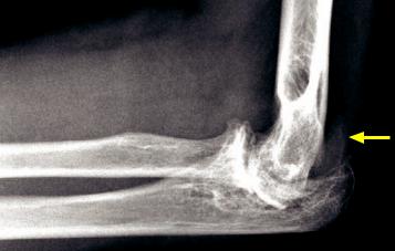
Hemophilic arthropathy
Hemophilic arthropathy: Lateral elbow radiograph shows severe joint space narrowing. Increased soft-tissue density surrounding the joint (arrow) is due to synovial hemosiderin deposition.
In this case, the radial head is not enlarged, but classically hemophilia features an enlarged radial head.
•Hemophilia is an X-linked inherited disorder of either factor VIII (hemophilia A) or IX (hemophilia B; Christmas disease) deficiency causing recurrent bleeding.
•Hemophilia most often affects the knees, elbows, and ankles. Recurrent hemarthrosis results in synovial hypertrophy and hyperemia. The hyperemia may cause epiphyseal enlargement and early fusion.
Characteristic appearance of the elbow is an enlarged radial head and widened trochlear notch.
Characteristic appearance of the knee is squaring of the patella and widened intercondylar notch.
359
•Secondary arthritis may lead to marked joint space narrowing.
•Deposition of iron in the synovium causes increased soft-tissue density around joints.
•Juvenile idiopathic arthritis (discussed below) also causes articular hyperemia and may have similar radiographic findings, especially in the knee (widened intercondylar notch) and elbow (enlarged radial head).
•Pseudotumor of hemophilia is a benign lesion caused by recurrent intraosseous or subperiosteal bleeding. The chronic cyclical bleeding leads to bony scalloping and pressure erosion, often with an associated soft-tissue mass.
On radiography, pseudotumor is benign-appearing, with well-circumscribed and sclerotic margins. Pseudotumor may have a complex MRI appearance due to different stages of blood products.
Juvenile idiopathic arthritis (JIA), previously called juvenile rheumatoid arthritis
•Juvenile idiopathic arthritis (JIA) is a spectrum of related chronic inflammatory arthropathies affecting children under 16 years of age.
•Monoarticular or pauciarticular (most common) JIA may affect either a single joint or a few joints including the knees, ankles, elbows, or wrists.
•Polyarticular JIA is a systemic disease affecting multiple joints including the hands, feet, and cervical spine in addition to the joints affected by mono/pauciarticular disease.
•AvariantofJIAisStilldisease,whichisasystemicdisorderaffectingchildrenyounger than5,featuringacutefebrileillness,rash,adenopathy,pericarditis,andmildarthralgias.
•Radiographic hallmarks of JIA are abnormal bone length or morphology due to hyperemia in a skeletally immature patient. Growth disturbances are more
commonly seen in early-onset disease. Abnormal morphology results from epiphyseal overgrowth and enlargement (ballooning) of the ends of bone. Affected joints demonstrate premature skeletal maturation and physeal fusion.
•In the hand, premature fusion of the growth plate may cause brachydactyly.
PA radiograph of the hand in a patient with juvenile idiopathic arthritis shows a shortened 5th metacarpal (arrow).
•In the knee, the characteristic appearance is a widened intercondylar notch, metaphyseal flaring, and uniform joint space narrowing. This appearance can be similar to hemophilia.
•In the elbow, there is characteristic enlargement of the radial head and trochlear notch, with uniform cartilage space narrowing. These findings can also be seen in hemophilia.
•In the hips, symmetrical cartilage space narrowing, protrusio deformity, and gracile appearance of the femoral shaft are characteristic.
•Ankylosis may occur in the wrist and zygapophyseal (facet) joints of the cervical spine. Ankylosis occurs much more commonly in juvenile idiopathic arthropathy compared to adult rheumatoid arthritis. The differential diagnosis of a child with cervical spine ankylosis is Klippel–Feil syndrome, which is failure of cervical segmentation.
360
Neuropathic arthropathy (Charcot joint)
•Neuropathic arthropathy, also called Charcot joint, is a destructive form of arthritis caused by neurosensory deficit. Lack of sensation ultimately causes severe degenerative changes with fragmentation of bone and cartilage.
•Neuropathic arthropathy clinically presents as a (usually) painless, swollen joint.
•Neuropathic arthropathy can be caused by any process that affects sensory nerves. The peripheral neuropathy of diabetes is implicated most frequently, typically affecting joints in the ankle and foot. Other causes include syringomyelia (usually affecting the upper extremity), chronic alcohol abuse, amyloid, spinal tumors, and very rarely syphilis or leprosy.
•Two forms of neuropathic arthropathy are hypertrophic (more common) and atrophic variants.
•The hypertrophic variant looks like anarchy in a joint, with destruction, dislocation (or subluxation), debris, disorganization, and no demineralization.
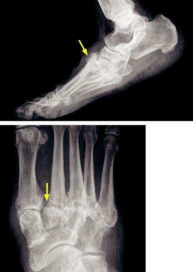
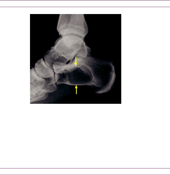
Neuropathic arthropathy:
Lateral radiograph of the foot demonstrates subluxation of the Lisfranc joint (yellow arrow). Hypertrophic degenerative changes are present at the tarsometatarsal joint
There was no history of trauma.
AP radiograph of the foot in the same patient shows divergent lateral subluxation of the Lisfranc joint in the same patient.
The yellow arrow points to the enlarged space between the first and second metatarsals. Offset of the second metatarsal base with the mid cuneiform may be an early finding of Lisfranc injury.
•The atrophic variant of neuropathic arthropathy occurs most commonly in the shoulder. It features a classic radiographic appearance of humeral head resorption with a sharp, surgical-like margin. Syringomyelia should be suspected in upper extremity neuropathic arthropathy and confirmed by cervical spine MRI.
361

Sarcoidosis
•Sarcoidosis is a multisystemic granulomatous disease. Lung findings, including adenopathy and parenchymal disease, are present in the majority of patients and are the primary manifestation of disease.
•Bony manifestations of sarcoid are rare. A characteristic finding in the hands is lacelike lytic lesions in the middle or distal phalanges.
•Sarcoidosismayalsomanifestasacuteorchronicpolyarthritis,althoughthereareno distinctiveradiographicpatterns.Ankleinvolvement,especiallyifbilateralorassociatedwith erythemanodosum,shouldraisesuspicionofsarcoidosisandpromptachestradiograph.
Puttingittogether:Evaluationofthehandsforarthritis:ABCDEs
A: Alignment
Subluxations
•Non-reducible subluxations are seen in rheumatoid arthritis.
•Reducible subluxations are typical for systemic lupus erythematosus (SLE) or Jaccoud arthropathy. In contrast to rheumatoid, erosions are not typically seen in SLE.
Dislocations
•Joint dislocations may be present in advanced rheumatoid arthritis.
B:Bone mineral density
Evaluation of bone mineral density
•Bone mineral density is evaluated by assessing the cortical thickness of the second metacarpal shaft. The cortical thickness should be at least 1/3 of the total width of the metacarpal shaft. Evaluation of bone density helps to distinguish between inflammatory and non-inflammatory arthropathies.
Diffuse osteopenia
•Diffuse osteopenia is associated with advanced rheumatoid arthritis.
•Note that generalized osteoporosis secondary to a medical condition can be seen in any arthropathy.
Periarticular osteopenia
•Periarticular osteopenia is a nonspecific finding that can be seen in rheumatoid arthritis or the early stage of any inflammatory arthropathy.
B:Bone creation
Osteophytosis
•An osteophyte is the result of endochondral bone formation that occurs at the margins of a joint and is caused by degeneration of the adjacent articular cartilage.
•Osteophyte formation is the hallmark of osteoarthritis, but can also be seen as a secondary finding in other conditions such as CPPD and hemochromatosis.
Periosteal new bone
•Periosteal new bone formation may be triggered as a reparative response to erosive change.
•Periosteal reaction is seen in psoriatic arthritis and reactive arthropathy, but is less common in adult rheumatoid arthritis.
362

Bony ankylosis
•Ankylosis is the bony fusion of a joint and is seen in aggressive arthropathies that destroy articular cartilage.
•Ankylosis of the wrist and cervical spine is a typical finding in advanced juvenile idiopathic arthropathy. In advanced rheumatoid arthritis, ankylosis may uncommonly occur in the wrists.
•Ankylosis of the DIPs is a typical finding in some cases of psoriatic arthritis.
C:Calcification
•Crystal deposition diseases known to cause arthritis are calcium pyrophosphate dihydrate (CPPD) deposition disease, hydroxyapatite deposition disease (HADD), and sodium urate monohydrate arthropathy (gout).
Chondrocalcinosis
•Chondrocalcinosis is typically due to deposition of calcium pyrophosphate dihydrate crystals in cartilage, which may be idiopathic or due to hyperparathyroidism or hemochromatosis.
Calcification of tendons
•Calcification of tendons is typically caused by deposition of hydroxyapatite crystals, and is also known as calcific tendinitis. Tendon calcification can also be seen in CPPD.
Soft-tissue calcification
•The radiolucent urate crystals that make up a gouty tophus may precipitate calcium.
•Soft-tissue calcifications can also be seen in scleroderma, dermatomyositis, polymyositis, and SLE.
C:Cartilage spaces
Preserved cartilage spaces
•Gout generally features preserved cartilage spaces. Focal narrowing may be present in the region of the gouty tophi and erosion.
Asymmetrical narrowing
•Asymmetrical cartilage space narrowing is typical of osteoarthritis and gout.
Symmetrical narrowing
•Symmetrical cartilage space narrowing is seen in the inflammatory arthropathies.
Increased cartilage spaces
•Acromegaly produces cartilage space widening early in the disease, prior to the development of secondary osteoarthritis.
D:Distribution
Hand: DIP and PIP
•Osteophytespresent:Osteoarthritis(noerosions);erosiveosteoarthritis(erosionspresent).
•No osteophytes: Psoriatic arthritis (erosions present).
Hand: MCP and PIP
•Without new bone formation: Rheumatoid arthritis.
Hand: MCP only
•With erosions: Rheumatoid arthritis.
•With osteophytes: CPPD or hemochromatosis.
363

Wrist: CMC
•Osteophytes without erosions: Osteoarthritis.
•Osteophytes with erosions: Erosive osteoarthritis.
•Erosions without osteophytes: Gout.
Wrist: diffuse (pan-carpal)
•Inflammatory arthropathy.
•Post-traumatic osteoarthritis can occur anywhere in the wrist.
E: Erosions
•Erosions are first seen in the “bare” area of bone just inside the joint at the edge of attachment of synovium. Erosions may subsequently spread further into the joint and even destroy the entire joint in severe cases.
Variable erosions
•Rheumatoid arthritis may feature erosions in certain characteristic locations, including the radial aspect of the second and third metacarpal heads, the bases of the proximal phalanges, and the ulnar styloid.
Pencil in cup erosion
•Psoriatic arthritis.
Gullwing erosions
•Erosive osteoarthritis.
Overhanging margin of cortex
•Gout. Caused by chronic erosion from a tophus remodeling the cortex.
S:Soft-tissue swelling
Symmetrical swelling around a joint
•A characteristic finding of rheumatoid arthritis is joint distension, which radiographically appears as soft-tissue swelling, although this finding can be seen in any inflammatory arthropathy.
Asymmetrical swelling around a joint
•In osteoarthritis, characteristic locations of nodular soft-tissue swelling are due to osteophytes and capsule–ligamentous thickening.
Heberden node: Swelling of the DIP.
Bouchard node: Swelling of the PIP.
Swelling of an entire digit
•“Sausage digit” in the hand: Psoriatic arthritis or reactive arthropathy.
•“Sausage digit” in the foot: More commonly reactive arthropathy.
Lumpy-bumpy soft-tissue swelling
•Lumpy-bumpy soft-tissue swelling is typically caused by infiltration with a foreign substance, such as gouty tophi, sarcoidosis, amyloid, or multicentric reticulohistiocytosis.
364
Bone tumors and tumor-like lesions: approach
Morphology
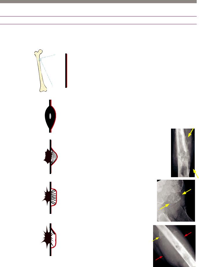
Periosteal reaction
The morphology of a bone lesion’s associated periosteal reaction gives an important to clue to the rate of growth, and hence the aggressiveness, of the lesion.
Normal periosteal anatomy
 Periosteum covers essentially the entire bone excluding the joint surfaces.
Non-agressive: Solid periosteal reaction
A slow-growing process, such as oseoid osteoma (radiolucent nidus), incites the periosteal cells
to lay down bone in a smooth, continuous manner.
Aggressive: Lamellated periosteal reaction
An irregularly growing process that grows in starts and stops produces a characteristic lamellated or onion-skinned appearance (arrows). When the lesion is growing quickly the periosteal cells don’t have enough time to lay down bone.
Very aggressive: Sunburst periosteal reaction
A very fast growing lesion pushes the periosteal cells outward as the lesion expands, with each periosteal cell leaving a
trail of bone formation that looks like hair on end or sunburst (arrows).
Very very aggressive: Codman triangle
A lesion that grows so aggressively that the periosteum does not even have a chance to lay down
visible calcification except at the periphery produces a characteristic Codman triangle (yellow arrow).
Note the associated soft tissue mass (red arrows).
365

Pattern of bone destruction: Margin analysis
Lodwick classification of bone destruction
•Analysis of a bone lesion’s margins (i.e., zone of transition from normal to abnormal bone) helps to characterize the bony destruction and stratify a lesion as aggressive or non-aggressive.
•A sharply marginated zone of transition usually denotes a less aggressive lesion with slower rate of growth. The faster the rate of growth, the more aggressive the lesion may be. A wide zone of transition suggests rapid growth and a more aggressive lesion.
•The Lodwick classification of bone destruction helps to stratify aggressiveness:
•Type 1: Geographic pattern: Margins have a thin zone of transition and may be sclerotic or well-
defined.
IA: Thin sclerotic margins. Almost always non-aggressive. 1B: Well-defined margins. Usually non-aggressive.
1C: Any part of the margin is indistinct.
•Type 2: Moth-eaten pattern: It is difficult to define any border at all. Aggressive.
•Type 3: Permeative: The permeative pattern is characterized by multiple tiny holes that infiltrate the bone. This pattern is very aggressive and is seen in lymphoma, leukemia, and Ewing sarcoma.
New matrix created by tumor
•Matrix produced by an osteoid lesion, such as malignant osteosarcoma, appears as fluffy, cloud-like bone.
•Matrix produced by a chondroid lesion, such as a benign enchondroma or malignant chondrosarcoma, has a ring and arc or popcorn-like appearance.
•A ground glass matrix describes blurring of the trabeculae and is seen in fibrous dysplasia, a benign fibroosseous lesion involving abnormal proliferation of intraosseous fibroblasts.
Unique features
•The fallen-fragment sign is seen in a simple (unicameral) bone cyst with pathologic fracture.
•An aneurysmal or expansile appearance suggests an aneurysmal bone cyst.
•Resorption of distal clavicles or tumoral calcinosis can suggest hyperparathyroidism, which can cause brown tumors.
Patient age
•The two most likely considerations of an aggressive lytic bone lesion in a patient over age 40 are metastasis or myeloma.
•Under age 20, an aggressive lytic lesion is most likely to represent eosinophilic granuloma, infection, or Ewing sarcoma.
Location within bone
Eccentric within bone
•Giant-cell tumor, chondroblastoma, aneurysmal bone cyst, non-ossifying fibroma, and the rare chondromyxoid fibroma are located eccentrically within the bone.
Central (in the middle of a long bone)
•Simple bone cyst, enchondroma, and fibrous dysplasia are located centrally within the bone.
366
Bone lesions organized by cell of origin
Bone forming (osteo-) lesions
Benign and incidental: Enostosis (bone island)
•Enostosis (commonly called a bone island) is an extremely common incidental finding of a small spiculated osteoblastic focus.
•A bone island is only clinically significant in that it may rarely be difficult to differentiate from an osteoblastic metastasis, osteoid osteoma, or a low-grade osteosarcoma.
•Agiantvariant(>2cm)maybemostdifficulttodifferentiatefromlow-gradeosteosarcoma.
•Bone scan of bone island is usually normal.
•Osteopoikilosis is an autosomal dominant syndrome of multiple bone islands and keloid formation.
•Osteopathia striata is a benign, asymptomatic sclerotic dysplasia characterized by linear bands of sclerosis in the long bones and fan-like sclerosis in the flat pelvic bones. Bone scan is typically normal.
Benign and incidental: Osteoma
•Osteoma is a slow-growing lesion that may arise from the cortex of the skull or the frontal/ethmoid sinuses.
•Gardner syndrome is an autosomal dominant syndrome of multiple osteomas, intestinal polyposis, and soft-tissue desmoid tumors.
•In contrast to a bone island, osteoma arises from the cortex rather than the medullary canal.
Benign: Melorheostosis
•Melorheostosis(alsocommonly spelledmelorrheostosis)isanonneoplasticproliferationofthickened andirregularcortexwithatypical candle-waxappearance.
•It clinically presents with pain, decreased range of motion, legbowing, and leg-length discrepancy.
•Melorheostosis may be associated with scleroderma-like skin lesions over the affected region.
•Melorheostosis is usually seen in a single lower limb, in the distribution of a single sclerotome. A sclerotome represents a zone supplied by a single sensory nerve.
•Melorheostosis features intense uptake on bone scan.
Osteoma: Axial head CT in bone window shows a densely sclerotic osteoma arising from the cortex of the frontal sinus.
Melorheostosis: Frontal radiograph of the right tibia and fibula shows thickened, irregular wavy cortex of the medial tibia (arrow).
Case courtesy Michael Callahan, MD, Boston Children’s Hospital.
367
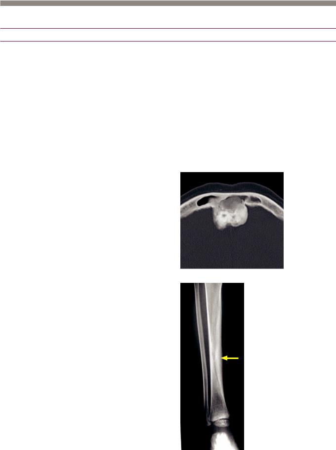
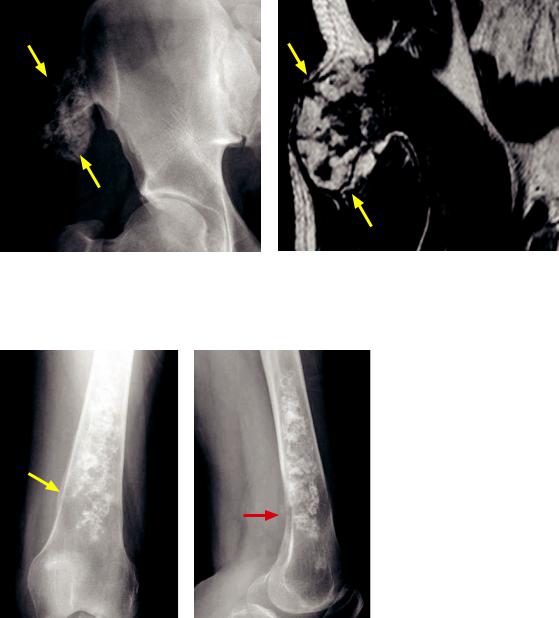
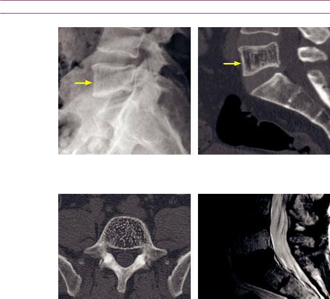
Benign: Osteoid osteoma
•Osteoid osteoma is a benign osteoblastic lesion characterized by a nidus of osteoid tissue surrounded by reactive bone sclerosis. The etiology is controversial. Inflammatory, vascular, and viral causes have been proposed.
•The classic clinical presentation is night pain relieved by aspirin in a teenager or young adult.
•Osteoid osteoma tends to occur in the diaphyses of the leg long bones (femur and tibia) most commonly. About 20% occur in the posterior elements of the spine. Spinal osteoid osteoma is an important cause of painful scoliosis.
•On radiography and CT, a lucent nidus is surrounded by sclerosis. There is often central calcification within the nidus. Bone scan will be positive, with the double density sign representing intense uptake centrally in the region of the nidus and adjacent reactive uptake corresponding to sclerosis. Osteoid osteoma can be difficult to see on MRI alone. The nidus is usually low-signal on T1-weighted images and reactive marrow edema can obscure the lesion on T2-weighted images.
Axial CT |
Sagittal CT |
T1-weighted MRI Tc-99m MDP bone scan (posterior projection) Osteoid osteoma: CT shows a radiolucent nidus in the left sacral ala (posterior element location) with a large central calcification (yellow arrows) with adjacent sclerosis (red arrows). The lesion is less conspicuous on MRI and has very low signal on the T1-weighted image (yellow arrow). The posterior bone scan shows increased radiotracer uptake (yellow arrow) of the nidus, with a double density sign (red arrow; corresponding to reactive sclerosis).
•Treatment of osteoid osteoma is interventional radiology radiofrequency ablation, surgical curettage, or resection.
368
Benign: Osteoblastoma
•Osteoblastoma is a benign osteoid-producing tumor that is histologically the same as an osteoid osteoma but is greater than 2 cm in size.
•Osteoblastoma is approximately four times less common than osteoid osteoma, although it also occurs in the adolescent/young adult age range and also presents with pain. Interestingly, the pain of osteoblastoma is not typically relieved by aspirin.
•Themostcommonlocationistheposteriorelementsofthespine,occurringanywherefrom thecervicalspinethroughthesacrum.Osteoblastomamayalsooccurinthefemurandtibia.
•The most common radiographic appearance of osteoblastoma is a lytic lesion with mineralization. Very rarely, osteoblastoma may be aggressive with a large soft-tissue mass, but lacking metastatic potential. Secondary aneurysmal bone cyst may be seen, especially when spinal in location.
•A lytic lesion in the posterior elements of a young person may represent an osteoblastoma or aneurysmal bone cyst. If any mineralization is present within the lesion, osteoblastoma should be favored.
Malignant: Osteosarcoma
•Osteosarcomarepresentsaheterogeneousgroupofmalignanttumorswheretheneoplastic cellsarederivedfromosteoidlineageandmostsubtypesproduceanosteoidmatrix.
•Osteosarcoma can be primary or secondary. Secondary osteosarcoma may arise from Paget disease or after radiation. Secondary osteosarcoma in Paget disease is extremely aggressive.
•General imaging hallmarks of osteosarcoma are bony destruction, production of osteoid matrix, aggressive periosteal reaction, and an associated soft-tissue mass. Early osteosarcoma may be only evident as subtle sclerosis.
•There are more than 10 primary subtypes. The four most important subtypes are conventional (most common), telangiectatic, and the two juxtacortical subtypes including parosteal (pronounced PAR-osteal) and periosteal (pronounced PERI-osteal).
•Conventional(intramedullary)osteosarcomarepresents75%ofosteosarcomasandoccurs inadolescents/youngadultsusuallyaroundthekneeinthemetaphysisofthefemurortibia.
Conventionalosteosarcomafeaturesanintramedullaryosteoidmatrix,bothintramedullaryand corticalbonedestruction,aggressiveperiostealreaction(sunburstorCodman),andasoft-tissuemass.
Radiograph |
T1-weighted MRI |
T2-weightedMRIwithfatsupp. |
Conventionalosteosarcoma:Radiograph(leftimage)showsaregionofsubtleheterogeneoussclerosis inthemedialproximaltibialmetaphysis(arrow).Noperiostealreactionisapparent,whichisunusual forosteosarcoma.T1-weightedMRIshowsawell-definedregionofmarrowreplacementbytumor (redarrows).T2-weightedMRIwithfatsuppressiondemonstratesdiffusemarrowedemainthetibia, witharegionofdecreasedT2signal(arrow)likelycorrespondingtothesclerosisseenonradiography.
369
•Telangiectatic osteosarcoma is an osteolytic destructive sarcoma, which may mimic a benign aneurysmal bone cyst on imaging.
The presence of solid nodular components on MRI helps to differentiate a telangiectatic osteosarcoma from a benign aneurysmal bone cyst.
Unlike other osteosarcomas, telangiectatic osteosarcoma does not produce any bony matrix. Pathologically, telangiectatic osteosarcoma is vascular with large cystic spaces filled with blood.
Although telangiectatic osteosarcoma is an aggressive lesion, new treatment options increase survival, which is now slightly improved compared to a conventional osteosarcoma.
•Parosteal (PAR-osteal) osteosarcoma is a juxtacortical osteosarcoma that arises from the outer periosteum. It most commonly occurs at the posterior aspect of distal femoral metaphysis and has a cauliflower-like exophytic morphology (mnemonic: parboil cauliflower before eating). A lucent line may be seen separating it from the cortex.
Patients are usually in their 3rd and 4th decades, older compared to other osteosarcoma subtypes.
Parosteal osteosarcoma is the least malignant of all osteosarcomas, with ~90% 5-year survival.
•Periosteal (PERI-osteal) osteosarcoma, the other juxtacortical osteosarcoma, is a rare osteosarcoma variant arising from the inner periosteum. It features cortical thickening, aggressive periosteal reaction, and a soft-tissue mass. Histologically, periosteal osteosarcoma may show chondroid differentiation.
The most common location of periosteal osteosarcoma is the diaphysis of the femur or tibia.
Patients tend to be younger than 20 years old.
•Regardless of subtype, osteosarcoma may metastasize to lungs, where the metastases typically calcify.
Frontal radiograph shows innumerable calcified osteosarcoma metastases
in the thorax and visualized portion of the upper abdomen, demonstrating fluffy osteoid matrix.
370
Cartilage-forming (chondro-) lesions
Benign: Synovial chondromatosis/osteochondromatosis
Synovial osteochondromatosis: Frontal external rotation (left image) and internal rotation (right image) radiographs of the shoulder show multiple small round calcifications tracking along the expected location of the long head of the biceps tendon sheath (arrows).
•Synovial chondromatosis is non-neoplastic synovial metaplasia characterized by the formation of intra-articular lobulated cartilaginous nodules, which may or may not ossify. It is usually a monoarticular disorder.
•The cartilaginous foci often ossify, in which case the term osteochondromatosis is used.
•Synovial proliferation tends not to directly cause arthropathy, although the intraarticular nodules may cause mechanical erosions and secondary osteoarthritis.
•The most common location is the knee. The shoulders, hip, and elbow may also be affected.
•Radiography shows multiple round intra-articular bodies of similar size and variable mineralization. The primary differential is intra-articular bodies from osteoarthritis; however, in osteoarthritis the bodies tend to be more varied in size and shape and fewer in number. Diagnosis can be difficult in the absence of calcification, especially if mechanical erosions are present.
•MRI appearances are variable, depending on the degree of ossification and the presence of chondroid matrix. When calcified or ossified, MRI will show multiple globular and rounded foci of low signal.
•The MRI finding of multiple intra-articular low-intensity foci is nonspecific and can also be seen in pigmented villonodular synovitis (PVNS). A radiograph will clearly show rounded calcified bodies in osteochondromatosis.
•Very rarely, malignant degenerate to chondrosarcoma may occur.
371
Benign: Enchondroma
•Enchondroma is a benign lesion of mature hyaline cartilage rests.
•Inthelongbones,enchondroma featurescharacteristicchondroid (popcornor ringandarc)calcifications.
•Thedifferentialdiagnosisof
enchondroma includes medullary bone infarct(whichproducesserpentine sclerosis)andchondrosarcoma.
•MRI is usually able to differentiate between infarct and enchondroma. Enchondroma has a characteristic lobulated hyperintense signal on T2-weighted images.
Radiograph of the distal femur shows an enchondroma with characteristic ring and arc chondroid-type calcification (arrow).
•When occurring in the hand, enchondroma typically does not produce visible matrix and appears as a geographic lytic lesion.
•Enchondroma may be complicated by pathologic fracture.
Enchondroma of the middle finger proximal phalanx (left image) complicated by pathologic fracture seen in a subsequent radiograph (right image). The enchondroma (arrow) has no perceptible chondroid matrix, which is a typical appearance of an enchondroma in the hand. Note the interval partial fusion of the physes occurring in the time interval between the two radiographs.
•Enchondroma may rarely undergo malignant transformation. Aside from Ollier and Maffucci syndrome (discussed on the following page), malignant transformation to chondrosarcoma is very rare, with the key finding being new pain in the absence of fracture. Other findings suggestive of malignant transformation include:
Soft-tissue mass.
Destruction of the cortex.
Thickening of the cortex.
•Treatment of enchondroma is curettage.
372
•Multiple enchondromas are seen in Ollier (multiple enchondromas only) and Maffucci (multiple enchondromas and venous malformations producing phleboliths) syndromes, the two familial enchondromatoses. Both syndromes carry an increased risk of malignant transformation to chondrosarcoma, with a higher risk in Maffucci syndrome.
Frontal radiograph of the foot in a patient with Maffucci syndrome shows numerous expansile enchondromas most prominently in the great toe (red arrows). Multiple phleboliths (yellow arrows) represent soft-tissue venous malformations.
Note the relative paucity of chondroid calcification, which is typical of the enchondromas seen in Maffucci syndrome.
Benign: Osteochondroma
•An osteochondroma is a benign cartilagecapped bony growth projecting outward from bone, often pedunculated. It is the most common benign bone lesion.
•Osteochondroma may present clinically as a palpable mass, which usually stops growing at skeletal maturity.
•Key features are the continuity of cortex of host bone with the cortex of the osteochondroma and communication of the medullary cavities. Osteochondroma arises from the metaphysis and grows away from the epiphysis.
•An uncommon complication is malignant transformation to chondrosarcoma.
Like enchondroma, the presence of pain in the absence of a pathologic fracture is a red flag.
An associated soft-tissue mass is usually present with malignant transformation.
A cartilage cap thickness >2 cm on MRI suggests malignant transformation to chondrosarcoma.
Osteochondroma: Frontal knee radiograph in a skeletally immature patient shows a pedunculated exostosis (arrow) of the tibial metaphysis, with characteristic continuity of
the cortex and communication of the medullary cavities. The lesion arises from the metaphysis and projects away from the epiphysis.
•Multiple osteochondromascanbeseeninfamilialosteochondromatosis(multiplehereditary exostoses),withincreasedriskformalignanttransformation.Familialosteochondromatosisis anautosomaldominantskeletaldysplasia,withthekneesmostcommonlyinvolved.
373
Benign: Chondroblastoma
•Chondroblastoma is a benign lesion located eccentrically in the epiphysis of a long bone in a skeletally immature patient. It most commonly occurs about the knee or proximal humerus.
•Calcified chondroid matrix is present on almost all CT studies, but is seen only ~50% of the time on radiographs.
•Chondroblastoma is unique amongst chondroid lesions in that it typically demonstrates low or intermediate signal on T2-weighted images. Most chondroid lesions are T2 hyperintense.
•Treatment is with curettage, cryosurgery, or radiofrequency ablation. There is a low risk of local recurrence. Chondroblastoma is very rarely malignant.
Benign: Chondromyxoid fibroma
•Chondromyxoidfibromaisaveryrare,benigncartilagetumorthatistypicallyeccentricin thetibialorfemoralmetaphysisabouttheknee.Itrarelydemonstrateschondroidmatrix.
•ItusuallyhasscleroticmarginsonradiographyandishighinsignalonT2-weightedMRI.
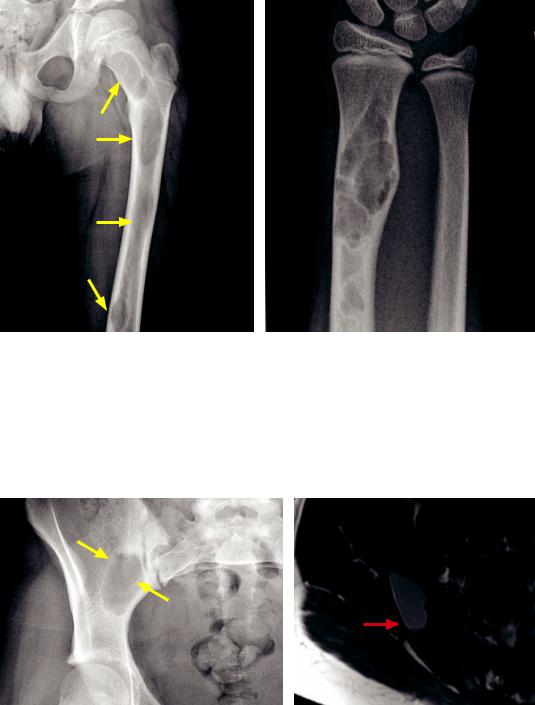
Malignant: Chondrosarcoma
Chondrosarcoma: Frontal radiograph of the pelvis (left image) and coronal T1-weighted MRI (right image) show an exophytic lesion (arrows) arising from the right iliac crest, which is continuous with the intramedullary cavity. The lesion demonstrates ring and arc chondroid-type calcification on radiography, and is heterogeneously hyperintense with a lobulated appearance on T2-weighted MRI (arrows).
Case courtesy Roger Han, MD, Brigham and Women’s Hospital.
Chondrosarcoma in a different patient:
Frontal and lateral radiographs of the distal femur show
an intramedullary lesion with ring and arc chondroid calcifications. Periosteal reaction (yellow arrow) and
cortical disruption (red arrow) suggest aggressive behavior.
Case courtesy Stacy Smith, MD, Brigham and Women’s Hospital.
374
•Chondrosarcoma is a malignant tumor of cartilage. Like osteosarcoma, there are multiple primary and secondary variants.
•Secondary forms arise from enchondroma (more commonly in the Maffucci and Ollier familial enchondromatoses), Paget disease, and osteochondroma (more common in familial osteochondromatosis).
An osteochondroma with a cartilage cap thickness of >2 cm is highly suggestive of chondrosarcoma.
•The conventional (intramedullary) chondrosarcoma subtype is most common. On imaging, chondrosarcoma is typically an expansile lesion in the medullary bone, with ring and arc chondroid matrix. The tumor causes thickening and endosteal scalloping of the cortex, and there is often an associated soft-tissue mass.
•The dedifferentiated subtype of chondrosarcoma is aggressive and may contain fibrosarcoma or osteosarcoma elements.
•Othersubtypesofchondrosarcomaincludetheraremesenchymalandclearcellvariants.
Lesions of fibrous origin
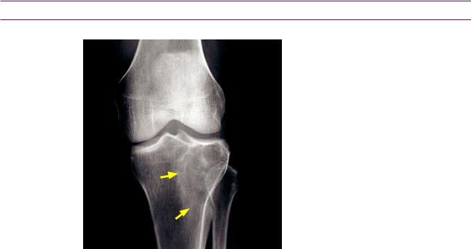
Benign: Nonossifying fibroma/fibrous cortical defect
Nonossifying fibroma: Frontal (left image) and lateral (right image) radiographs of the proximal lateral tibia in a 17-year-old male show a well-defined lucent lesion in the medial tibial metaphysis. The lesion features a faint sclerotic rim in continuity with the thinned lateral cortex
(arrow).
•Nonossifying fibroma (sometimes called a fibroxanthoma) is an asymptomatic and common incidental radiolucent lesion in the long bones (especially the leg) in children and adolescents. Nonossifying fibroma and fibrous cortical defect are thought to represent the same lesion; the term nonossifying fibroma is generally reserved for larger (>2 cm) or symptomatic lesions. They are the same histologically. These lesions are believed to arise from the periosteum.
•Theradiographicappearanceisusuallydiagnosticanddemonstratesalucentlesionwitha narrowzoneoftransition,scleroticmargin,andnomatrixcalcification.CTorMRImayshow corticaldisruptionorthinning,representingreplacementofthecortexbyfibroustissue.
•Mostlesionsundergospontaneousscleroticinvolutionasthepatientreachesadulthood.
Malignant: Malignant fibrous histiocytoma (MFH)
•Malignantfibroushistiocytoma(MFH)isacontroversialwaste-basketdiagnosismore recently renamed as undifferentiatedpleomorphicsarcomanototherwisespecified.Despite thecontroversyandrecentrenaming,thetermMFHisstillincommonusebyradiologists.
•The term “fibrous” refers to the microscopy appearance of MFH, not to the cell of origin. In fact, no definite cell of origin has been determined.
•MFHisthemostcommonadultsoft-tissuesarcoma.Itusuallyoccursinmiddle-agedor olderadultsinthethighortheretroperitoneum,butitmayoccurinanyextremity.Less commonly,MFHmayoccurinthebones,whereitappearsasanaggressive,lyticlesion.
375
Fibrous dysplasia
•Fibrous dysplasia is a benign congenital non-neoplastic condition of children and young adults characterized by replacement of normal cancellous bone by abnormal fibrous tissue.
•Fibrous dysplasia can affect one bone (monostotic) or multiple bones (polyostotic). When polyostotic, it tends to be unilateral.
•The most frequent complication is pathologic fracture, commonly at the femoral neck.
•Fibrous dysplasia of the long bones tends to be central and metadiaphyseal, often causing a bowing deformity such as the extreme varus of the shepherd’s crook.
Frontal radiograph of the hip shows multiple lucent lesions in the metaphysis and diaphysis of the femur in a skeletally immature patient (arrows). The lesions feature the characteristic ground glass internal matrix of fibrous dysplasia, with faint peripheral sclerosis.
Frontal radiograph of the forearm demonstrates a multiseptated lucent lesion of the central metadiaphysis of the distal radius in a different skeletally immature patient. The differential of this appearance includes fibrous dysplasia and aneurysmal bone cyst.
•In the ribs or long bones, the matrix is typically indistinct and ground glass.
•In the pelvic bones, fibrous dysplasia is often cystic.
Alucentlesionoftherightiliacbone(yellowarrows)showsintermediatetohyperintensesignalon proton-density-weightedMRI,withasubtlefluidlevel(redarrow).Thedifferentialdiagnosisofacystic supra-acetabularlesioninayoungadultincludescysticfibrousdysplasiaandunicameralbonecyst.
376
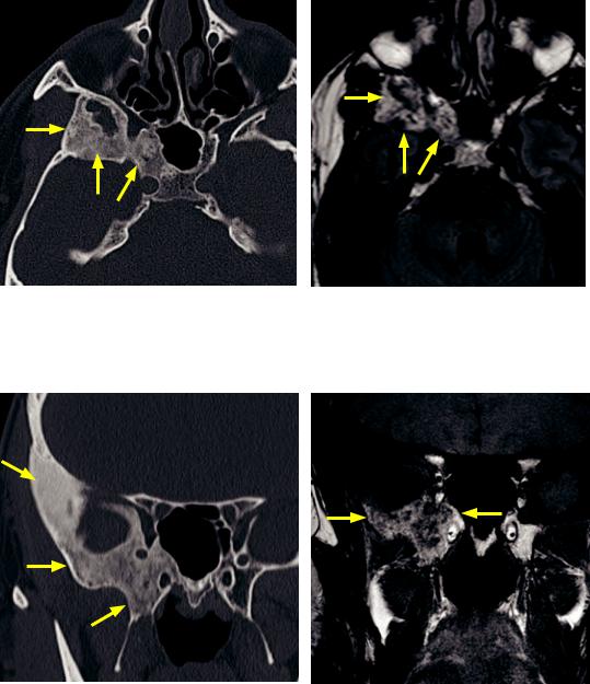
•In the skull base, fibrous dysplasia is typically expansile and can look highly unusual on MRI. The primary differential of an expansile skull base lesion is Paget disease, but the age of the patient is the key: fibrous dysplasia occurs in children and young adults, while Paget occurs in older adults.
Axial CT shows the typical appearance of fibrous dysplasia of the skull base, with expansile lesions primarily of the right sphenoid bone that have a hazy, ground-glass matrix (arrows).
FLAIR MRI correlated to the same region as the CT to the left shows the highly heterogeneous signal of fibrous dysplasia (arrows).
Coronal CT and T1-weighted MRI in the same patient show the true extent of the abnormality (arrows).
Case courtesy Mary Beth Cunnane, MD, Massachusetts Eye and Ear Infirmary, Boston.
•McCune–Albright syndrome is polyostotic fibrous dysplasia, precocious puberty, and cutaneous café au lait spots.
•Mazabraud syndrome is fibrous dysplasia and intramuscular myxomas, which tend to occur in the same region of the body.
377
Lesions of vascular origin
Benign: Hemangioma
Lateral radiograph of the lumbosacral spine shows mild loss of height of the L5 vertebral body with a subtle lace-like striated trabecular appearance (arrow).
Axial CT through L5 demonstrates the typical polkadot sign of the hemangioma, which involves the complete medullary cavity.
Sagittal CT shows coarsened, vertically oriented trabeculae with a typical corduroy appearance in L5 (arrow).
Sagittal T2-weighted MRI demonstrates high signal intensity of the L5 vertebral body.
•Hemangioma is a benign lesion that typically occurs in the vertebral body, characterized by vascular channels lined by endothelial cells.
•Although usually incidental, rarely a hemangioma may be associated with a soft-tissue mass that can cause neurologic compromise.
•Hemangioma causes reactive trabecular thickening in response to bony resorption by vascular channels.
•On MRI, high signal intensity on both T1and T2-weighted images is from fat contained within the hemangioma. On radiography and CT, corduroy striations are typical. The polka-dot sign demonstrates thickened trabeculae imaged in crosssection.
Malignant: Angiosarcoma of bone
•Angiosarcoma looks and acts aggressively. Lung metastases are often seen.
378
Lesions of hematopoietic origin
(Usually) benign: Giant cell tumor (osteoclastoma)
Giant cell tumor:
Frontal radiograph of the knee in a skeletally mature individual shows an eccentric lucent lesion (arrows) in the lateral tibial epiphysis and metaphysis extending to the articular surface.
Case courtesy Scott Sheehan, MD, Brigham and Women’s Hospital, Boston.
•Giant cell tumor is an epiphyseal lucent lesion located eccentrically at the articular end of long bones in skeletally mature patients between ages 20 and 40. It arises from the metaphysis but crosses the closed epiphyseal plate to involve the epiphysis. The cell of origin is a multinucleated giant cell, similar in appearance to an osteoclast.
•Most giant cell tumors are benign. Approximately 5% are malignant, but it is impossible to differentiate behavior based on the appearance of the primary lesion.
•Multifocal giant cell tumors can be seen in Paget disease or hyperparathyroidism.
•Treatment is typically curettage or wide resection.
Benign: Eosinophilic granuloma (Langerhans cell histiocytosis)
•A disorder of immune regulation, Langerhans cell histiocytosis (LCH) is caused by an abnormal proliferation of histiocytes. LCH is primarily seen in children 5–10 years old and is discussed more in depth in the pediatric imaging section.
•In the skull, the classic appearance of LCH is a lytic lesion with a beveled edge.
•Inthemandibleormaxilla,LCHmaycauseafloatingtoothfromresorptionofalveolarbone.
•In the spine, LCH may cause vertebra plana, which is complete collapse of the vertebral body.
•In the long bones, LCH may appear as a destructive radiolucent lesion with aggressive (often lamellated) periosteal reaction that may look like lymphoma or Ewing sarcoma.
Malignant: Ewing sarcoma
•Ewingsarcomaisahighlymalignantsmallroundcelltumor(similartoPNET)affecting childrenandadolescentswithamalepredominance.Theclinicalpresentationisnonspecific. Ewingsarcomausuallypresentswithpain.Systemicsymptomsincludingfeverareoften present,makingthedistinctionbetweenEwingsarcomaandosteomyelitisdifficult.
•Ewing sarcoma is the second most common pediatric primary bone tumor (following osteosarcoma).
•Radiographic features are of an aggressive lesion, with permeative bone destruction, aggressive periosteal reaction, and often an associated soft-tissue mass.
•In addition to Ewing sarcoma, the differential of an aggressive lytic lesion in a child
includes osteomyelitis, eosinophilic granuloma, and metastatic neuroblastoma.
379
Malignant: Multiple myeloma (MM)/Plasmacytoma
Multiple myeloma: Frontal and lateral radiographs of the skull (top images) show innumerable tiny lytic lesions. AP (bottom left image) and lateral (bottom right image) radiographs of the femur show a permeative appearance (yellow arrow), with focal cortical thinning anteriorly best seen on the lateral view (red arrow).
•Multiple myeloma is the most common primary malignant bone tumor in patients over 40.
•By far the most common presentation of myeloma is multiple lytic lesions, with the most severe form being diffuse myelomatosis with endosteal scalloping.
•Sclerosing myelomatosis is an uncommon variant, associated with POEMS syndrome:
Polyneuropathy.
Organomegaly (liver/spleen).
Endocrine disturbances (amenorrhea/gynecomastia).
Monoclonal gammopathy.
Skin changes (hirsutism and hyperpigmentation).
•The main differential of multiple lytic lesions in an adult is metastatic disease. Multiple myeloma originates from the red marrow and usually does not involve regions where there is minimal red marrow, such as the pedicles in the spine. Multiple myeloma may be negative on bone scan, unlike most metastases.
•A solitary tumor is a plasmacytoma. Most patients with plasmacytoma will get fullblown multiple myeloma within 5 years.
380
Malignant: Lymphoma
•Primary bone lymphoma is very rare and tends to occur in adults over 40.
•Bone lymphoma appears as an aggressive lytic lesion, but may also be an important differential consideration for an ivory (diffusely sclerotic) vertebral body.
•Lymphoma is often associated with an adjacent soft-tissue mass.
Fat (lipo-) lesions
Benign: Lipoma
Lateral radiograph of the ankle shows a nonspecific
circumscribed lucent lesion in the calcaneus (arrows) with a thin rim of peripheral sclerosis.
The differential for this lesion would include instraosseous lipoma, simple bone cyst, or aneurysmal bone cyst.
If central or ring-like calcification were present, that would more strongly favor intraosseous lipoma.
•Intraosseous lipoma is an uncommon benign neoplasm. The most common sites are the calcaneus, subtrochanteric region of the femur, distal tibia/fibula, and metatarsals. Imaging can be variable depending on the degree of fat, calcification, fibrous tissue, and peripheral sclerosis. Central or ring-like calcification is often present.
•Soft-tissue lipoma is the most common soft-tissue tumor. It is important to note that up to one third of lipomas may contain some nonadipose tissue. The presence of minimal nonadipose tissue does not suggest malignant transformation.
Malignant: Liposarcoma
•The primary differential consideration of a soft-tissue lipoma containing nonadipose tissue is a well-differentiated liposarcoma, also called an atypical lipoma.
•Features suggesting well-differentiated liposarcoma include large size (>10 cm), thick septations, globular or nodular soft tissue, or a composition consisting of <75% fat.
Notochordal lesions
Malignant: Chordoma
•Chordoma is a malignant lesion deriving from a notochord remnant. It arises in the midline of the axial skeleton, either in the spheno-occipital region, body of C2, or sacrococcygeal location.
•Chordoma is a highly destructive lesion with irregular scalloped borders.
•Calcifications may be seen within the lesion. These calcifications are due to necrosis, not bone formation.
381
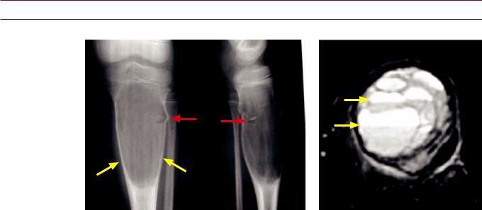
Lesions of unknown cell origin
Benign: Simple bone cyst (SBC), also called unicameral bone cyst (UBC)
Solitary bone cyst: Frontal and lateral radiographs of the proximal tibia in a skeletally immature patient show a central, expansile lesion of the proximal tibia (yellow arrows). A pathologic fracture is present with a displaced fragment (red arrows), representing the fallen fragment sign. T2-weighted MRI with fat suppression (right image) shows multiple fluid levels (yellow arrows). The differential diagnosis includes an aneurysmal bone cyst.
Case courtesy Michael Callahan, MD, Boston Children’s Hospital.
•Simple bone cyst/unicameral bone cyst is the result of a benign local disturbance of bone growth in children and adolescents.
•Thelesioniseitherholloworfluidfilledandtypicallyfoundintheproximaldiaphysisofthe humerusorfemur.Otherlocationsincludethecalcaneus,iliacbone,andtibia.
•SBC lacks periosteal reaction (in contrast to aneurysmal bone cyst), as long as there is no fracture.
•Upto66%ofcasesarecomplicatedbypathologicfracture.Thefallen fragment sign representsacorticalfracturefragmentthathasfallenintothecyst.Itispathognomonic forSBCwhenseeninayoungpatientinalesioninanappropriatelocationforSBC.
•SBC is always located centrally within a bone.
•Fluid–fluid levels are seen on MRI. Fluid levels are nonspecific and can also be seen in aneurysmal bone cyst, giant cell tumor, and telangiectatic osteosarcoma.
•One nonsurgical treatment option is the intra-lesional injection of methylprednisolone, which induces osteogenesis.
Benign: Aneurysmal bone cyst (ABC)
•Aneurysmal bone cyst (ABC) is an expansile or “aneurysmal” multicystic lesion seen in children and adolescents.
•Histologically, ABC is made of blood-filled sinusoids and solid fibrous elements. ABC may arise secondarily within a pre-existing tumor. This is important to consider if an aggressive lesion is biopsied and the pathology returns ABC, as this may imply that the true lesion was not sampled.
•UnlikeSBC,ABCcanbelocatedanywhereinbone(centraloreccentric).Itmayoccurinthe posteriorelementsofthespine.UnlikeSBC,ABCoftenfeaturesa“buttressing”smooth periostealreactionattheproximalanddistalaspects.LikeSBC,fluidlevelsarepresent.
Malignant: Adamantinoma
•Adamantinoma is a very rare, low-grade malignant tumor occurring in the tibia with a soap-bubble appearance. It may be difficult to differentiate from fibrous dysplasia.
382
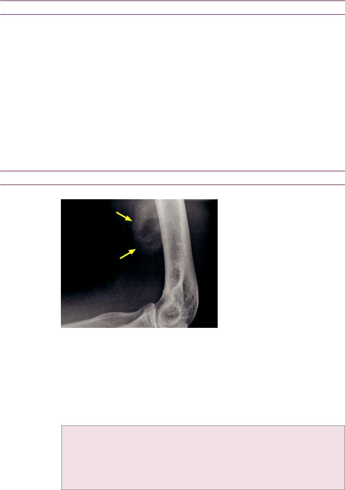
Osseous metastasis
•Osseous metastases are over ten times more common than primary bone tumors.
•Most metastases occur in the red bone marrow, most commonly in the axial skeleton.
•It can be difficult to differentiate a pathologic vertebral body fracture (associated with an underlying tumor) from an osteoporotic fracture, but involvement of the pedicle or posterior vertebral body suggests metastasis.
•A fracture of the lesser trochanter should raise concern for pathologic fracture.
•Asolitarysternallesionishighlypredictiveofmetastasisinapatientwithbreastcancer.
Lytic osseous metastases |
Blastic (sclerotic) osseous metastases |
||
• Lung (most common purely lytic). |
• Breast (lytic or blastic). |
||
• Breast (lytic or blastic). |
• |
Prostate, seminoma. |
|
• |
Thyroid. |
• |
Transitional cell carcinoma. |
• |
Kidney. |
• |
Mucinous tumors. |
• Stomach, colon (lytic or blastic). |
• |
Carcinoid. |
|
Benign tumor mimics
Myositis ossificans
Myositis ossificans:
Lateral radiograph of the elbow shows peripheral osteoid within a mass abutting the anterior humeral cortex. The peripheral ossification (arrows) suggests myositis ossificans, at least 5 weeks old.
The main differential diagnosis is parosteal osteosarcoma; however, the peripheral ossification more strongly suggests myositis ossificans. This case was followed and confirmed by close-interval follow-up imaging.
•Myositisossificans(MO)isheterotopicboneformationintheskeletalmusclesecondaryto trauma,althoughthehistoryoftraumamaynotberememberedandthetraumamaybe minor.MOmostcommonlyoccursaroundtheelbowandthigh,whicharepronetotrauma.
•Myositis ossificans may mimic parosteal osteosarcoma. Parosteal osteosarcoma is usually more heavily calcified centrally, while myositis ossificans is typically more calcified peripherally.
•If myositis ossificans is suspected, a biopsy should not be performed. A biopsy can lead to unnecessary surgery, as the histology may resemble sarcoma.
•The appearance of myositis ossificans evolves over a period of weeks to months:
Weeks 1–2: Soft-tissue mass only.
Weeks 3–4: Formation of amorphous osteoid matrix. May cause periosteal reaction of adjacent bone. This stage is most suggestive of an early osteosarcoma.
Weeks 5–8: The periphery of the lesion matures into compact bone. Up to 6 months: Ossification continues to mature.
>6 months: Typically decreases in size.
383
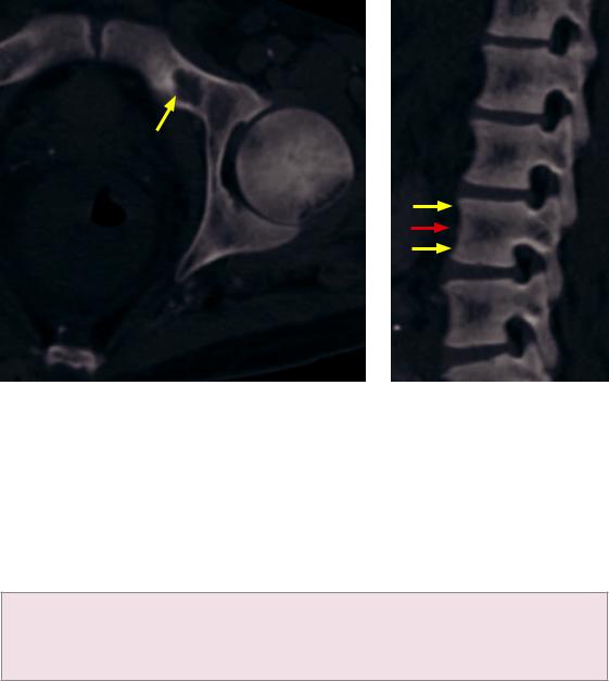
Brown tumor
Brown tumor: Axial CT (left image) shows a nonspecific lytic lesion with a faint sclerotic margin in the left superior pubic ramus (arrow) in a patient with renal osteodystrophy and secondary
hyperparathyroidism. Note the rugger jersey spine on the sagittal CT with alternating bands of sclerosis (yellow arrows) and relative central lucency (red arrow).
•A brown tumor is a benign lytic lesion seen in patients with hyperparathyroidism, caused by increased osteoclast activation.
•Brown tumor does not have any specific imaging features and may be difficult to differentiate from a giant cell tumor both radiologically and pathologically.
•Associated features of hyperparathyroidism are usually present, including:
Osteopenia.
Subperiosteal bone resorption (especially of the radial aspect of the 2nd and 3rd middle phalanges and the acromial ends of clavicles).
Soft-tissue calcifications.
•Lab abnormalities associated with hyperparathyroidism (in addition to elevated PTH level) include elevated calcium and decreased phosphorous.
•If hyperparathyroidism is secondary to renal failure, secondary findings of renal osteodystrophy may be seen as well, such as rugger jersey spine.
Osteomyelitis
•Osteomyelitis, discussed on the following page, may cause a permeative or lytic bony lesion that can be indistinguishable from an aggressive malignancy.
384
Musculoskeletal infection
Osteomyelitis
Overview of osteomyelitis
•Literallymeaning“inflammation(-itis)ofthebone(osteo-) marrow(-myelo-),” osteomyelitisisinfectioninbone.Osteomyelitishasproteanclinicalandimaging manifestations,withanendpathwayofchronicprogressivebonydestructionifuntreated.
•Osteomyelitis can be classified by route of spread (hematogenous, contiguous spread, or direct inoculation) or by chronicity (acute or chronic), although any route of spread can lead to either acute or chronic disease.
•The anatomic distribution and clinical presentation of osteomyelitis are highly dependent on the age of the patient, the specific organism, and the presence of any underlying disorders, such as vascular insufficiency or compromised immunity.
In adults, osteomyelitis typically arises from either contiguous spread or direct inoculation, such as in diabetic foot infections, open fractures, or as a complication of surgery. Infection is usually polymicrobial.
Inchildren,hematogenousspreadofinfectionismorecommon,usuallyfromStaphylococcus aureus.
Terminology of osteomyelitis
•Osteitis is inflammation/infection of the cortex.
•Periostitis is inflammation/infection of the periosteum.
•A sequestrum is a piece of necrotic bone that is separated (sequestered) from viable bone by granulation tissue. A sequestrum is a surgical lesion that can chronically harbor living organisms and function as a nidus for recurrent infection if not resected.
•An involucrum is living bone surrounding necrotic bone.
•A cloaca is an opening on the involucrum.
•A sinus tract is an opening from the infection to the skin surface.
•A Brodie abscess is a form of subacute osteomyelitis characterized by central lucency and peripheral sclerosis. The radiographic differential diagnosis of a Brodie abscess is an osteoid osteoma.
Hematogenous osteomyelitis
•Acute hematogenous osteomyelitis is usually seen in infants and children. The highly vascularized metaphyses of long bones are most commonly affected. Metaphyseal venules have sluggish flow, which facilitate bacterial invasion.
•In infants up to 12 months old, infections can involve the metaphysis, epiphysis, and joint due to the presence of bridging vessels that cross the physis. In older children, infection tends to be isolated to the metaphysis. In adults, the physes are closed but hematogenous metaphyseal infection is uncommon.
infant |
child |
adult |
385
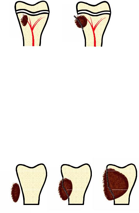
Hematogenousinfectionistypicallycausedbyasingleorganism.Ifbloodculturesare positivethenthatspecificorganismcanbetargetedandbiopsyisgenerallynotneeded.
Hematogenous osteomyelitis occurs from the inside out, beginning with infection of the medullary cavity. Secondary involvement of the cortex occurs as bacteria spread through Haversian and Volkmann canals into the periosteum and subsequently the soft tissues. In infants and children, the periosteum is loosely adherent to the bone, causing prominent lifting of the periosteum by infection. This manifests
radiographically as exuberant periostitis. In contrast, in adults the periosteum is more tightly adherent to bone and periostitis is less prominent.
hematogenous osteomyelitis
|
|
|
|
initial infection is |
infection spreads through |
||
intramedullary |
the cortex and uplifts the periosteum |
||
n adults, hematogenous osteomyelitis most commonly affects the spine.
Chronic osteomyelitis may produce infected nonviable tissue (sequestrum). An involucrum is living bone that surrounds the sequestrum. The involucrum may be perforated by cloacae, which can open to the skin to form a sinus tract. Chronic drainage of a sinus tract predisposes to squamous cell carcinoma
nitial subtle radiographic changes in hematogenous osteomyelitis include focal softtissue swelling, regional osteopenia, and obliteration of the soft-tissue fat planes.
The radiographic appearance of medullary infection is a focal, ill-defined lucent metaphyseal lesion. The lucency may cross the epiphysis in young children, where epiphyseal bridging vessels may be still patent. Periosteal reaction may appear aggressive and lamellated, mimicking Ewing sarcoma
Contiguous focus osteomyelitis
Osteomyelitis in adults is most commonly from contiguous spread of infection, characterized by penetration of the periosteum and cortex and subsequent medullary nvasion
contiguous focus osteomyelitis
soft-tissue infection |
periosteal and cortical |
medullary invasion |
|
penetration |
|
One of the most common causes of adult osteomyelitis is contiguous spread of a diabetic foot ulcer to the bones. Diabetic neuropathy leads to development of foot ulcers due to unrecognized trauma and impaired vascular reserve.
386
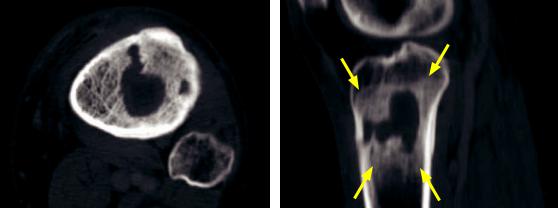
•The primary differential diagnosis of diabetic foot osteomyelitis is a neuropathic joint, a common sequela of diabetic neuropathy.
Neuropathic arthropathy usually affects the midfoot and features polyarticular involvement, absence of contiguous soft-tissue infection, and absence of an associated ulcer/sinus tract. The bony cortex is intact.
In contrast, diabetic foot osteomyelitis is almost always associated with a cutaneous ulcer and a sinus tract to the bone. The cortex of the involved bone is often disrupted.
Abnormal marrow signal is seen in both neuropathic arthropathy and diabetic foot osteomyelitis.
•In contrast to hematogenous osteomyelitis, contiguous focus osteomyelitis is typically polymicrobial and biopsy is generally warranted to ensure proper treatment.
Subacute osteomyelitis
Brodie abscess: Axial (left image) and sagittal unenhanced CT of the proximal tibia demonstrates a central, intramedullary circumscribed lucent lesion. Peripheral sclerosis (arrows) is better demonstrated on the sagittal image. Case courtesy Stacy Smith, MD, Brigham and Women’s Hospital.
•Brodie abscess is a characteristic lesion of subacute osteomyelitis, consisting of a walled-off intraosseous infection surrounded by granulation tissue and sclerotic bone. A Brodie abscess is essentially an intraosseous bone abscess.
Chronic osteomyelitis
•Chronicosteomyelitisisanindolentinfectionlastinggreaterthan6weeks.Devascularized, necroticboneleadstoasequestrumsurroundedbygranulationtissueandinvolucrum.
•Chronic osteomyelitis can cause a mixed lytic and sclerotic appearance, with a thickened cortex.
•Itcanbedifficulttodifferentiatebetweenactiveandinactivechronicosteomyelitis.Serial radiographsinactivechronicosteomyelitismayshowdevelopmentofperiostealreaction.
•Sclerosing osteomyelitis, also known as osteomyelitis of Garré, is an uncommon form of chronic osteomyelitis characterized by sclerosis and thickening of bone. The differential diagnosis of sclerosing osteomyelitis includes lymphoma, sclerotic metastasis, and osteoid osteoma.
Specific organisms causing osteomyelitis
•Staphylococcus:Staph.aureusisthemostcommoncauseofhematogenousosteomyelitis.
•Salmonella: Osteomyelitis from Salmonella is typically seen in patients with sickle cell disease, with a propensity to affect the diaphysis. It may be difficult to distinguish between diaphyseal bone infarct and osteomyelitis in sickle cell patients.
•Pseudomonas: Osteomyelitis from Pseudomonas aeruginosa is classically caused by a puncture wound of the foot through a sneaker.
•Tuberculosis: The most common musculoskeletal infection with Mycobacterium
tuberculosis is infection of the spine, also known as Pott disease.
387
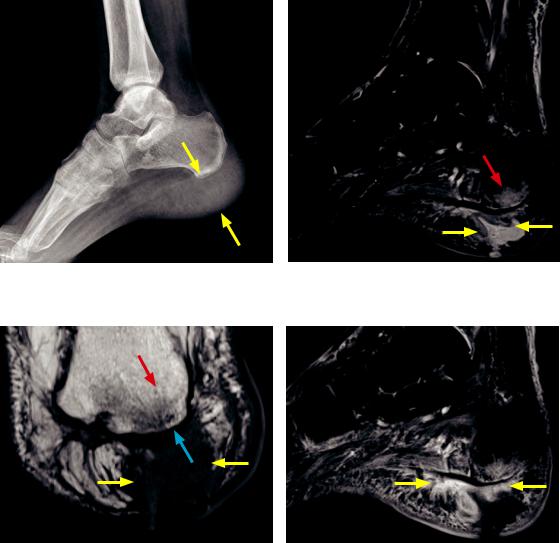
Specific locations
•Hand and finger: The metacarpals and phalanges may become infected after a bite wound.
•Toe: A stubbed great toe with nail-bed injury is at risk for osteomyelitis of the distal phalanx, due to the location of periosteum immediately adjacent to the nail bed.
•Spine: Vertebral osteomyelitis/discitis in an adult may be caused by hematogenous arterial spread, spinal surgery, or spread of a genitourinary infection through the epidural venous Batson plexus. Infection begins in the subendplate region of the vertebral body and subsequently spreads to the endplate, intervertebral disc, and adjacent vertebral body.
In contrast, in children discitis is thought to represent direct hematogenous seeding of the persistently-vascularized disc.
Imaging osteomyelitis
Calcaneal osteomyelitis due to foreign body: Radiograph shows marked thickening of the heel pad (arrows). No bony changes are evident.
Coronal T1-weighted MRI shows the low-signal fluid collection in the heel pad (yellow arrows), regional decreased T1 marrow signal (red arrow), and irregularity of the calcaneal cortex (blue arrow).
SagittalSTIRMRIshowscalcanealbonemarrow edema(redarrow)andafluidcollection(yellow arrows)withintheheelpad,contiguouswiththeskin.
Sagittal T1-weighted post-contrast MRI with fat suppression shows marked enhancement (yellow arrows) surrounding the abscess and enhancement of the surrounding soft tissues.
Case courtesy Marie Koch, MD, Brigham and Women’s Hospital, Boston.
•Radiographsaretypicallythefirstmodalitytoevaluatesuspectedosteomyelitis,although ittypicallytakesbetween10and14daysforradiographicchangestobeevident.
388

•Radiographic findings depend on the route of spread. Early radiographic changes of hematogenous osteomyelitis include focal osteopenia due to reactive hyperemia, followed by a lucent medullary lesion. In contiguous focus osteomyelitis, the first radiographic sign may be soft-tissue swelling and periosteal reaction, followed by erosion of the cortex.
•Scintigraphy and MRI imaging are more sensitive to detect early osteomyelitis.
•Three-phase Tc-99m MDP bone scan becomes positive within 24–48 hours after the onset of symptoms. Acute osteomyelitis is positive on all three phases (flow, blood pool, and delayed).
In contrast, cellulitis is positive on flow and blood pool phases, and negative on delayed.
Although highly sensitive, Tc-99m bone scan is less specific than leukocyte scintigraphy and MRI.
•Combining WBC and sulfur colloid scintigraphy adds specificity in the evaluation of osteomyelitis because bone marrow is replaced by infection and white cells. Actively infected bone marrow will show discordantly increased uptake on the WBC scan and reduced activity on sulfur colloid.
Note that WBC scan imaging is not sensitive for spinal osteomyelitis, thought to be due to the inability of leukocytes to migrate into an encapsulated infection.
WBC scan can be performed with either Indium-111-WBC or Tc-99m-HMPAO-WBC. Indium WBC scan has higher radiation dose, takes 24 hours to perform, and the image has more noise. The disadvantage of Tc-99m-HMPAO is its tendency to dissociate, leading to genitourinary excretion of radiotracer.
•MRI is highly sensitive to detect early osteomyelitis within 3–5 days. MRI has similar sensitivity to radionuclide studies, but greater specificity. MRI can better delineate the extent of infection, any fluid collections that must be treated surgically, sinus tracts, and skin ulcers.
•The hallmark of MRI imaging of osteomyelitis is replacement of the normal fatty marrow signal. Edema and exudates cause high marrow signal on T2-weighted images and low signal on T1-weighted images. Increased signal of the cortical bone, normally low signal on all sequences, signifies infectious involvement. MRI has very high negative predictive value: A negative MRI essentially excludes osteomyelitis. Gadolinium is helpful to delineate any fluid collections and to evaluate for the presence of nonenhancing necrotic bone (sequestrum).
Soft-tissue infections
Septic arthritis
•Septic arthritis is infection of a joint. The gold standard for diagnosis of septic arthritis is joint aspiration.
•The imaging and clinical hallmark of septic arthritis is a joint effusion.
•In children, the hip is a common site of septic arthritis caused by contiguous extension of proximal femoral metaphyseal osteomyelitis. The proximal femoral metaphysis is within the hip joint capsule.
•If untreated, septic arthritis can lead to rapid joint destruction and eventual ankylosis.
•Intravenous drug abusers are susceptible to infection of the sacroiliac and acromioclavicular joints.
389
Necrotizing fasciitis
Necrotizing fasciitis:
Unenhanced CT through the proximal thigh shows several tiny locules of gas (arrow) in the medial subcutaneous tissues.
•NecrotizingfasciitisisanextremelyaggressivesofttissueinfectioncausedbyClostridium orothergram-positiverods.Itisasurgicalemergency,requiringimmediatedebridement.
•The characteristic radiographic and CT finding of necrotizing fasciitis is gas bubbles in the soft tissues.
Diffuse bone disease
Multifactorial bone disease
Osteoporosis
•Osteoporosis is the most common metabolic bone disease and is defined as a T score of <-2.5, where a T score of -1 is a bone density one standard deviation below the mean for healthy young women.
TheZscoreisthestandarddeviationsfromage-matchedwomen.Itisnotusedtodetermineosteoporosis.
•Osteopeniamaybesecondarytonumerousnutritional,endocrine,andotheretiologies.
•Vitamin or nutritional deficiencies can cause osteoporosis, including:
Osteomalacia (Looser zone).
Alcoholism.
Hypophosphatemia.
Scurvy (Wimberger sign).
•Endocrine disturbances can impact calcium metabolism, causing osteoporosis, including:
Hyperparathyroidism (subperiosteal resorption).
Cushing disease or any increase in endogenous/exogenous steroids.
•Diffuse malignancy, such as myelomatosis, can cause diffuse bony demineralization.
•Genetic causes of osteoporosis include:
Osteogenesis imperfecta.
Gaucher disease.
Anemia, including sickle cell and thalassemia.
•Focal osteopenia has a more limited list of causes, including:
Immobility/disuse.
Reflex sympathetic dystrophy.
Transient regional osteoporosis of the hip.
390
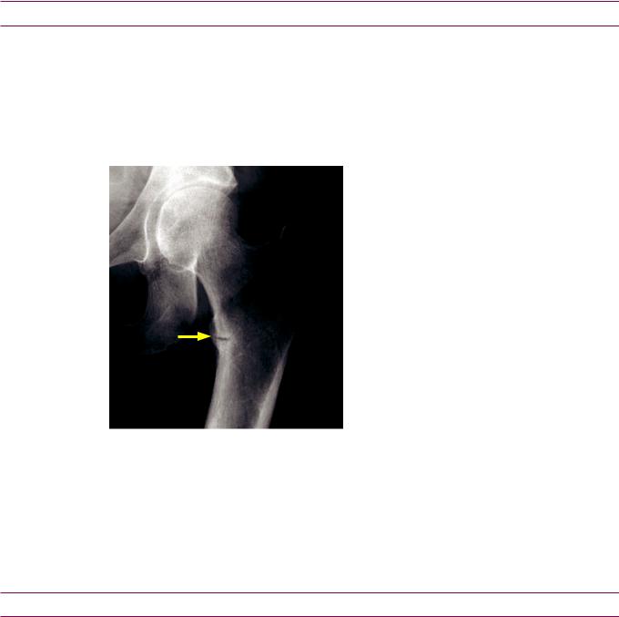
Vitamin-deficiency bone disease
Scurvy
•Scurvy (hypovitaminosis C) causes generalized osteopenia because osteoblasts require vitamin C to form mature osteoid tissue.
•Other signs of scurvy include the Wimberger ring sign, which is increased epiphyseal sclerosis due to disorganized epiphyseal ossification, and a Pelkin’s fracture, which is a metaphyseal corner fracture. A dense metaphyseal line may be present.
Osteomalacia
Osteomalacia: Frontal radiograph of the hip demonstrates an incomplete linear lucency (arrow) through the medial (weight-bearing) proximal femur, representing a Looser zone.
Case courtesy Stacy Smith, MD, Brigham and Women’s Hospital.
•Osteomalacia is faulty mineralization of bone matrix caused by vitamin D deficiency. The same process is called osteomalacia in adults and rickets in children. Rickets is discussed in the pediatric imaging section.
•Osteomalaciamanifestsasdiffuseosteopenia;however,aLooser zone (pseudofracture) ishighlyspecific.ALooser zone isacorticalstressfracturethatisfilledwithabnormal, poorlymineralizedosteoidandappearsasaradiolucencythroughthecortex.Common sites for Looser zones arethemedialproximalfemurs,distalscapulae,andpubicbones.
•Osteomalacia may be complicated by insufficiency fracture.
Endocrine bone disease
Acromegaly
•Acromegaly is the clinical syndrome caused by excess growth hormone. When the growth hormone excess occurs when the physes are open, the bones will grow longitudinally, leading to gigantism. However, when the hormone excess occurs once the physes have closed then the bones cannot lengthen and instead obtain a characteristic appearance of acral (distal) growth and widening.
•In the head there is enlargement of frontal sinuses, thickening of cranial bones, and an enlarged jaw.
•Inthehands,beak-likeosteophytesofthemetacarpalheadsandspade-likeovergrowths ofthedistalphalangesarecharacteristic.Initially,jointspacesarewidened,butmay becomenarrowedlaterinthediseaseduetosecondaryosteoarthritis.
•Inthefeet,acharacteristicfindingisincreasedheel-padthicknessgreaterthan24mm.
24mmisanormalheel-padthicknessfora150lb.person,withapproximately1mmallowedfor eachadditional25lb.inbodyweight.Forinstance,a250lb.personmayhaveaheel-padof28mm.
391
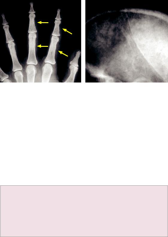
Hyperparathyroidism (Recklinghausen disease of bone)
Hyperparathyroidism: Radiograph of the hand (left image) shows subperiosteal resorption and scalloping of the radial margins of the 2rd and 3rd proximal and middle phalanges (arrows). Lateral skull radiograph in a different patient shows the classic salt-and-pepper appearance due to trabecular resorption.
Cases courtesy Stacy Smith, MD, Brigham and Women’s Hospital.
•Hyperparathyroidism is excess parathyroid hormone (PTH). PTH is normally secreted in response to hypocalcemia and maintains serum calcium levels by stimulating osteoclasts.
•Primary hyperparathyroidism is autonomously increased secretion from an overactive parathyroid gland, typically due to a parathyroid adenoma. Serum calcium is high and phosphate is low.
•Secondary hyperparathyroidism is increased secretion of PTH by a normal gland in response to hypocalcemia secondary to renal failure. Serum calcium is usually normal.
•Tertiary hyperparathyroidism is seen after prolonged secondary hyperparathyroidism, where the parathyroid glands escape regulatory control and oversecrete PTH.
•The radiological hallmarks of hyperparathyroidism are diffuse bony demineralization and subperiosteal and subligamentous bone resorption.
Skull: The classic salt-and-pepper skull is due to trabecular resorption.
Hands: Subperiosteal resorption of the radial aspects of the 2rd and 3rd middle phalanges is specific for hyperparathyroidism.
Clavicle: Subperiosteal resorption of the clavicle tends affect the acromial (distal) aspect. Knee: Subperiosteal bone resorption can be seen in the medial proximal tibial metaphysis. Teeth: Loss of lamina dura surrounding the tooth socket can occasionally be seen.
Anywhere: Brown tumors are more common in primary hyperparathyroidism. Everywhere: Diffuse osteopenia results from continued osteoclast activation.
•Calcium pyrophosphate dihydrate deposition (CPPD) may occur, which is more common in primary hyperparathyroidism.
•Soft-tissue calcification, periosteal reaction, and sclerosis are seen more commonly in secondary hyperparathyroidism.
•Complications of hyperparathyroidism include insufficiency fracture and increased propensity for ligaments and tendons to rupture.
392
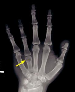
Renal osteodystrophy
•RenalosteodystrophyisduetothecombinedeffectsofabnormalvitaminDmetabolism (osteomalacia)andsecondaryhyperparathyroidismfromprolongedrenalfailure.
•In addition to renal osteodystrophy, patients with renal failure have an increased risk of additional bone disorders including osteomyelitis, avascular necrosis (if on steroids), and amyloidosis (from chronic dialysis).
•Unlike osteomalacia, Looser zones are uncommon in renal osteodystrophy.
•Thecharacteristicruggerjerseyspinedescribessclerosisofthevertebralbodyendplates.
•Soft-tissue and vascular calcifications are often present.
Hypothyroidism
•In children, hypothyroidism causes a delay in skeletal and dental maturity.
•Hypothyroidismmaycausebullet-shapedvertebralbodiesandWormianbonesintheskull.
•Hypothyroidism is associated with slipped capital femoral epiphysis.
Hypoparathyroidism
•Hypoparathyroidism is most commonly a consequence of parathyroid gland resection for hyperparathyroidism.
•Hypoparathyroidism causes metastatic deposition of calcium. In particular, calcium is deposited in the subcutaneous tissues and in the basal ganglia.
Pseudohypoparathyroidism and pseudo-pseudohypoparathyroidism
•Pseudohypoparathyroidismisduetoadefectoftheparathyroidhormone(PTH)receptor.
•Inpseudo-pseudohypoparathyroidism,thePTHlevelisnormalandPTHreceptorisnormal, butthepatient’sphenotypeisindistinguishablefromthatofpseudohypoparathyroidism.
•Both entities clinically present with obesity, round facies, short stature, and brachydactyly (short fingers).
•The classic radiographic finding is a short metacarpal of either the 4th or 5th digit or the thumb.
Pseudohypoparathyroidism: Radiograph of the hand shows a markedly shortened 4th metacarpal (arrow).
In addition to pseudohypoparathyroidism and pseudo-pseudohypoparathyroidism, an additional differential consideration for a short 4th metacarpal includes Turner syndrome.
Hyperthyroidism
•Hyperthyroidismcausesacceleratedbonymaturationinchildren.Thyroidacropachyisa rarecauseofdiaphysealperiostealreactionofmultiplebonesincludingthemetacarpals, metatarsals,andphalanges,seeninthesettingoftreatedhyperthyroidism.
393
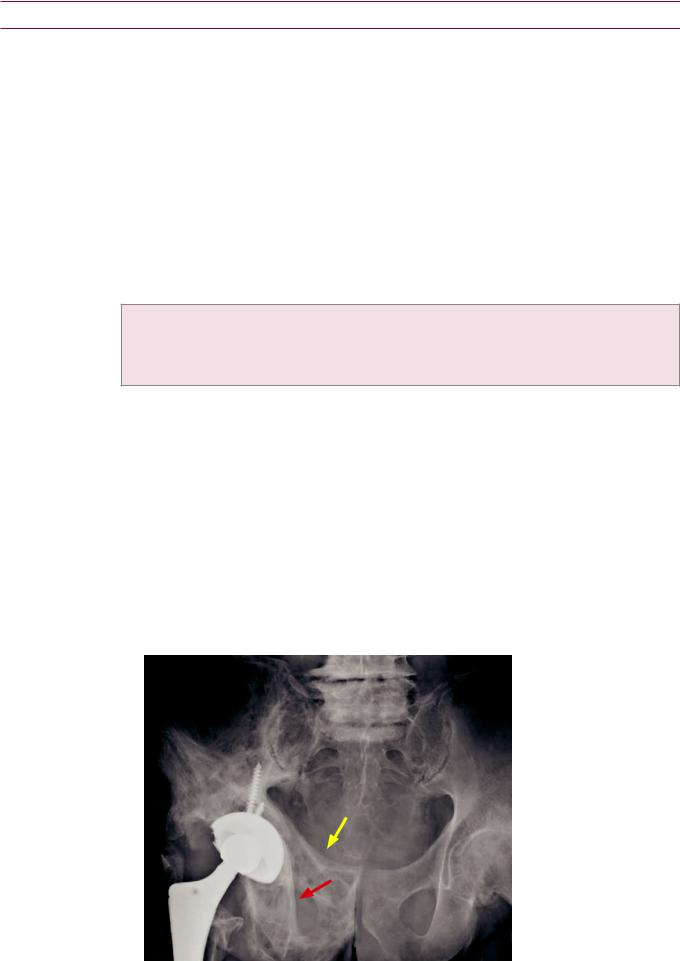
Idiopathic bone disease
Paget disease
•Paget disease is a progressive disturbance of osteoclastic and osteoblastic regulation seen in older adults. It may be seen in up to 10% of patients over 80 years old,
but it is rare in patients under age 40. The cause of Paget disease is unknown, but paramyxovirus may be involved.
•Symptoms of Paget disease include bone pain, osseous enlargement or bowing, compression of cranial nerves, and high output cardiac failure. Weakened subchondral bone commonly leads to osteoarthritis.
•Paget disease affects the skull, spine, and pelvis most commonly, followed by the articular ends of the femur, proximal tibia, and proximal humerus. Involvement of the ribs and scapula can occur but is uncommon.
•Three sequential phases are described, although there is often overlap between the phases and the disease may not always progress through the three stages in order.
Phase I: Osteolytic phase is due to hyperactivated osteoclasts and resultant active bone resorption.
Phase II: Mixed lytic and sclerotic phase is continued bony resorption and new bone formation. Phase III: Sclerotic phase is dominated by increased bone density and sclerosis.
•Chronic Paget disease shows thickening of cortex, coarse irregular trabecular pattern, and expansion of bone. This appearance has been called a caricature of normal bone.
•Paget of the skull: In the osteolytic phase (phase I), the typical appearance is a sharply marginated geographic lytic region called osteoporosis circumscripta. In the mixed phase (phase II), the classic appearance is the cotton wool skull, which looks as though an artist dabbed paint on the skull with a piece of cotton.
•Pagetofthevertebralbodies:Themixedphase(phaseII)canproduceeithertheclassic pictureframevertebralbody(withincreaseddensityoftheperipheryofthevertebralbody, asopposedtotheruggerjerseyspineofrenalosteodystrophy,whichfeaturessclerosisof justthesuperiorandinferiorendplates)oranivory(diffuselysclerotic)vertebralbody.
•Paget of the pelvis: When Paget disease involves the pelvis, the typical appearance is asymmetric coarsened trabecular thickening, with thickening of the iliopectineal and ilioischial lines. Acetabular protrusio may result.
Paget disease of the right hemipelvis:Frontalradiograph ofthepelvisshowsexpansion oftheinferiorandsuperior pubicramiwithcoarsened trabecularpatternandcortical thickening.Thereisthickening ofboththeiliopectineal (yellowarrow)andilioischial (redarrow)lines.
A right total hip replacement is present, secondary to pathologic fracture of the proximal femur. Acetabular protrusio is present on the right.
394
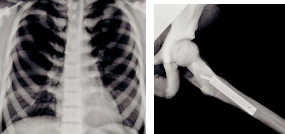
•Paget of the long bones: The lytic phase (phase I) progresses from the proximal articular end of bone into the diaphysis with a sharply marginated border. This border has been called the blade of grass or flame-shaped margin and allows differentiation from a neoplasm. As Paget disease progresses through its phases, it may cause bowing of the long bones and coxa vara deformity of the proximal femur.
•ComplicationsofPagetdiseaseincludepathologicfracture,malignantdegeneration(most commonlyosteosarcoma),secondarygiantcelltumor,andsecondaryosteoarthritis.
Hereditary hyperphosphatasia (juvenile Paget)
•Hereditaryhyperphosphatasia,alsocalledjuvenilePagetdisease,isanautosomal recessivediseaseofinfantsandyoungchildren.Thereiscorticalthickening,trabecular thickening,osteopenia,andbowingofallbonesinthebody,resultinginseveredeformity.
•Aside from the young age of the patients and diffuse involvement, the radiography of juvenile Paget otherwise appears very similar to adult Paget disease. There may be sparing of the articular ends of bones (epiphyseal sparing) in juvenile Paget disease.
Osteopetrosis
Osteopetrosis: Chest radiograph shows dramatic |
Radiograph of the femur in the same patient |
sclerosis of all the visualized bones in the thorax. |
shows a fracture of the proximal femur, status |
|
post ORIF. |
•Osteopetrosis is an extremely rare deficiency of osteoclastic carbonic anhydrase, which leads to inability of osteoclasts to resorb bone.
•Osteopetrosis is characterized by diffuse, marked sclerosis of the entire skeleton. The bones are extremely brittle, which typically leads to multiple fractures.
•The vertebral bodies may have a sandwich or rugger jersey appearance similar to renal osteodystrophy, but diffuse bony sclerosis is more marked in osteopetrosis.
Gaucher disease
•Gaucherdiseaseiscausedbyanautosomalrecessivedefectinglucocerebrosidase,leading toabnormalcerebrosidedepositioninalteredmacrophagescalledGauchercells.Gaucher cellscauseboneinfarctions,medullaryexpansion,andhepatosplenomegaly.
•The classic Erlenmeyer flask deformity of long bones is a result of abnormal modeling.
•Characteristic H-shaped vertebral bodies are due to endplate avascular necrosis.
H-shaped vertebral bodies are not specific to Gaucher disease, and are in fact seen more commonly in sickle cell disease. The size of the spleen can differentiate between sickle cell and Gaucher disease: Sickle cell causes splenic auto-infarction, while patients with Gaucher disease have splenomegaly.
395
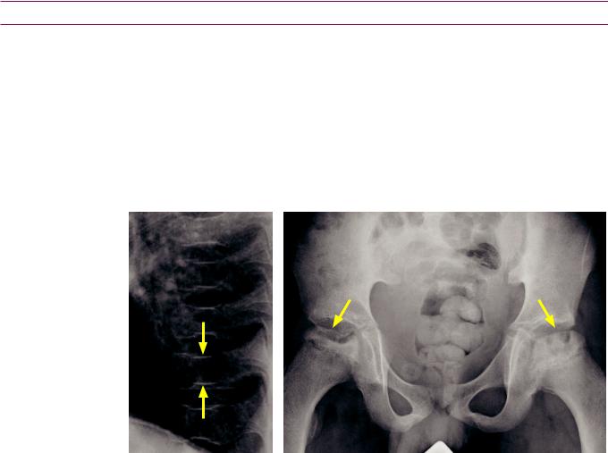
Hematologic bone disease
Sickle cell disease
•Sickle cell disease is an autosomal recessive defect in the beta-chain of hemoglobin. Red cells are sickle-shaped and cause microvascular occlusion. The three osseous manifestations of sickle cell disease are bone infarcts, dramatically increased risk of osteomyelitis, and marrow expansion/hyperplasia.
•Sickle cell bone infarcts: Infarcts and avascular necrosis are caused by microvascular occlusion from the sickled red cells. Avascular necrosis of vertebral body endplates causes the characteristic H-shaped or Lincoln-log vertebral bodies. Other common locations for infarction include the femoral heads and the diaphyses and metaphyses of long bones.
H-shaped vertebral bodies: Lateral thoracic spine radiograph shows characteristic central endplate depressions of the vertebral bodies (arrows) representing vertebral body
avascular necrosis.
In advanced sickle cell, patchy sclerosis from multiple bone infarctions may become nearly confluent, producing diffusely sclerotic bones.
Sickle cell dactylitis (also called hand–foot syndrome) is a special consideration in very young children who have persistent hematopoietic marrow in the digits of the hands and feet. Microvascular occlusion exacerbated by cold temperature causes dactylitis, which is evident on radiographs as soft-tissue swelling and periosteal reaction.
•Sickle cell osteomyelitis: Patients with sickle cell have an approximately 100x increased risk of osteomyelitis, most frequently with Salmonella species.
•Sickle cell marrow expansion: The radiographic findings of marrow expansion are more pronounced in thalassemia, but similar findings can be seen in sickle cell.
MRI of the bone marrow shows diffusely low signal on T1-weighted images, reflecting diffuse red (cellular) marrow.
In the long bones, chronic stimulation of red cell production causes widening of the medullary spaces and coarsening of the trabeculae, which results in osteopenia and increased risk of insufficiency fracture.
Intheskull,marrowexpansioncancausehair-on-end striationsandwideningofthemedullarycavity.
Involvement of the facial bones and extramedullary hematopoiesis are seen more commonly in thalassemia.
396
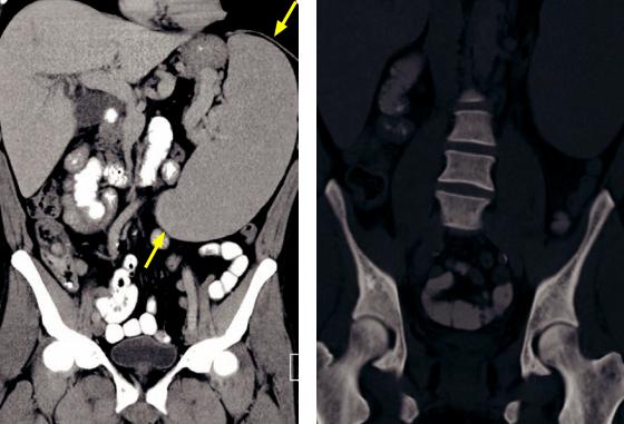
Thalassemia
•Thalassemia is an autosomal recessive congenital anemia that is caused by a defect in the alpha or beta hemoglobin subunits.
•Similar to sickle cell disease, the osseous hallmarks of thalassemia are bone infarcts, marrow hyperplasia, and infection. However, in contrast to sickle cell, the marrow expansion is much more prominent as the anemia can be more severe.
•In the hand, marrow expansion can cause widening and squaring of the phalanges and metacarpals.
•In the skull, the hair-on-end striations are a classic manifestation of marrow expansion.
•In the facial bones, marrow expansion may obliterate the paranasal sinuses, causing the typical rodent-like facies.
•In the long bones, the marrow expansion may cause an Erlenmeyer flask deformity.
Myelofibrosis
Myelofibrosis: Coronal CT in soft-tissue (left image) and bone windows show splenomegaly (arrows) and diffusely dense bones.
•Myelofibrosis is progressive fibrosis of bone marrow in older patients, leading to anemia and splenomegaly. Bone marrow fibrosis leads to diffusely sclerotic bones.
Mastocytosis
•Mastocytosis is abnormal proliferation of mast cells. It may result in either diffuse osteoporosis or sclerosis from the reaction of the marrow to the infiltrating mast cells.
397
Differential diagnosis of common bone lesions
Di usely dense (sclerotic) bones
Renal osteodystrophy
Sickle cell disease
Myelo brosis
Di use osteoblastic metastases
Osteopetrosis
Pyknodysostosis
Mastocytosis
Fluorosis
Normal variant − Athlete
sclerotic) vertebral body
Osteoblastic metastasis
Lucent epiphyseal
equivalent) lesion include apophyses,
calcaneus
mnemonic: GEIChA
Giant cell tumor
 Eosinophilic granuloma
Infection
Chondroblastoma
Aneurysmal bone cyst
Expansile or lytic rib lesion
mnemonic: FAMEB
Fibrous dysplasia
Aneurysmal bone cyst
Metastasis/Multiple myeloma/
Plasmacytoma
Paget disease |
Enchondroma |
|
Lymphoma |
Brown tumor |
398
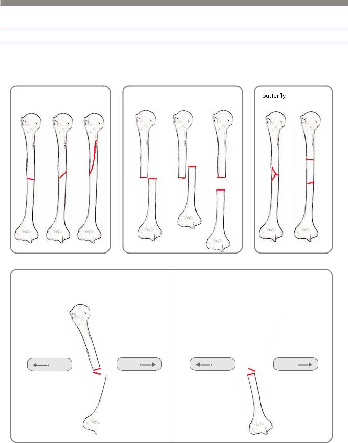
Basics of trauma
Fractures
Terminology
fracture direction |
|
position of distal fragment |
special types of |
|
|
|
comminuted fractures |
transverse oblique |
spiral |
displaced foreshortened distracted |
segmental |
|
|
and displaced |
|
angulation of distal fragment
apex medial angulation |
apex lateral angulation |
(lateral angulation of the distal fragment) |
(medial angulation of the distal fragment) |
lateral |
medial |
lateral |
medial |
•Whendescribingalong-bonefracture,thefracturelocation(proximal,mid-shaft,distal), directionoffracture,angulation,andpositionofthedistalsegmentshouldbedescribed.
•A comminuted fracture is defined as >2 fracture fragments.
•It is essential to mention if a fracture extends to the articular surface of a joint.
399
•A stress fracture may be a fatiguefracture(abnormalrepetitivestressonanormalbone) or an insufficiencyfracture(normalstressonanabnormalbone;e.g.,inthesettingof demineralizationduetoosteoporosisormetabolicbonedisease).Thetermstress fracture is oftenusedinterchangeablywithfatiguefracture,althoughthesetermsarenotsynonyms.
•Apathologicfractureiscausedbynormalstressonaboneweakenedbyanunderlying lesion(typicallytumor,butalsoincludinginfection,fibrousdysplasia,andPagetdisease).
Basics of MR
Tendons
•A normal tendon is dark on all MRI sequences.
•A normal tendon may have artifactually increased signal due to the magic angle phenomenon. Because tendons have fibers coursing along a single direction (demonstrating anisotropy), tendons may demonstrate artifactually increased signal on short TE sequences when oriented 55 degrees relative to the bore of the magnet. This phenomenon is called the magic angle artifact.
Short TE sequences include T1-weighted images, proton density, and GRE. T2-weighted sequences are generally not susceptible to the magic angle artifact.
Unlike true tendon pathology, the magic angle artifact disappears with a long TE and the tendon will otherwise have a normal morphology.
Tenosynovitis
•Tenosynovitis is inflammation surrounding a tendon. Tenosynovitis may be secondary to repetitive motion, inflammatory arthritis, or infection.
•On MRI, fluid completely surrounds the tendon circumferentially.
•A potential pitfall is that fluid can track along tendon sheaths that communicate directly with an adjacent joint (such as the long head of the biceps tendon in the shoulder). This should not be confused with tenosynovitis. Similarly, a small amount of fluid in the synovial sheath of the posterior tibialis tendon in the foot is also normal.
•A variant form, called stenosing tenosynovitis, features several loculated collections of fluid in the tendon sheath.
Stenosing tenosynovitis can be seen surrounding the flexor hallucis longus tendon at the medial ankle in os trigonum syndrome and about the wrist in de Quervain’s stenosing tenosynovitis.
Myxoid degeneration (tendinosis)
•Agingoroveruseoftenleadstomyxoiddegeneration,whichissynonymouswithtendinosis.
The word tendinitis should not be used as myxoid degeneration is not due to inflammation.
•MRI of myxoid degeneration/tendinosis will show intermediate intra-substance (within the tendon) signal. The tendon may be either normal in size or enlarged. If fluidintensity signal is seen within a tendon, concern should be raised for a partial tear, although MR cannot always reliably differentiate between tendinosis and partial tear.
Partial tear
•A partial tendon tear represents incomplete disruption of the fibers and can have a varied MRI appearance. The tendon may be thickened, thinned, or contain intrasubstance fluid.
Complete tear
•A complete disruption of the tendon will appear as complete discontinuity of the tendon. There is often retraction of the tendon remnants.
400
Foot and ankle
Forefoot
Anatomy (blue) and overview of common fractures (red)
phalanges
Freiberg’s infraction
metatarsals
|
|
5th metatarsal fractures |
|
|
diaphyseal fracture |
|
|
Jones fracture |
|
1st (medial) cuneiform |
peroneal brevis avulsion |
|
2nd (middle) cuneiform |
|
|
3rd (lateral) cuneiform |
cuboid fracture |
|
|
|
cuboid |
navicular |
|
|
|
|
calcaneus |
talus |
|
(anterior process) |
|
Fifth metatarsal fractures
•Metatarsal base avulsion (zone 1) is a fracture at the most proximal base of the 5th metatarsal.
Treatment is conservative, with a boot.
The peroneus brevis and lateral aspect of the plantar aponeurosis attach at the 5th metatarsal base.
•Jones fracture (zone 2) is a fracture of the metaphyseal–diaphyseal junction.
A Jones fracture carries a worse prognosis compared to an avulsion fracture due to reduced blood supply at the metaphyseal– diaphyseal junction. Treatment is variable and may require surgery.
•The metatarsal shaft (zone 3) is a common location for stress fracture.
Treatment may be surgical.
Freiberg’s infraction
great toe distal phalanx fracture most common phalangeal fracture
turf toe injury |
forefoot |
Lisfranc injury
|
|
|
es |
|
|
tur |
|
|
ac |
|
|
fr |
|
|
|
eiform |
|
|
|
cun |
|
|
|
midfoot
Zone 3 (shaft fracture)
Zone 2 (Jones fracture) metaphyseal-diaphyseal junction
Zone 1 (avulsion fracture)
•Freiberg’s infraction is avascular necrosis of the second metatarsal head. It is caused by repetitive stress or poorly fitting shoes (such as high-heels) and usually occurs in young women.
401
Metatarsal stress fracture
Second metatarsal stress fracture: Frontal (left image) and oblique (right image) radiographs of the foot show a minimally displaced mid-diaphyseal fracture (arrows) of the second metatarsal. There is minimal associated callus formation.
Case courtesy Barbara N. Weissman, MD, Brigham and Women’s Hospital.
•The first radiographic sign of a metatarsal stress fracture is a barely perceptible linear cortical lucency. Usually, stress fractures are not apparent on radiographs until periostitis and callus have begun to form.
Sesamoid fracture
•Fracture of the great toe sesamoid bones is typically caused by extreme hyperextension or dorsal dislocation (which may be transient) of the first metatarsophalangeal joint.
The flexor hallucis brevis attaches to the sesamoids; the medial head attaches to the tibial (medial) sesamoid and the lateral head attaches to the fibular (lateral) sesamoid.
•A bipartite sesamoid is a normal variant that may simulate a sesamoid fracture; however, a bipartite sesamoid will be round in shape and its margins will be completely corticated. Additionally, the sum of the parts of the bipartite sesamoid will be larger in size than the other sesamoid.
•The term turf toe has been used to describe a wide range of injuries at the first MTP joint including sesamoid fracture.
Lisfranc fracture-dislocation
Frontal radiograph of the foot shows a homolateral dislocation at the Lisfranc joint, with fracture of the base of the second metatarsal (arrow).
402
The tarsometatarsal joint is the Lisfranc joint, named after the French surgeon in the Napoleonic wars who performed amputations at this joint. The stability of the joint depends on multiple ligaments, with the Lisfranc ligament being the most important. The Lisfranc ligament is an interosseous ligamentous complex attaching the medial cuneiform to the second metatarsal base.
Lisfranc ligament
1st cuneiform to 2nd metatarsal
Lisfranc joint tarsometatarsal joint
A Lisfranc fracture-dislocation is a fracture-dislocation of the tarsometatarsal joint.
The treatment of a Lisfranc injury is surgical. A missed or untreated Lisfranc injury can
ead to debilitating osteoarthritis and flattening of the longitudinal arch.
Subtle malalignment of the tarsometatarsal joint may signal serious ligamentous injury. Weight-bearing radiographs are the most sensitive. Careful evaluation of the alignment of the Lisfranc joint must always be performed, as in this normal example below.
frontal |
oblique |
cuboid
 1st 3nd 2nd
cuneiforms
lateral base of 1st metatarsal should be aligned with lateral aspect of 1st cuneiform medial base of 2nd metatarsal should be aligned with medial aspect of 2nd cuneiform medial base of 3rd metatarsal should be aligned with medial aspect of 3rd cuneiform medial base of 4th metatarsal should be aligned with medial aspect of cuboid
easier to see on oblique
the dorsal pro le of the tarsometatarsal (Lisfranc) joint should be uninterrupted
403
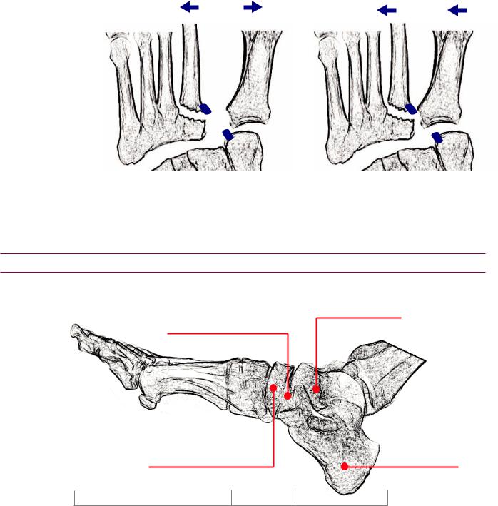
•Lisfranc injuries are classified into homolateral and divergent based on the direction of dislocation of the first metatarsal. In a divergent Lisfranc injury, the first metatarsal is medially dislocated and the 2nd through 5th metatarsals are laterally dislocated. In homolateral injury, all metatarsals will dislocate laterally.
divergent |
homolateral |
|
Navicular osteonecrosis
•Osteonecrosis of the navicular is called Kohler disease in childhood and Müeller– Weiss disease in adults. Kohler disease is typically self limited and occurs more commonly in boys, while Mueller–Weiss disease is more severe in course and occurs more commonly in adult women.
Midfoot and hindfoot trauma
Overview of common fractures
|
talar neck fracture |
Chopart fracture-dislocation |
classi cation: Hawkins |
|
|
talonavicular and calcaneocuboid joints |
|
Kohler disease (children) |
calcaneal fractures |
multiple classi cations: |
|
Müeller−Weiss disease (adults) |
Essex−Lopresti |
avascular necrosis of the navicular |
Sanders (CT-based) |
|
forefoot |
midfoot |
hindfoot |
Chopart fracture-dislocation
•The Chopart joint is formed by the talonavicular and calcaneocuboid joints.
•Chopart fracture-dislocation is typically caused by high-impact trauma. Associated fractures of the calcaneus, cuboid, and navicular bones are often present.
404
Calcaneal fracture
The calcaneus is the most commonly fractured tarsal bone.
Traumatic fractures of the calcaneus are typically the result of a high-impact injury, such as a fall from height, in which case the fracture is known as the lover’s fracture. n such cases, there is a high association with other serious injuries including lumbar spine fractures, traumatic aortic rupture, and renal vascular pedicle avulsion. If a traumatic calcaneal fracture is identified, further imaging of the lumbar spine and/or abdomen is recommended.
Subtle fractures may not be directly visible; however, a decrease in the Boehler angle to less than 20 degrees is diagnostic of a calcaneal fracture.
superior aspect of |
|
anterior process of |
|
calcaneus |
Boehler angle
normal is 20−40˚
<20˚ is diagnostic of calcaneal fracture
posterior tuberosity of calcaneus
superior aspect of posterior subtalar facet
The Boehler angle is formed by drawing two lines:
uperior aspect of posterior tuberosity of calcaneus to the superior aspect of posterior subtalar facet uperior aspect of anterior process of calcaneus to superior aspect of posterior subtalar articular surface
A Boehler angle <20˚ is diagnostic of a calcaneal fracture.
A normal Boehler angle (20−40˚) does not exclude a calcaneal fracture.
The Essex–Lopresti classification divides calcaneal fractures into those sparing the
subtalar joint (25%) and fractures extending into the subtalar joint (75%).
The Sanders classification is based on the number of comminuted fragments as seen
on coronal CT.
Calcaneal stress fracture
A stress fracture of the calcaneus typically appears as a fluffy band of sclerosis within the calcaneus, with an intact cortex. Boehler’s angle is normal.
Talar fracture
Fractures of the talus can be divided anatomically into lateral process, posterior process, head, body, and neck fractures.
Fractures of the talar neck are classified by the Hawkins classification. A talar neck fracture may disrupt the blood supply to the talus and predispose to osteonecrosis.
TheHawkinssigndescribesasubchondrallucentbandseenonthefrontalankle radiograph6–8weeksafterankleimmobilization.Thelucentbandrepresentsincreased bonereabsorptionfromactivehyperemia.ThepresenceoftheHawkinssignimpliesan intactbloodsupplytothetalardomeandisagoodprognosticindicatorintalarneck fractures.AbsenceoftheHawkinssignsuggestsavascularnecrosis.
405
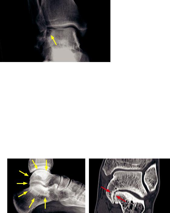
Osteochondral lesion of the talus
Talar osteochondral lesion:
Frontal radiograph of the ankle shows crescentic lucency (arrow) of the lateral talar dome with underlying sclerotic change.
•Osteochondral lesion of the talus is an umbrella term including osteochondrosis dissecans from chronic repetitive microtrauma and acute traumatic osteochondral fracture.
Osteochondrosis dissecans is preferred over the older terminology of osteochondritis dissecans.
The suffix -itis is misleading as this entity is not inflammatory.
•Osteochondral lesions and their staging are discussed in the knee section.
•In the talus, an osteochondral lesion appears on radiography as a crescentic lucency of the talar dome. MRI imaging is similar to MRI imaging of knee osteochondral lesions.
Hindfoot (tarsal) coalition
•Abnormal fusion of two tarsal bones may be a cause of hindfoot pain. Hindfoot coalitions often present in adolescence as the coalition begins to ossify. A coalition may be fibrous, cartilagineous, or osseous.
•The vast majority of hindfoot coalitions are either talocalcaneal (fusion of the talus and calcaneus) or calcaneonavicular (fusion of the calcaneus and navicular).
•Talocalcaneal coalition occurs most commonly at the middle subtalar facet.
Talocalcaneal coalition: Lateral radiograph of the ankle (left image) shows the classic C sign (yellow arrows) representing the fused medial talus and sustentaculum tali. A fibrous coalition (red arrows) is confirmed on the coronal CT (right image), with sclerosis and subchondral cysts of the middle subtalar facet.
The C sign on the lateral radiograph describes a continuous C-shaped contour of the fused middle subtalar facet (medial talus fused with the sustentaculum tali). The C sign is not sensitive for the detection of talocalcaneal coalition, but may be helpful when seen.
The talar beak sign on the lateral radiograph describes a triangular-shaped beaking at the anteriorsuperior aspect of the talus.
•Calcaneonavicular coalition is fusion between the anterior process of the calcaneus and the navicular.
The anteater sign represents the elongated anterior process of the calcaneus, which looks like an anteater’s nose on the lateral view.
406
Ankle tendon anatomy
tibialis anterior tendon extensor hallucis longus
extensor digitorum longus
anterior extensor tendons
(Tom, Harry, and Dick from medial to lateral)
|
tibialis posterior |
|
|
|
|
|
|
|
|
|
|
|
|
|
|
|
|
|
|
|
|
bula |
|
|
|
|
|
|
|
|
|
|
|
|
|
|
|
|
|
|
|
|
|
||||
|
exor digitorum longus |
|
|
|
|
|
|
|
|
|
|
|
|
|
|
|
|
|
|
|
|
||
|
|
|
|
|
|
|
|
|
|
|
|
|
|
|
|
|
|
|
|
||||
exor hallucis longus tendon |
|
|
|
|
|
|
|
|
|
|
|
|
|
|
|
|
|
|
|
|
|
||
peroneus longus tendon |
|||||||||||||||||||||||
|
|
|
|
|
|
|
|
|
|
|
|
|
|
|
|
|
|
|
|
||||
|
(and adjacent muscle) |
|
|
|
|
|
|
|
|
|
|
|
|
|
|
|
|
|
|
|
|
||
|
|
|
|
|
|
|
|
|
|
|
|
|
|
|
|
|
|
|
|
|
|||
|
|
|
|
|
|
|
|
|
|
|
|
|
|
|
|
|
|
|
|
|
|
peroneus brevis tendon |
|
|
posterior exor tendons |
|
|
|
|
|
|
|
|
|
|
|
|
|
|
|
|
|
|
|
|
||
|
|
|
|
|
|
|
|
|
|
|
|
|
|
|
|
|
|
|
|
|
|
||
|
|
|
|
|
|
|
|
|
|
|
|
|
|
|
|
|
|
|
|
|
|||
|
|
|
|
|
|
|
|
|
|
|
|
|
|
|
|
|
|
|
|
|
|||
|
|
|
|
|
|
|
|
|
|
|
|
|
|
|
|
|
|
|
|
|
|||
|
(Tom, Dick, and Harry |
|
|
|
|
|
|
|
|
|
|
|
|
|
|
|
|
|
|
|
|
peroneus brevis muscle |
|
|
|
|
|
|
|
|
|
|
|
|
|
|
|
|
|
|
|
|
|
||||
|
from medial to lateral) |
|
|
|
|
|
|
|
|
|
|
|
|
|
|
|
|
|
|
|
|
||
|
|
|
|
|
|
|
|
|
|
|
|
|
|
|
|
|
|
|
|
|
|
||
|
posterior tibial artery |
|
|
|
|
Achilles tendon |
|||||||||||||||||
|
|||||||||||||||||||||||
|
|
|
|
|
|
|
|
|
|
|
|
|
|
|
|
|
|
|
|
|
|
||
|
|
|
|
|
|
|
|
|
|
|
|
|
|
|
|
|
|
|
|
|
|
||
Lateral ankle tendons and ligaments
ATFL |
|
(anterior talobular ligament) |
|
most commonly torn ligament |
bula |
navicular
medial  talus cuneiform
1st
metatarsal
cuboid
5th metatarsal  peroneus brevis tendon
attaches to base of 5th metatarsal
PTFL
(posterior talobular ligament)
very strong, rarely torn
calcaneo bular ligament
next most commonly torn
peroneus longus tendon
runs under foot, attaches to medial cuneiform and rst metatarsal
(only medial cuneiform attachment shown)
407
Medial ankle ligaments
deltoid ligament complex
posterior tibiotalar (deep) tibiocalcaneal (super cial) tibionavicular (super cial) anterior tibiotalar
sustentaculum tali (part of calcaneus)
spring ligament complex: not typically individually resolved on MRI
(calcaneonavicular ligaments)
 superomedial calcaneonavicular ligament
 medioplantar oblique calcaneonavicular ligament
  inferoplanatar longitudinal calcaneonavicular ligament
along with the posterior tibial tendon* (not drawn), the spring ligament complex functions to maintain the arch of the foot
*most common cause of acquired at foot is posterior tibial tendon tear (especially in diabetic patients)
Medial ankle tendons
contents of tarsal tunnel, from anterior to posterior: Tom, Dick, ANd Harry
posterior Tibial tendon
exor Digitorum longus tendon
posterior tibial Artery
posterior tibial Nerve
exor Hallucis longus tendon
 medial malleolus
navicular tuberosity
medial cuneiform
exor retinaculum
exor hallucis longus tendon
runs between (but does not connect with) sesamoids inserts on base of great toe distal phalanx
exor digitorum longus tendon
inserts on base of 2nd-5th distal phalanges
(only insertion on base of second distal phalanx is drawn)
posterior tibial tendon (only main component is drawn)
main component inserts on navicular tuberosity and medial cuneiform plantar component inserts onto bases of 2nd-4th metatarsals recurrent component inserts on sustentaculum tali of calcaneus
Anterior extensor tendons
Arranged from medial to lateral, the anterior extensor tendons can be remembered
with the mnemonic “Tom, Harry, and Dick”:
Tibialis anterior.
Extensor H allucis longus.
Extensor Digitorum longus.
The anterior tibial artery runs anteriorly with the extensor tendons.
408
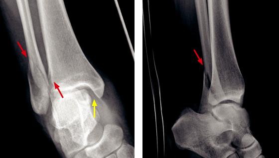
Ankle fractures
*
* = syndesmosis
Weber B (Lauge–Hansen supination–external rotation) ankle fracture:
Oblique(leftimage)andlateralankleradiographsshowanobliquefracture(redarrows)throughthedistal fibulaatthelevelofthesyndesmosis(bluestar).Thereiswideningofthemedialclearspace(yellowarrow).
•Ankle sprains and fractures are the most common of all joint injuries.
•TheDanis–Weberclassificationisthesimplestclassificationofanklefracturesanddivides anklefracturesbasedonthelevelofthefibularfractureandresultantsyndesmoticinjury.
The syndesmosis is a fibrous ligamentous complex that connects the distal tibia and fibula, where the distal fibula fits into a groove in the distal tibia. It is formed by the anterior and posterior tibiofibular ligaments. The interosseous membrane connects the lengths of the tibia and fibula, superior to the syndesmosis.
•Danis–Weber A is a distal fibular fracture with an intact syndesmosis.
•Danis–Weber B is a more proximal transsyndesmotic fracture, usually associated with partial syndesmotic rupture.
•Danis–Weber C is a high fibular fracture above the level of the syndesmosis, usually associated with total syndesmotic rupture and subsequent ankle mortise instability.
•The Lauge–Hansen classification divides ankle fractures based on the directional mechanism of trauma. A supination–adduction injury correlates to Weber A; supination–external rotation correlates to Weber B; and pronation–external rotation correlates to Weber C.
Named ankle fractures
•Tillaux fracture is a Salter Harris III fracture of the lateral tibial epiphyses, typically seen in adolescents.
•Triplane fracture (also known as a Marmor–Lynn fracture) represents three distinct fractures through the distal tibia: A vertical epiphyseal fracture, horizontal physeal fracture, and oblique metaphyseal fracture.
•Wagstaffe–LeFort fracture is an avulsion of the anterior distal fibula at site of insertion of the anterior tibiofibular ligament.
•Maisonneuvefractureisahigh(proximal)fibularfractureassociatedwithaninterosseous membranetearormedialmalleolarfracture.Isolatedimagingoftheanklemayonlydetect subtlewideningoftheanklemortise,whichshouldpromptamoreproximalradiograph.
409
•Pilon fracture is a comminuted vertically oriented fracture of the distal tibia, with resultant disruption of the tibial plafond. A pilon fracture is caused by axial loading.
Pilon fracture: Frontal (left image) and lateral (right image) radiographs of the ankle show comminuted oblique and vertically oriented fractures (yellow arrows) of the distal tibia with disruption of the ankle mortise and tibial plafond (red arrow).
Achilles tendon injury
Achilles tendon rupture:
Lateral radiograph shows a subtle disruption of the contour of the Achilles tendon (arrow).
There is also increased soft tissue and fluid in Kager’s fat pad. Kager’s fat pad is a radiolucent triangle bounded by the flexor hallucis longus anteriorly, the calcaneus inferiorly, and the Achilles tendon posteriorly.
•The Achilles tendon is the most commonly injured ankle tendon.
•Achillestendinosiscansometimesbeevidentonradiographyasthickeningofthetendon.
Achilles thickening is not specific for tendinosis. Thickening of the Achilles tendon can also be caused by xanthomas in patients with a hyperlipidemia syndrome.
•Compete Achilles rupture is usually evident on clinical exam. Radiography shows discontinuity of the tendon contour, with increased soft tissue in Keger’s fat pad.
•The differential diagnosis of increased soft tissue in Kager’s fat pad includes:
Achilles tendon injury.
Accessory soleus muscle (most common soft-tissue mass seen in Kager’s fat pad).
Haglund disease, which is retrocalcaneal bursitis associated with thickening of the distal Achilles tendon. Poorly fitting shoes may be a cause, leading to the nickname pump bumps.
410
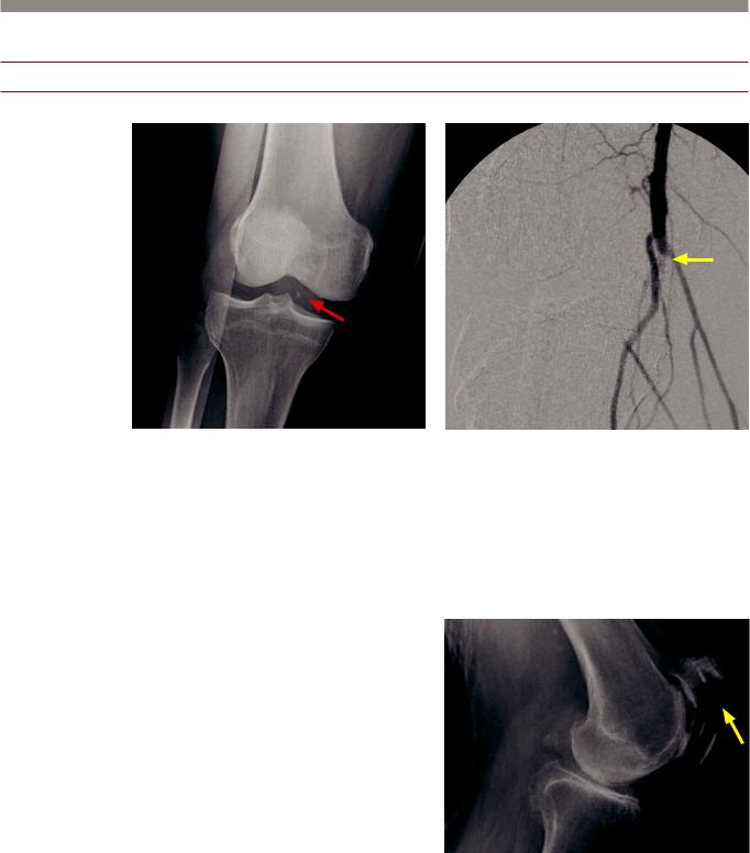
Knee and knee MRI
Knee trauma
Knee dislocation
Posterior dislocation with vascular injury: Frontal radiograph of the knee (left image) shows marked widening of the joint space with slight lateral displacement of the tibia. The widened joint space suggests a dislocation. An ossific fragment projects over the joint space (red arrow). Intraoperative digital subtraction angiogram (right image) demonstrates a complete traumatic occlusion of the popliteal artery (arrow) just above the knee.
•The knee may dislocate in any direction, although anteriorly is the most common.
•There is a high risk for vascular injury to the popliteal artery, and a vascular injury may be present even if the distal pulses are still intact. CT angiography is therefore typically recommended for all knee dislocations.
Patellar fracture
•Patellar fracture may be caused by a direct blow (usually causing a
comminuted fracture) or extreme sudden tension of the extensor mechanism
(usually causing a distracted fracture).
•It is important to distinguish a fracture from a bipartite or multipartite patella, which are normal variants and will have well corticated margins. A dorsal defect of the patella is another normal variant, appearing as a round lucency at the superior articular (dorsal) aspect of the patella.
Tibial plateau fracture
Lateral radiograph of the knee shows a distracted patellar fracture (arrow), suggestive of an injury caused by extreme tension of the extensor mechanism.
•Tibial plateau fractures are classified by the Schatzker classification.
•Lateral tibial plateau fractures are more stable than medial tibial plateau fractures.
411
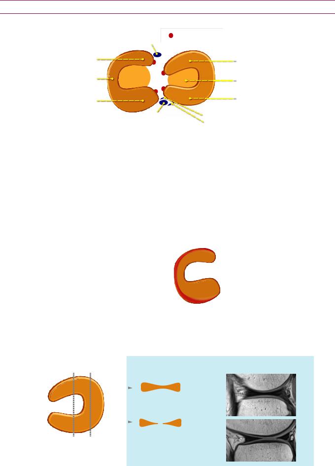
Menisci
Normal anatomy
|
|
|
|
ACL |
|
|
|
|
|
|
|
|
||
|
= meniscal root |
|
|
|
||||||||||
|
|
|
|
attachment |
|
|
|
|||||||
|
|
|
|
|
|
|
|
|
|
|
|
|||
anterior horn of |
|
|
|
|
|
|
|
|
|
|
|
|
|
anterior horn of |
|
|
|
|
|
|
|
|
|
|
|
|
|
||
medial meniscus |
|
|
|
|
|
|
|
|
|
|
|
|
|
lateral meniscus |
meniscal body |
|
|
|
|
|
|
|
|
|
|
|
|
|
articular surface of tibia |
|
|
|
|
|
|
|
|
|
|
|
|
|
||
|
|
|
|
|
|
|
|
|
|
|
|
|
||
posterior horn of |
|
|
|
|
|
|
|
|
|
|
|
|
|
posterior horn of |
|
|
|
|
|
|
|
|
|
|
|
|
|
||
medial meniscus |
|
|
|
|
|
|
|
|
|
|
|
|
|
lateral meniscus |
PCL |
anterior meniscofemoral ligament (Humphry) |
|
posterior meniscofemoral ligament (Wrisberg) |
||
attachment |
||
|
The medial and lateral menisci are crescent-shaped fibrocartilaginous structures that
form a discontinuous figure-eight to stabilize the knee and disperse the axial loading.
ach meniscus consists of an anterior and a posterior horn.
The posterior horn of the medial meniscus is larger than its anterior horn.
Both horns of the lateral meniscus are the same size.
ach meniscus is attached to the tibial plateau by an anterior and posterior meniscal root. Trauma to the meniscal root may cause meniscal extrusion.
There are two meniscofemoral ligaments, which extend from the posterior horn of the lateral meniscus to the medial femoral condyle. It is important not to mistake a meniscofemoral ligament (which are variably present) for a meniscal tear.
The ligament of Humphry is anterior to PCL; the ligament of Wrisberg is posterior to the PCL.
The peripheral third of each meniscus is relatively vascular and is called the red zone The central portion of the meniscus is considered avascular.
Meniscal injuries confined to the red zone may heal spontaneously or after surgical repair, while more central meniscal injuries
generally do not heal.
Normal appearance of menisci on MR
axial or arthroscopic view |
sagittal schematic |
||
|
|
|
|
|
|
|
|
The peripheral 10-30% of the meniscus is the red zone, which is more highly vascularized than the central portion.
sagittal MRI
A normal meniscus shows the classic “bowtie” appearance when imaged through the body.
Case courtesy Nehal Shah, MD, Brigham and Women’s Hospital.
412
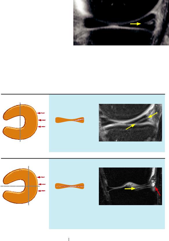
Myxoid degeneration versus tear
Myxoiddegenerationisincreased signalwithinthemeniscusthat does not extend to the meniscal surface.Itisnotthoughttocause symptomsandisnottreated.
n contrast, a meniscal tear is seen as a linear band of increased signal that does extend to the articular surface. Themorphology of the meniscus is commonly abnormal. MRI is >90% sensitive and specific for the diagnosis of meniscal tears.
Meniscus myxoid degeneration: Sagittal fluid-sensitive
MRI demonstrates increased globular signal (arrow) in the peripheral meniscus that does not extend to the articular surface.
Case courtesy Nehal Shah, MD, Brigham and Women’s
Hospital
Oblique/horizontal tear
oblique meniscal tear
axial or arthroscopic view sagittal schematic sagittal MRI
horizontal meniscal tear
axial or arthroscopic view sagittal schematic coronal MRI
Since the cleavage plane is parallel to the tibial plateau, the surface of the meniscus will appear normal.
A true horizontal tear cleaves the meniscus in a plane parallel to the tibial plateau. The plane of cleavage appears similar
in both the sagittal (as in the schematic) and coronal views. Note the associated parameniscal cysts (red arrow).
Case courtesy Nehal Shah, MD, Brigham and Women’s Hospital.
The terms oblique and horizontal are sometimes used interchangeably when referring to meniscal tears. Strictly speaking, however, a horizontal tear remains parallel to the tibial plateau, while an oblique tear may be at a slight angle to the tibial plateau.
An oblique/horizontal tear is the most common meniscal tear, typically occurring in the posterior horn of the medial meniscus.
Oblique and horizontal tears are more often degenerative, rather than due to trauma.
413
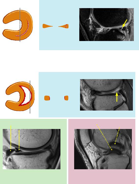
Vertical/longitudinal tear
axial or arthroscopic view sagittal schematic sagittal MRI
Nehal Shah, MD
A vertical (longitudinal) tear is present (arrow) demonstrating complete superior-to-inferior surface extension.
The vertical (synonymous with longitudinal) tear is a curved tear that follows the contour of the meniscus. On serial sagittal images, the tear will be in a vertical orientation and will remain a fixed distance from the edge of the meniscus.
Bucket-handle tear
axial or arthroscopic view |
sagittal schematic |
sagittal MRI |
|
|
|
Nehal Shah, MD |
|
|
Absent bow tie sign: No normal bow tie is seen posteriorly (arrow). |
|
native anterior meniscus |
displaced meniscal fragment |
||
|
displaced bucket handle fragment |
normal PCL |
|
|
|||
|
|
|
|
|
|
|
|
|
|
|
|
|
|
|
|
|
|
|
|
Nehal Shah, MD
The double delta sign is seen if the displaced fragment flips anteriorly.
Nehal Shah, MD
The double PCL sign is seen if the displaced fragment flips centrally into the intercondylar notch.
Abucket-handletearistheresultofanextensiveverticaltear,occurringwhenthefreeinner
edgeofthemeniscus(medialmeniscusmostcommonly)getsdisplacedorflippedover.
The most sensitive imaging finding of a bucket-handle tear is the absent bow tie sign, seen when there are fewer than two “bow ties” on adjacent 4 mm-thick sagittal slices.
The most common location of the displaced meniscal fragment is the intercondylar
notch, but the meniscal fragment may also displace anteriorly or posteriorly.
Anteriordisplacementproducesthe doubledeltasignonsagittalMRI(eithermeniscus).
Posteriordisplacementproducesthe doublePCLsignonsagittalMRI(medialmeniscusonly).
414
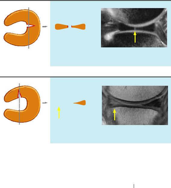
Radial/transverse tear
radial tear of meniscal body
axial or arthroscopic view sagittal schematic sagittal MRI
Radial tear is a vertically oriented tear extending from the free edge of the meniscus to the periphery.
radial tear of meniscal horn
axial or arthroscopic view sagittal schematic sagittal MRI
Ghost meniscus sign: No meniscal tissue is seen when the plane of imaging is directly through the radial tear (arrows).
Cases courtesy Nehal Shah, MD, Brigham and Women’s Hospital.
A radial tear (also known as a transverse tear) is a vertically oriented tear that is perpendicular to the arc of the meniscus. In contrast, the previously discussed vertical/longitudinal tear follows the curve of the meniscus.
On a single image, a radial tear may be indistinguishable from a vertical/longitudinal tear. On serial sagittal images, however, the radial tear will change position relative to the edge of the meniscus. This phenomenon is the marching cleft sign
Depending on the orientation of the tear in relation to the scan plane, a radial tear may also show the ghost meniscus sign. The ghost meniscus is typically described as absence of the posterior horn of the meniscus on sagittal images (although it may also occur with absence of the anterior horn, as seen above). It occurs in tears adjacent to the intercondylar notch.
Radial tears are associated with a high rate of meniscal extrusion, which may lead to osteoarthritis if not treated.
Meniscal cyst
A meniscal cyst is loculated fluid adjacent to the meniscus, thought to result from
extension of joint fluid through a meniscal tear.
Meniscal cysts are most commonly associated with horizontal cleavage tears.
t is important to mention the presence of a meniscal cyst because the meniscal cyst may continue to cause symptoms even if the meniscus is resected. Not all cysts are able to be visualized by arthroscopy.
415
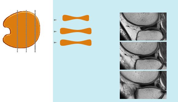
Discoid meniscus
axial or arthroscopic view |
sagittal schematic |
sagittal MRI |
|||
|
|
|
|
|
|
|
|
|
|
|
|
|
|
|
|
|
|
A discoid meniscus has a bow tie appearance
in the sagittal plane for 12 mm (or three or more slices, assuming 4 mm-thick slices).
Case courtesy Nehal Shah, MD, Brigham and Women’s Hospital.
Discoid meniscus is a spectrum of congenital malformations of the meniscus that ranges from increased thickness of the meniscus to a complete disc-like shape. Discoid meniscus is defined by having a portion of the meniscus extending to the central tibial plateau
Discoid meniscus typically affects children and is a common cause of joint-line pain, clicking, or locking in a child or adolescent. Discoid meniscus is prone to undergo cystic degeneration and tear. Even in the absence of a tear, a discoid meniscus may be symptomatic
The lateral meniscus is more commonly affected.
On MRI, a meniscus is discoid if an oval or bow tie-shaped meniscus is seen on three or more contiguous sagittal slices with standard 4 mm-thick sagittal slices
(representing 12 mm in medial–lateral thickness).
416
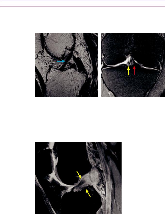
Knee Ligaments
Anterior cruciate ligament (ACL)
•The anterior cruciate ligament (ACL) arises from the femoral intercondylar notch and attaches on the anterior tibial plateau, directly lateral to the tibial spine. The tibial attachment of the ACL is stronger than its femoral origin. The ACL is composed of two fiber bundles, a small anteromedial band and larger posterolateral band.
Normal ACL: Sagittal proton-density (left image) and coronal proton-density fat-sat (right image) MRI shows the normal course of the ACL fibers with the typical striated appearance (blue arrow) on the sagittal image. The coronal image demonstrates the smaller anteromedial (yellow arrow) and larger posterolateral (red arrow) fiber bundles in the intercondylar notch.
Case courtesy Nehal Shah, MD, Brigham and Women’s Hospital.
ACL tear
•ACL tear is typically caused by an acute injury.
•MRI of ACL tear may show either frank discontinuity of the ligament fibers or abnormal course and signal of the ligament.
ACL tear: Sagittal fluidsensitive MRI demonstrates frank rupture of the ACL (arrows), with discontinuity of the fibers and increased signal.
Case courtesy Nehal Shah, MD, Brigham and Women’s Hospital.
•ACLinjuryisoftenassociatedwithmeniscalandMCLtears(O’Donoghue’striad). PosterolateralcornerkneeinjuriesarealsofrequentlyseeninassociationwithACLinjury.
417
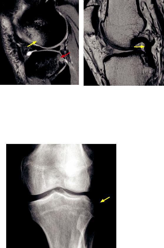
•There are multiple secondary findings of ACL injury. A characteristic bone contusion pattern with bone marrow edema in the lateral femoral condyle and the posterolateral tibial plateau is highly suggestive of ACL injury. Secondary buckling of the posterior collateral ligament (PCL) may also be seen due to anterior displacement of the tibia (positive drawer sign).
Secondary findings of ACL tear: Sagittal fluidsensitive MRI image shows bone marrow edema in the lateral femoral condyle (yellow arrow) and posterolateral tibial plateau (red arrow), a pattern characteristic of ACL injury.
Secondary findings of ACL tear in a different patient: Sagittal proton-density MRI shows buckling of the PCL (arrow), suggestive of an ACL injury.
Cases courtesy Nehal Shah, MD, Brigham and Women’s Hospital.
•A clue to ACL injury on radiography is a Segond fracture, which is an avulsion fracture of the lateral tibia plateau. Segond fracture is associated with both ACL injury and iliotibial (IT) band injury. The IT band inserts on Gerdy’s tubercle of the tibia.
The Segond fracture is thought to represent detachment of the lateral capsular ligament.
Segond fracture: Frontal knee radiograph shows an avulsion fracture of the lateral tibial plateau (arrow).
These fractures can be easily missed on radiography, but are important to diagnose.
MRI should be recommended if a Segond fracture is seen.
418
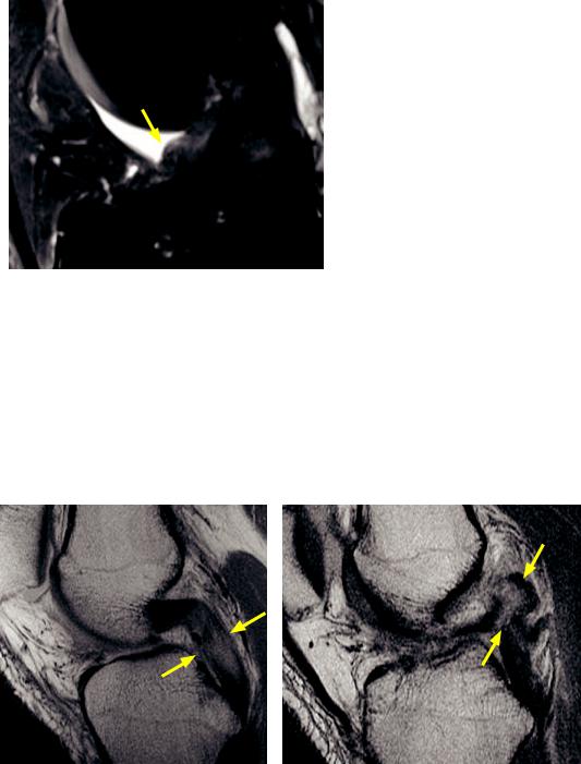
•After ACL reconstruction, the ACL graft should run parallel to the intercondylar notch, behind Blumensaat’s line (a line drawn on radiography along the intercondylar roof).
If the reconstructed ACL is too steep graft can be impinged by the femur with leg extension. If the reconstructed ACL is too lax graft may not provide enough stability.
AcomplicationofACLreconstructionisthecyclops lesion,whichisnodularscarringinHoffa’sfatpad.
Cyclops lesion post ACL reconstruction: Fluid-sensitive sagittal MRI shows nodular soft-tissue (arrow) anterior to the reconstructed ACL.
Case courtesy Nehal Shah, MD, Brigham and Women’s Hospital.
Posterior cruciate ligament (PCL)
•The posterior cruciate ligament (PCL) is a stronger, thicker ligament than the ACL. It also arises from the femoral intercondylar notch (more anteriorly than the ACL), but inserts on the posterior tibial plateau.
The PCL is normally low signal on all sequences.
The PCL is much less commonly injured than the ACL.
PCL tear
•In contrast to an ACL tear, a complete disruption of the PCL fibers is uncommon.
•The typical MRI appearance of PCL injury is increased laxity of the PCL, with or without abnormal high signal on T2-weighted images.
PCL interstitial tear: Sagittal proton-density MRI shows a PCL interstitial tear (arrows) with increased thickness, abnormal signal, and increased laxity of the PCL.
PCL complete tear (in a different patient): Sagittal proton-density MRI shows complete rupture of the PCL with frank discontinuity of the fibers (arrows).
Cases courtesy Nehal Shah, MD, Brigham and Women’s Hospital.
•PCL tears tend to be less frequently repaired compared to ACL tears.
419
Medial collateral ligament (MCL)
The medialcollateralligament(MCL)arisesfromtheposterioraspectofthemedial
femoralcondyleandattachestothemedialtibialmetaphysis,deeptothepesanserinus.
The MCL fibers are interlaced with the joint capsule and the medial meniscus.
The MCL is an extrasynovial structure and is not visualized by arthroscopy.
MCL injury
MCL injuries are commonly divided into three grades:
Grade 1 (MCL sprain): High signal in the medial soft tissues with normal signal of the ligament.
Grade 2 (severe sprain/partial tear): High signal or partial disruption within the MCL fibers.
Grade 3 (complete tear): Complete disruption of the MCL.
The Pellegrini–Stieda lesion is post-traumatic calcification medial to the medial femoral condyle, which may be secondary to MCL avulsion injury.
Lateral collateral ligament (LCL) complex
The lateral collateral ligament (LCL) complex is composed of many structures, three of
which are routinely evaluated by MRI. From posterior to anterior, these structures are:
Biceps femoris tendon (most posterior), LCL, and iliotibial band (most anterior).
biceps femoris tendon most posterior
LCL
iliotibial band most anterior
The biceps femoris tendon and the
LCL both attach on the lateral bular head, where they often have a conjoined tendon.
The iliotibial band attaches to Gerdy’s tubercle on the proximal lateral tibia.
LCL injury
LCL injury is less common than MCL injury and is usually associated with injury to other structures in the posterolateral corner. Injury to the posterolateral corner is a surgical near-emergency as there is poor outcome without prompt intervention.
The posterolateral corner of the knee is a complex region consisting of:
LCL complex, consisting of the biceps femoris tendon, LCL, and the iliotibial band.
Arcuate ligament, a Y-shaped ligament connecting the fibula to the lateral femur.
Popliteofibular ligaments, which are important lateral stabilizers.
Popliteus tendon, which inserts on the lateral femur.
Iliotibial band syndrome
Fluid surrounding (both superficial and deep to) the iliotibial band may represent iliotibial (IT) band syndrome, which is caused by friction between the anterolateral femur and the tendon of the iliotibial band.
Clinically, IT band syndrome may be a cause of anterolateral knee pain and is seen commonly in runners.
tisimportanttodistinguishITbandsyndromefromalateralmeniscustear.Bothmay presentwithlateralkneepain,buttreatmentofiliotibialbandsyndromeisnonsurgical.
420
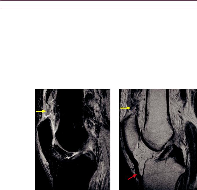
Extensor mechanism and patella
Normal anatomy of the extensor mechanism
•The extensor mechanism is composed of the conjoined quadriceps tendon (attaches to the patella), the patella, and the patellar tendon (attaches to the tibial tuberosity).
The patella is a sesamoid bone and is enclosed within the quadriceps tendon.
•Three of the quadriceps muscles (vastus intermedius, medialis, and lateralis) originate from the femur. The rectus femoris arises from the anterior inferior iliac spine (AIIS).
Patella alta and patella baja
•Patella alta is an abnormally high patella and patella baja is an abnormally low patella.
•TheInsall-Salvatiratiocanhelpdeterminethecraniocaudalalignmentofthepatella,based onasagittalMRIorlateralradiograph(ideallywith30degreesofflexion).TheInsall-Salvati ratioisdefinedastheratioofthepatellartendonlengthtothepatellarlength.AnInsallSalvatiratio>1.2issuggestiveofpatellaalta,andaratio<0.8issuggestiveofpatellabaja.
Quadriceps tendon tear
Quadriceps tendon tear: Sagittal proton density fat-saturated (left image) and proton density (right image) MRI shows complete disruption of the distal quadriceps tendon (yellow arrows) with surrounding marked edema. There is secondary buckling of the patellar tendon (red arrow).
Case courtesy Nehal Shah, MD, Brigham and Women’s Hospital.
•Tear of the quadriceps tendon clinically presents with loss of knee extension.
•Quadriceps tendon tear most commonly occurs within 2 cm of the patella and may be evident on radiography due to the resultant patella baja (patella too low).
Patellar tendon injury
•Similar to quadriceps tendon tear, patellar tendon tear clinically presents with loss of knee extension. In contrast to quadriceps tendon tear, tear of the patella tendon causes superior displacement of the patella (patella alta).
•Patellar tendinosis is seen on MRI as thickening of the patellar tendon. This is known as jumper’s knee.
421
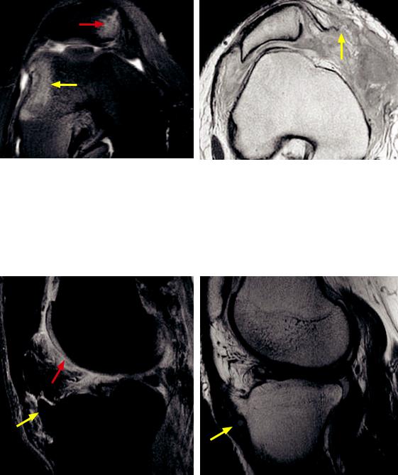
Lateral patellar dislocation
•Traumaticlateralpatellardislocationleavesbehindadistinctivepatternofinjury,evenifthe patellareducesbacktothenormalposition.Bonemarrowcontusionsofthelateralfemoral condyleandthemedialpatellarfacetaretypical,withtearingofthemedialretinaculum.
Lateral patellar dislocation: Axial fluid-sensitive MRI demonstrates the typical bone marrow edema pattern, with contusions of the lateral femoral condyle (yellow arrow) and medial patellar facet (red arrow).
The patella has relocated to its anatomic position.
Lateral patellar dislocation in a different patient: Axial proton-density MRI shows persistent lateral subluxation of the patella, with complete tearing of the medial retinaculum (yellow arrow).
Cases courtesy Nehal Shah, MD, Brigham and Women’s Hospital.
Osgood–Schlatter disease
Osgood–Schlatter disease: Sagittal fluid-sensitive (left image) and proton density (right image) MRI shows an enlarged, fragmented tibial tubercle (yellow arrows). There is associated edema anterior to the tibial tubercle and within Hoffa’s fat pad (red arrow).
Case courtesy Nehal Shah, MD, Brigham and Women’s Hospital.
•Osgood–Schlatter disease is an osteochondrosis of the tibial tubercle, thought to be caused by repetitive microtrauma. It occurs in adolescents.
•Radiographsshowanenlargedtibialtuberositywithfragmentationandadjacentedema.
•MRI shows increased signal of the distal patellar tendon, marrow edema within the tibial tubercle, and edema within Hoffa’s fat pad.
422
Knee cartilage
Overview and anatomy
The cartilageofeachofthethreemajorcompartments(patellofemoral,medial tibiofemoral,andlateraltibiofemoral)shouldberoutinelyevaluatedoneverykneeMRI.
Further subdividing the patellofemoral cartilage, there are three facets of patellar cartilage (medial, lateral, and odd facets; the medial and lateral facets are divided by the median ridge) and three segments of trochlear cartilage (medial, central, and ateral). The trochlea is the femoral groove where the patella lies.
Cartilage thinning or injury
Cartilage thinning is reported as <50%, 50%, or >50% thinning.
Cartilageinjurymaymanifestassurfaceirregularity,fissuring,ordelamination.
Delaminationisadissecting-typedetachmentofthecartilageundersurfacefrombone.
Osteochondrosis dissecans (OCD)
Osteochondrosis dissecans (OCD) is an osteochondral (affecting both articular cartilage and bone) injury thought to be caused by repetitive trauma, which tends to occur in adolescents.
The term osteochondritis dissecans should be avoided as there is no inflammation involved.
TheetiologyofOCDisthoughttobeduetocartilagedehydrationandsubsequentstiffening,
whichtransmitsgreaterforcetothesubchondralboneeachtimethejointisloaded.
The most common joints affected are the knee, ankle, and elbow, in typical locations:
Knee (most common): Lateral aspect of medial femoral condyle.
Ankle (second most common): Posteromedial or anterolateral talar dome.
Elbow (in gymnasts or throwing athletes): Anterolateral capitellum.
An “unstable” osteochondral fragment means the lesion is unattached to bone. An unstable lesion may develop secondary osteoarthritis if not treated. The most important goal of MRI is to determine stability of an osteochondral fragment. A
curvilinear region of high signal on a fluid-sensitive sequence interposed between the fragment and the underlying bone suggests that the fragment is unstable.
MRI staging of OCD:
stage 1 OCD: intact articular cartilage radiograph: normal T2-weighted image (T2WI):
bone marrow edema
stage 2 OCD: articular cartilage defect radiograph: semicircular fragment T2WI: cartilage defect; low-signal line
surrounding lesion
= unstable (stages 3 and 4
stage 3 OCD: displaceable fragment radiograph: semicircular fragment T2WI: high-signal line
surrounding fragment
stage 4 OCD: displaced fragment radiograph: defect or loose body T2WI: defect and/or loose body
Osteochondralinjury isanacute(notrepetitive)traumaticinjuryofcartilageandbone.
423
Synovium
Pigmented villonodular synovitis (PVNS)
PVNS: Lateral knee radiograph shows a dense knee effusion (red arrows) and a soft-tissue mass posterior to the knee (yellow arrows).
Axial proton-density MRI in the same patient shows low signal masses (arrows) posterior to the knee.
Sagittal gradient echo MRI shows the extremely |
At arthroscopy, multiple synovial-based masses are |
dark masses accentuated by susceptibility artifact |
evident. |
(arrows). |
|
•Pigmented villonodular synovitis (PVNS) is a benign hyperplastic proliferation of the synovium within the joint, now thought to represent a tumor. The same process outside of the joint is called giant cell tumor of the tendon sheath.
•The knee is by far the most common location for PVNS.
•PVNS presents as knee pain and swelling, with recurrent dark brown effusions from prior hemorrhage.
•PVNS most commonly diffusely affects a joint, but it may also be focal. Focal PVNS can clinically mimic internal derangement.
•Recurrent hemorrhage leads to residual intra-articular hemosiderin, which is paramagnetic. The paramagnetic effects of hemosiderin cause T1 prolongation and T2 shortening, resulting in dark signal on both T1and T2-weighted images, with blooming on gradient-echo sequences.
424
Lipoma arborescens
Lipoma arborescens is overgrowth of intracapsular synovial fatty tissue, causing
obulated and globular intra-articular fatty masses.
Lipoma arborescens is treated with synovectomy to prevent premature osteoarthritis.
Knee soft tissues
Baker’s cyst
Baker’s cyst is a popliteal cyst caused from a ball-valve type communication with the knee joint.
The typical location is between the semi embranosus tendon and edial head of the gastrocnemius muscle (mnemonic: Baker’s M & M ).
An important differential consideration is a popliteal aneurysm.
Tennis leg
Tears of either the plantaris tendon or the medial head of the gastrocnemius have
both been called tennis leg.
Hip and hip MRI
Hip anatomy
Bony acetabulum
The acetabulum is formed by the fusion of three bones: The ilium superiorly, the pubis
anteriorly, and ischium posteriorly.
ediatric radiographs of the hip clearly show the separation of these three bones, which meet at the triradiate cartilage to form the acetabulum.
Cartilage, labrum, and ligaments
The articular surface of the acetabulum is lined by a horseshoe-shaped thin articular cartilage. The center of the acetabulum, called the pulvinar, is not covered by cartilage.
The labrum forms a fibrocartilaginousring approximately270degreesaround theperipheryoftheacetabulum
Thelabrumblendswiththe transverseligamentanterior/ inferiorly.Notethatthereisno labrum at the anterior/inferior aspectoftheacetabulumwhere thetransverseligamentlies.
labrum
articular |
cartilage |
pulvinar
transverse ligament
ligamentum teres
The transverse ligament gives off fibers that form the ligamentum teres, which
connects to the femoral head at its central fovea.
425
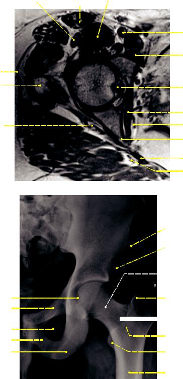
Cross-sectional anatomy of the hip musculature
rectus femoris |
sartorius |
iliopsoas |
|
|
|
|
|
|
|
|
|
|
|
|
|
|
|
|
|
tensor fascia lata 

gluteus minimus 
 gluteus medius

iliotibial band |
|
|
greater trochanter |
femoral |
|
head |
|
|
|
|
superior gemellus 
 sciatic nerve
gluteus maximus 

Normal radiographic anatomy of the hip
common femoral vasculature
pectineus
ligamentum teres
obturator internus
obturator vessels
ischial spine
sacrospinous ligament
inferior gluteal artery and vein
anterior superior iliac spine origin of sartorius
anterior inferior iliac spine origin of rectus femoris
subcapital region
(underneath the head)
fovea capitis superior pubic ramus
obturator foramen inferior pubic ramus
ischial tuberosity origin of hamstrings
femoral
head femoralneck 

greater trochanter insertion of gluteus medius and gluteus minimus
basicervical region
(base of the neck) intertrochanteric crest
lesser trochanter insertion of iliopsoas
femoral diaphysis
The femoral head is mostly covered by articular cartilage except at the fovea capitis
(central fovea), which is a central depression and attachment site to the ligamentum teres
The greater and lesser trochanters are apophyses. The greater trochanter is the insertion site for the gluteus medius, gluteus minimus, obturator internus and externus, and the piriformis. The lesser trochanter is the insertion site for the iliopsoas.
A lesser trochanteric fracture in an adult is considered pathologic until proven otherwise.
426
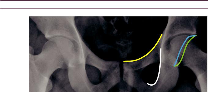
Trauma
Acetabular fracture
|
|
|
|
|
|
|
|
i |
n |
e |
|
|
|
|
|
|
|
|
|
|
|||
|
|
|
|
|
|
|
l |
|
|
|
|
|
|
|
|
|
|
l |
|
|
|
|
|
|
|
|
|
|
|
a |
|
|
|
|
|
|
|
|
|
|
e |
|
|
|
|
|
|
|
|
|
|
n |
|
|
|
|
|
||
|
|
|
|
i |
|
|
|
|
|
e |
|
|
|
|
t |
|
|
|
|
|
|
||
|
|
|
c |
|
|
|
|
|
|
|
|
|
|
e |
|
|
|
|
|
|
|
|
|
|
l p |
|
|
|
|
|
|
|
|
i |
|
|
i io |
|
|
|
|
|
|
|
|
|
|
anterior column |
|
|
|
|
|
|
|
|
|
|
n |
|
|
|
|
|
|
|
|
|
|
l |
|
|
|
|
|
|
|
|
|
|
|
|
l |
of acetabulum |
|
|
|
|
|
|
|
|
|
|
a |
|
|
|
|
|
|
|
|
|
i |
||
|
|
|
|
|
|
|
|
|
|
h |
|
|
|
|
|
|
|
|
|
|
|
c |
|
|
|
|
|
|
|
|
|
|
s |
||
|
|
|
|
|
|
|
|
|
i |
||
|
|
|
|
|
|
|
|
|
o |
||
|
|
|
|
|
|
|
|
i |
|||
|
|
|
|
|
|
|
|
|
l |
|
|
posterior column |
|
i |
|
||||||||
|
|
|
|
|
|
||||||
of acetabulum
|
e |
t |
a |
|
|
||
|
|
|
|
c |
|
|
|
a |
|
|
|
r |
|
|
|
o |
|
|
|
i |
|
|
|
r |
|
|
|
e |
|
|
|
t |
|
|
|
n |
|
|
|
a |
|
|
|
po |
|||
s
|
|
|
|
m |
|||
|
|
u |
|
|
|
||
|
l |
|
|
|
|
|
|
b |
u |
|
|
|
|
|
m |
|
|
|
|
|
|
||
|
|
|
|
|
|
u |
|
|
|
|
|
|
l |
|
|
|
|
|
a |
b |
u |
|
|
|
|
|
|
|
|
||
|
|
|
|
|
|
|
|
|
|
t |
|
|
|
|
|
|
|
e |
|
|
|
|
|
|
c |
|
|
|
|
|
|
|
a |
|
|
|
|
|
|
|
r |
|
|
|
|
|
|
|
o |
|
|
|
|
|
|
i |
|
|
|
|
|
|
|
r |
|
|
|
|
|
|
|
e |
|
|
|
|
|
|
|
t |
|
|
|
|
|
|
|
•Discontinuity of either the iliopectineal or ilioischial lines may represent a fracture of the anterior or posterior column of the acetabulum.
The iliopectineal line (also called iliopubic line) represents the border of the anterior column of the acetabulum. The ilioischial line represents the border of the posterior column of the acetabulum.
•The anterior and posterior walls of the acetabulum can usually be directly seen projecting over the femoral head. The posterior wall is more lateral.
•Classification of acetabular fractures is complex and multiple classifications have been introduced. The most widely used classification scheme is the Judet–Letournel classification, which describes 10 types of acetabular fractures.
•The Judet–Letournel classification describes five elementary patterns: fractures of the posterior wall, posterior column, anterior wall, anterior column, and transverse fractures. Five combination patterns are also described. The Judet-Letournel types are not related to fracture complexity or prognosis.
•The posterior wall of the acetabulum is the most commonly fractured element, alone or in combination with other injuries.
Overview of proximal femur fractures
•A “hip fracture” is a nonspecific term encompassing a range of fractures of the proximal femur, including femoral head, neck, intertrochanteric, and subtrochanteric fractures.
•Femoral head and neck fractures are intracapsular. In contrast, intertrochanteric and subtrochanteric fractures are extracapsular. Extracapsular fractures are usually caused by direct injury, such as a fall, and tend to affect older individuals.
•Animportantcomplicationofanintracapsular(headorneck)fractureisosteonecrosis.The majorityofthebloodsupplytothefemoralheadisfromthecircumflexfemoralarteries, whichcreatearingatthebaseofthefemoralneckandsendoffascendingbranchestothe femoralhead.Thisbloodsupplymaybedisruptedinanintracapsularfracture.
Only minimal blood supply to the femoral head arises from small arteries in the ligamentum teres.
•Incontrasttointracapsularfractures,extracapsular(intertrochantericand subtrochanteric)fracturesrarelyleadtoosteonecrosis.Themedialfemoralcircumflex arteryanditsbranchesareusuallynotdisruptedwithanextracapsularfracture. Additionally,thereisrobustbloodsupplyfromthemusclesthatattachatthetrochanters (gluteusmediusandminimusggreatertrochanter;iliopsoasglessertrochanter).
427
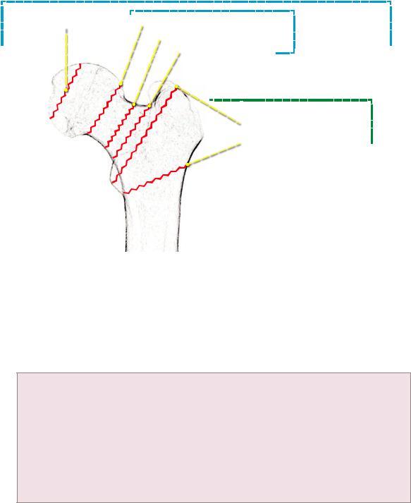
Femoral head fracture-dislocation (intracapsular)
Femoral head fractures are associated with posterior hip dislocations. Complications of femoral head fracture include avascular necrosis and post-traumatic arthritis.
Orthopedists use the Pipkin classification to characterize femoral head fracturedislocations.
Type I: Fracture inferior to the central fovea. There is disruption of the ligamentum teres.
Type II: Fracture superior to the central fovea. The ligamentum teres remains attached to the
fracture fragment.
Type III: Type I or II with associated femoral neck fracture.
Type IV: Type I or II with associated superoposterior acetabular rim fracture.
Femoral neck fracture (intracapsular)
|
|
intracapsular fractures |
|
femoral head fracture |
|
subcapital fracture (most common) |
femoral neck |
|
|||
|
|
|
|
transcervical fracture |
|
|
fractures |
|
||||
|
|
|
|
||||||
|
|
|
|
||||||
|
|
|
|
|
|
|
|||
|
basicervical fracture |
|
|||||||
|
|
||||||||
|
|
|
|
|
|
|
|
||
|
|
|
|
extracapsular fractures |
|
||||
|
|
|
|
|
|||||
|
|
|
|
|
|||||
|
|
|
|
intertrochanteric fracture |
|
||||
|
|
|
|
|
|||||
|
|
|
|
subtrochanteric fracture |
|
|
|
||
|
|
|
|
|
|
||||
Femoralneckfracturescanbedividedintothreecategoriesbasedonlocation:Subcapital
(directlybelowthefemoralhead),transcervical(throughtheneck),andbasicervical(at thebaseofthefemoralneck).Subcapitalfracturesarethemostcommon.
The Garden classification of subcapital femoral neck fractures (diagrammed on the following page) is based on the degree of valgus displacement and is commonly used by radiologists. Note that is defined as an angle with apex pointed laterally and valgus is defined as an angle with apex pointed medially (mnemonic: “fair” Lady and
“vulgar” Mouth).
Stage Impacted or incomplete femoral neck fracture, with valgus orientation of the femoral head/neck trabeculae and varus orientation of the femoral head and acetabulum. The femoral shaft is externally rotated. The medial trabeculae of the femoral head and neck form an angle
>180 . This fracture is stable and has a good prognosis.
Stage I Complete, nondisplaced fracture. The trabeculae of the femoral head and neck are in
mild varus and form an angle of approximately 160 . This fracture also has a good prognosis.
Stage II Complete, partially displaced fracture. There is more marked varus alignment of the trabecular pattern of the femoral head and neck, usually forming an angle <160

Stage IV Complete, displaced fracture. The trabecular pattern of the femoral head is aligned with that of the acetabulum, which is the normal anatomic alignment.
428
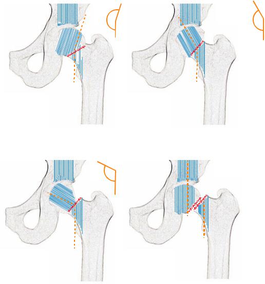
Garden stage I:
impacted or incomplete fracture
trabeculae of femoral head and neck are in valgus (>180˚)
>180˚
Garden stage III:
complete, partially displaced fracture trabeculae of femoral head and neck are in
more marked varus, usually forming an angle <160˚
<160˚
Garden stage II:
complete, nondisplaced fracture
trabeculae of femoral head and neck are in varus, usually forming an angle of approximately 160˚
~160˚
Garden stage IV:
complete, fully displaced fracture
trabeculae of femoral head , neck, and acetabulum are aligned
Intertrochanteric fracture (extracapsular)
The Boyd–Griffin and Evans classifications are used to describe intertrochanteric
fractures by orthopedists. These classifications are rarely used by radiologists.
Subtrochanteric fracture (extracapsular)
Fractures at or inferior to the lesser trochanter are subtrochanteric fractures.
Subtrochanteric fractures are classified using the Fielding or Seinsheimer
classifications by orthopedists.
429
Hip MRI technique
Imaging of metallic hip arthroplasties
•It is possible to image metallic hip arthroplasties by optimizing MRI technique. In general, field strength should be as low as reasonable and fast spin echo sequences should be used instead of GRE.
•The receiver bandwidth should be increased as much as possible. This will increase the amount of noise, so the number of acquisitions (NEX) will need to be proportionally increased.
•Artifacts can be directed in the superior–inferior plane so the region directly medial and lateral to the implant (where loosening would be seen) is not obscured.
•Voxel size should be decreased by decreasing slice thickness and increasing the matrix size.
Indications for hip MRI
Occult traumatic fracture
•Radiographs are relatively insensitive for the detection of a nondisplaced femoral neck fracture.
•The MRI appearance of a fracture is a hypointense fracture line on T1and T2weighted images, associated with hyperintense edema signal on T2-weighted images.
•MRI may be able to suggest an alternative etiology for the patient’s pain. For instance, a sacral fracture may clinically present as hip pain.
Occult stress fracture
•Stress fractures can be categorized as fatigue fractures (due to abnormal stress on normal bone) and insufficiency fractures (due to normal stress on demineralized bone).
•Radiographs are relatively insensitive for detection of hip stress fractures. When visible on a radiograph, a stress fracture typically appears as a band of sclerosis.
•MRIappearanceofastressfractureissimilartoanacutetraumaticfracturewitha hypointensefracturelineassociatedwithhyperintenseedemasignalonT2-weightedMRI.
•A classic location of a stress fracture is the inferomedial femoral neck.
•Less commonly, a stress fracture may occur in the superior femoral head.
Superior femoral head stress fractures may be difficult to differentiate from avascular necrosis (AVN), especially if large field of view imaging of both hips is acquired.
Both AVN and fracture are characterized by a subchondral hypointense line on T1-weighted images; however, it has been suggestedthat the shape of the hypointense line in the coronal plane may be able to differentiate between AVN and subchondral insufficiency fracture. The hypointense line of AVN is typically smooth and concave to the articular surface, while subchondral insufficiency fracture usually produces an irregular low-intensity fracture line convex to the articular surface. Additionally, a history of corticosteroid use or alcohol abuse is typically present with AVN.
•Atypical locations of femoral stress fractures, such as lateral femoral diaphyseal and transverse diaphyseal fractures, may be secondary to bisphosphonate use.
•Treatment of stress fractures is typically conservative and a follow-up MRI in 8 weeks should show resolution of the edema. Persistent edema on the follow-up study suggests treatment failure.
•In addition to the proximal femur, stress fractures may occur at several sites in the pelvis, including the superior and inferior pubic rami, sacrum, and acetabular roof.
430

Avascular necrosis (AVN)
•Avascular necrosis (AVN) is focal ischemia of the epiphyseal region or subchondral bone. AVN leads to trabecular necrosis, which increases susceptibility to stress fracture, eventual collapse, and secondary osteoarthritis if untreated. The more general term osteonecrosis indicates necrosis of bone and marrow elements.
•The femoral head is especially susceptible to AVN because of precarious blood supply. The blood supply to the femoral head is primarily through the medial femoral circumflex artery, which enters the femoral neck and gives off posterosuperior epiphyseal branches that course along the femoral neck to supply the femoral head.
•As previously discussed, intracapsular proximal femur fractures have a high risk of AVN, including femoral neck fractures in the elderly and transient subluxation in athletes.
•Nontraumatic causes of AVN are usually systemic, including red-cell abnormalities (e.g., sickle cell disease), abnormalities of marrow packing (e.g., Gaucher disease), or secondary effects of medications including steroids, alcohol, or immunosuppression from transplant. Systemic causes of AVN tend to be bilateral.
A clue to the presence of Gaucher disease is splenomegaly with AVN.
A clue to the presence of sickle cell is a small calcified spleen with AVN.
•The most common site of AVN is the proximal femur. The second most common site is the proximal humerus.
•The Ficat staging system is based on radiographic appearance.
Stage 1: Normal radiograph, with signs of AVN visible on MRI (discussed below) or bone scan (reduced tracer uptake in the femoral head). Treatment is core femoral head drilling.
Stage 2: Cystic and sclerotic changes. Treatment is same as stage I.
Stage 3: There is loss of the normal spherical shape of the femoral had due to collapse of subchondral bone. A subchondral lucent line representing the crescent sign may be visible on radiographs. Treatment is variable.
Stage 4: Flattening of the femoral head and secondary osteoarthritis. Treatment is joint replacement.
•Several systems have been proposed for the MRI classification of AVN, including a modified Ficat system and the Mitchell system. The Mitchell system characterizes the changes in MRI signal only. It has been suggested that the modified Ficat system is more clinically useful as it accounts for the progression in ischemia that results in collapse of the femoral head.
•The most common appearance of AVN on MRI is a geographic subchondral lesion outlined by a serpentine low signal rim on T1-weighted images. The subchondral anterior-superior portions of the femoral head, at 10 o’clock and 2 o’clock on coronal images, are typical locations.
•The double line sign is seen on T2-weighted MRI and is comprised of a peripheral lowintensity rim and an inner high-intensity band. The double line sign is thought to be pathognomonic for AVN.
•Bone marrow edema is present in 50% and clinically correlates with pain.
•The complete description of AVN should include the presence of collapse (CT is most sensitive, followed by radiographs), the size of the T1 signal abnormality (as percentage of involved areas of the femoral head), and presence of secondary involvement of the acetabulum.
431
Transient bone marrow edema (TBME)
•Transient bone marrow edema (TBME), also called transient osteoporosis of the hip, is a controversial entity that is a diagnosis of exclusion and may actually reflect a combination of disorders including sympathetic nervous system overactivity, stress fracture, or transient ischemia.
•The traditional teaching is that TBME is a self-limited clinical entity lasting a few months. It is characterized by severe hip pain in young to middle-aged adults (more commonly men, although it was first described in women in the third trimester of pregnancy).
•Radiographs show regional osteopenia in the femoral head without any secondary signs of arthritis. The cartilage space is preserved and there are no erosions or morphologic abnormalities of the acetabulum.
•T1-weighted MRI shows diffuse low signal corresponding to diffusely high signal on T2-weighted images. The signal abnormality may extend from the femoral head into the femoral neck.
•As TBME may be a wastebasket diagnosis, it is essential to always evaluate for subtle lesions to explain these findings. For instance, a subchondral hypointense line on T1weighted images may represent a fracture, or a small focus of AVN may be apparent.
Labral injury
•The acetabular labrum is analogous to the glenoid labrum in the shoulder. The acetabular labrum is a ring of fibrocartilage that functionally deepens the acetabulum. Unlike the shoulder, however, the femoroacetabular joint is a ball-in-socket joint that is inherently stable and does not rely on the extensive ligamentous and muscular support that maintains active and passive stability in the shoulder.
•Some authors advocate that hip labral injuries be called detachments rather than tears, but the use of this suggested terminology is inconsistent.
•Although labral detachment may be due to acute trauma, labral injuries are more commonly due to chronic repetitive microtrauma. Femoroacetabular morphologic abnormalities, such as developmental dysplasia of the hip (discussed in the pediatric imaging section) and femoral acetabular impingement may predispose to labral injury.
•Labral injuries are best diagnosed with MR arthrography, which shows focal contrast undermining a portion of the labrum.
•The most common location for labral injury is anterosuperior. There is no labrum at the inferior aspect of the hip joint.
432
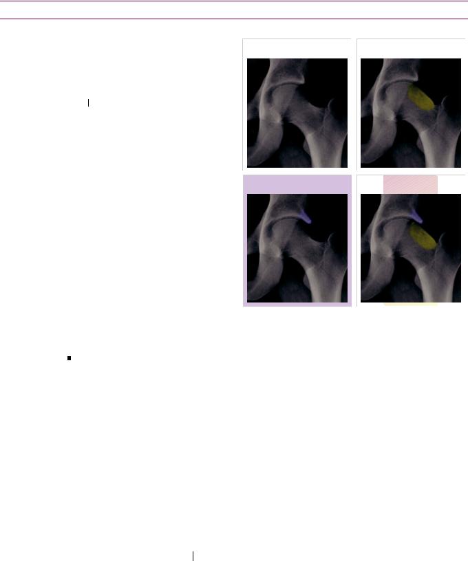
Femoroacetabular impingement (FAI)
Overview and controversy of femoroacetabular impingement (FAI)
emoroacetabular impingement
(FAI) is abnormal abutment of the femur with the acetabulum at extremes of range of motion.
Two types of impingement have been described, depending on whether the femoral head/ neck junction (cam-type) or the acetabulum (pincer-type) is abnormal.
Chronicmicrotraumaofthe femoralheadabuttingagainstthe acetabulum may cause acetabular abraltearsinyoungathletic individualsandmayacceleratethe developmentofhiposteoarthritis.
normal |
|
cam-type FAI |
|
|
|
pincer-type FAI |
mixed-type FAI |
While FAImaybeanimportant causeofhiposteoarthritisinyoung patients,thereiscontroversy
astowhetheradiagnosisof impingementcanbemadebased ontheimagingfindingsinthe absenceoftheclinicalsyndrome.
mpingement is primarily a clinical diagnosis and there is a significant subset of patients with abnormal morphology of the acetabulum or femoral head/neck junction who are asymptomatic.
Labral injury secondary to FAI (typically occurring in the anterosuperior labrum) is often associated with underlying chondral injury as well.
Regardlessofthecontroversy,FAIisanimportantconcepttobeawareofintheyoungadult.
AgeneralconsensusisthatimagingfindingssuggestiveofFAIshouldbedescribedwiththe
disclaimerthatthesefindingsmaypredisposetoimpingementinthepresenceofpain.
Cam-type impingement
An abnormal bump at the femoral head–neck junction may be due to a variety of causes, such as childhood slipped capital femoral epiphysis, Legg–Calve–Perthes disease, sequela of developmental hip dysplasia (which ironically is characterized by acetabular undercoverage), and malunited fracture.
Cam-type impingement is most common in young athletic males.
The pistol-grip deformity describes the abnormal morphology of the femoral head–neck junction that resembles an old-time pistol, with the femoral head/neck representing the handle and the femoral diaphysis the barrel.
The alpha angle defines the degree of abnormal femoral bump. An alpha angle is
measured on oblique axial MRI or CT. An alpha angle of >55 degrees is abnormal.
To measure the alpha angle, a line through the femoral neck bisecting the center of the femoral head is interfaced with a second line connecting the center of the femoral head to the most proximal abnormal contour of the femoral head/neck.
Cam-type impingement is treated with femoral osteoplasty.
433
Pincer-type impingement
Pincer-type FAI is caused by overcoverage of the acetabulum.
Pincer-type impingement is most common in middle-aged females.
The |
|
sign (also called cranial acetabular retroversion) is seen in pincer-type |
|
FAI and represents the superior aspect of the anterior acetabular wall crossing over the posterior acetabular wall on a frontal radiograph.
normal anterior overcoverage: crossover sign
anterior acetabulum
 posterior acetabulum

Treatment of pincer-type FAI is surgical trimming of the acetabular rim.
Mixed-type impingement
Mixed-type impingement is the most common form of FAI. It features abnormalities of both the femoral head/neck junction and the acetabulum.
Lumbar/thoracic spine
Lumbar/thoracic spine trauma
Three column concept
The three column concept of functional spinal anatomy helps to evaluate the stability of thoracolumbar spinal trauma. A spinal injury where the integrity of two of the three columns is preserved is generally considered stable, while a severe injury to all three columns is most likely to be unstable. Two column injuries may be stable or unstable.
The anterior column is comprised of the anterior two thirds of the vertebral body, including the anterior longitudinal ligament.
The middle column consists of the posterior third of the vertebral body and the posterior longitudinal ligament.
The posteriorcolumn representstheposteriorelementsandtheposteriorligaments
(consistingofthesupraspinousandinfraspinousligaments,andtheligamentumflavum).
Chance fracture
A Chance fracture, also known as a seat belt fracture, is a flexion distraction injury
caused by acute forward flexion of the spine during sudden deceleration.
There is horizontal splitting of the vertebra, beginning posteriorly in the spinous
process or lamina and extending anteriorly through the vertebral body.
Spondylolysis
Spondylolysis is a fracture of the pars interarticularis, which may be secondary to
chronic stress or an acute fracture.
Onoblique radiographs,spondylolysisisseenasadefectintheneckofthe“scottydog.”
Spondylolysis leads to spondylolisthesis, which is anterior–posterior subluxation of
one vertebral body relative to another.
434
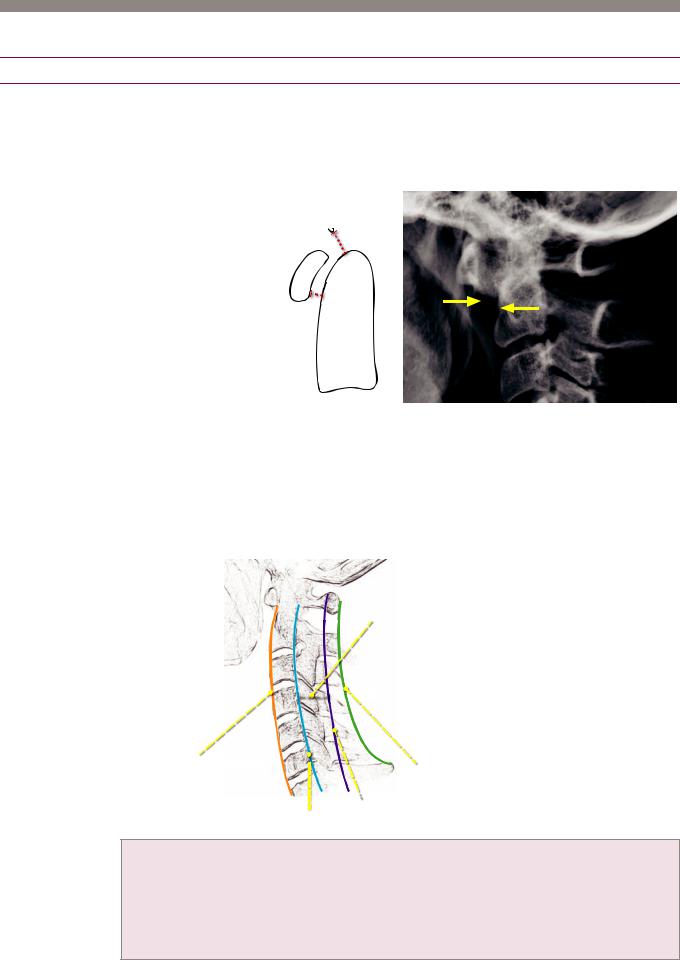
Cervical spine
Cervical spine trauma
ntervals: The basion-dental interval and atlanto-dental interval (also known as the atlanto-axial interval) should be evaluated in every lateral cervical spine radiograph.
An increased basion-dental interval >12 mm suggests craniocervical dissociation. An increased atlanto-dental interval >2.5 mm (some authors suggest 3 mm as a cutoff) can be seen in ligamentous laxity and resultant subluxation.
basion-dental interval should be <12 mm
C1
(atlas)
atlanto-dental interval (atlanto-axial interval) should be <2.5 mm in adults should be <5 mm in children
measured at the bottom of the C1−C2 articulation
 basion
dens of C2 (axis)
The atlanto-dental interval (ADI), also known as the atlanto-axial interval, should <2.5 mm in adults. Lateral cervical spine radiograph shows widening of the ADI (arrows) to >1 cm, indicative of anterior subluxation.
The C1 vertebral body is known as the atlas (who carried the globe on his shoulders) and does not have a body; instead it provides structural support via its lateral masses. C2 is known as the axis
(allows rotation).
Lines: Every lateral cervical spine radiograph should be evaluated for continuity and smoothness of the four primary osseous contours.
spinal canal (space between
posterior vertebral line and spinolaminar line)
|
|
|
|
|
|
spinous process line |
anterior vertebral line |
|
|
|
|
|
|
|
|
|
|
|
||
|
|
|
|
|
 spinolaminar line posterior vertebral line
Anterior vertebral line Traces the anterior margin of the vertebral bodies. The prevertebral soft
tissues are located anterior to the anterior vertebral line.
Posteriorvertebralline Tracestheposteriormarginofthevertebralbodiesandtheanteriormarginof thespinalcanal.Thespinalcanalisthespacebetweentheposteriorvertebralandspinolaminarlines.
Spinolaminar line Traces the posterior margin of the spinal canal.
Spinous process line Traces the posterior tips of the spinous processes.
435
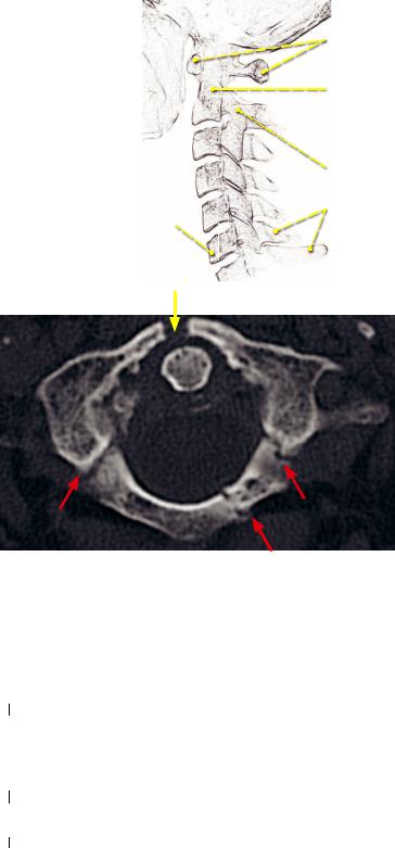
Overview of named cervical spine fractures
C1: Je erson fracture
axial force causes symmetrical fractures of anterior and posterior arches
C2: odontoid fracture
type 1: distal dens (usually stable)
type 2: base of the dens (usually unstable) type 3: dens extending into C2 vertebral body
(variable, may be stable or unstable)
C2: hangman’s fracture
hyperextension injury causes fracture of the arches of C2
variable: clay-shoveler’s fracture
variable: exion teardrop fracture |
hyper exion injury causes spinous process fracture |
most unstable cervical spine fracture associated with disruption of
posterior vertebral and spinolaminar lines
Jefferson fracture
Jefferson fracture: Axial CT demonstrates a fracture of the anterior arch of C1 near the midline (yellow arrow) and bilateral posterior arch fractures (red arrows).
A Jefferson fracture results from acute axial blow to the vertex of the head, causing
symmetrical fractures of the anterior and posterior arches of C1.
On an open-mouth odontoid radiograph, the lateral masses of C1 are displaced
aterally. Jefferson fracture is best seen on axial CT, however.
Odontoid fracture
Fractures of the odontoid process of C2 are usually caused by flexion injuries.
Type I odontoid fracture is a fracture of the distal tip of the dens. Type I odontoid fractures are considered stable and treatment is generally conservative. The differential diagnosis of a Type I odontoid fracture is an os odontoideum, a normal variant secondary ossification center located at the tip of the dens.
Type I odontoid fracture is a fracture of the base of the dens. It is considered an unstable injury and treatment is typically surgical fusion.
Type II odontoid fracture is a fracture of the base of the dens extending into the
C2 vertebral body. Type III fractures have been described as stable and unstable, depending on the reference.
Hangman’s fracture
Hangman’s fracture is traumatic spondylolysis of C2. It results from hyperextension and distraction, causing bilateral fractures through the pedicles of C2.
The characteristic radiographic finding is disruption of the spinolaminar line.
A hangman’s fracture with >3 mm displacement of C2 on C3 is likely unstable.
436
Burst fracture
•A burst fracture is similar in mechanism (axial loading) and appearance to a Jefferson fracture, but occurs lower in the cervical spine, at C2–C7.
Flexion teardrop fracture
•Flexion teardrop fracture is the most severe cervical spinal injury that is potentially compatible with life. It is caused by a hyperflexion and compression injury and results in posterior displacement of the affected vertebral body into the spinal canal.
•The key radiographic finding is an avulsed bony fragment from the anterior-inferior aspect of the vertebral body, caused by anterior longitudinal ligament avulsion. There is retropulsion of the affected vertebral body, which is often rotated and appears shorter in anterior–posterior dimension than the adjacent vertebrae.
•Flexion teardrop fracture can cause anterior cord syndrome due to compression of the pyramidal tracts and anterior spinothalamic tracts. This clinically presents as complete paralysis and loss of pain and temperature sensation. The dorsal column usually remains intact, preserving touch and vibration sense.
Extension teardrop fracture
•In contrast to a flexion teardrop fracture, an extension teardrop fracture is a stable fracture that usually occurs higher in the cervical spine, at C2 or C3.
•On the lateral radiograph, an anterior/inferior avulsion fragment is seen, similar to flexion teardrop. Unlike a flexion teardrop fracture, there is no subluxation and the spinolaminar line is not disrupted.
Clay-shoveler’s fracture
•A clay-shoveler’s fracture is a displaced avulsion fracture of a spinous process. It is caused by forced flexion.
•Clay-shoveler’s fracture occurs in the lower cervical/upper thoracic spine, from C6–T1.
Bilateral interfacetal dislocation (locked facets)
•Bilateral interfacetal dislocation, also called bilateral locked facets, is a hyperflexion injury caused by complete disruption of all spinal ligaments and resultant anterior dislocation of the affected vertebra by >50%.
•Bilateral interfacetal dislocation is unstable with a high risk of cord damage.
•Bilateral interfacetal dislocation typically occurs in the lower cervical spine.
•The naked facet sign is seen when the superior articular facets are not covered by the adjacent inferior articular facets.
•Perched facets are a less severe variation of locked facets. The inferior articular facets of the superior vertebral body are subluxated (but not displaced) and perched upon the superior articular process of the inferior vertebra. Perched facets feature anterior subluxation of >3 mm but less than the 50% seen in a complete dislocation.
Unilateral interfacetal dislocation
•Unilateral interfacetal dislocation is dislocation of a single facet joint, caused by hyperflexion with rotation opposite the site of the dislocation.
•Unilateral interfacetal dislocation is considered a stable fracture.
Grisel syndrome
•Grisel syndrome is nontraumatic rotatory subluxation of the atlantoaxial joint (C1–C2) secondary to an inflammatory mass, most commonly occurring as a sequela of pharyngitis or retropharyngeal abscess.
437
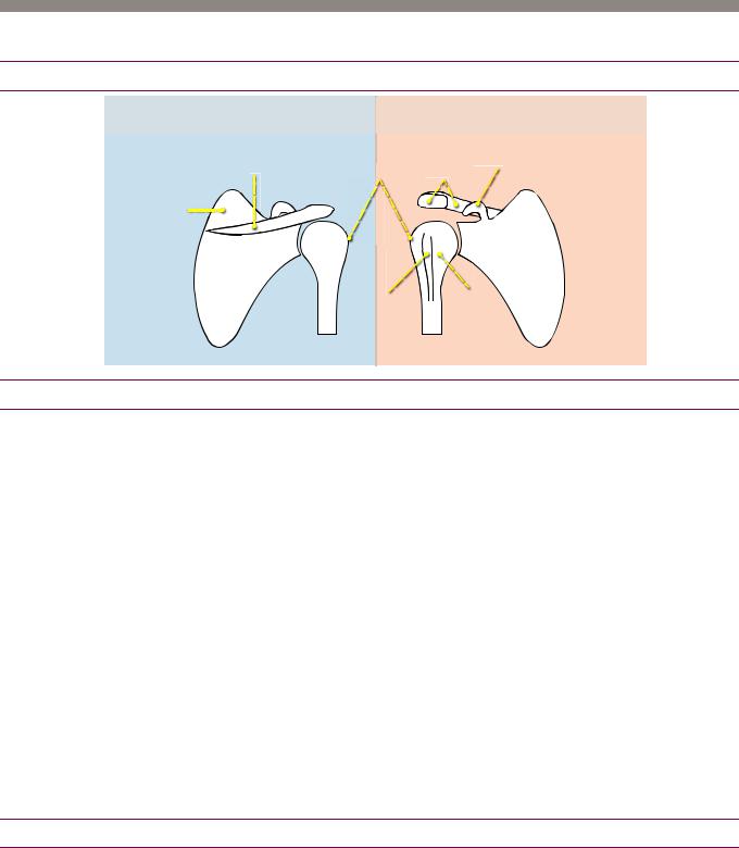
Shoulder and shoulder MRI |
|
|
|
Bony anatomy of the shoulder |
|
|
|
posterior |
|
anterior |
|
|
greater |
coracoid |
|
spine of scapula |
process |
||
tuberosity |
|||
acromion |
|||
|
|||
supraspinatus |
|
|
|
fossa |
|
|
|
infraspinatus |
|
subscapular |
|
|
fossa |
||
fossa |
|
|
|
|
groove |
lesser |
|
|
for |
tuberosity |
|
|
biceps |
|
|
|
tendon |
|
Acromioclavicular joint
Acromioclavicular (AC) joint separation
njuries to the acromioclavicular (AC) joint typically occur in young athletic adults, most commonly from a downward blow to the lateral shoulder. AC joint injuries range from an AC ligament sprain (grade I) to complete disruption of the AC joint capsule and associated coracoclavicular (CC) ligaments (grades III–VI).
ThenormalACjointspaceshouldbe<5mmandthenormalCCdistanceis<11–13mm,
withawiderangeofnormalvariation.Anasymmetryof>50%side-to-sideisabnormal.
Passivetraction,withthepatientholdingaweight,maybenecessarytounmaskaninjury.
Displacement of the acromion and clavicle determines AC ligament integrity, while the coracoclavicular interspace is assessed to evaluate the CC ligaments.
Grade I: AC ligament sprain. Radiographs are normal.
Grade II: Disruption of the AC ligament with intact CC ligaments.
Grade III: AC joint and CC ligamentous disruption. CC interspace is widened 25–100%
relative to the contralateral side.
GradeIV:Distalclavicledisplacedposteriorlyintotrapezius(seenonCToraxillaryview).
Grade V: Severe grade III injury, with >100% displacement relative to the other side.
Grade VI: Inferior dislocation of the distal clavicle.
Glenohumeral dislocation
Anterior glenohumeral dislocation
Anterior dislocation of the humerus with respect to the glenoid is by far the most common type of shoulder dislocation. Anterior dislocation is usually caused by direct force on the arm. The humeral head usually dislocates in an antero-inferior direction.
Diagnosis of anterior dislocation is best made on an axillary view.
As the humeral head recoils from the dislocation force, the posterolateral humeral head strikes the anterior-inferior glenoid. This impaction may fracture the humeral head, the glenoid, or both.
438
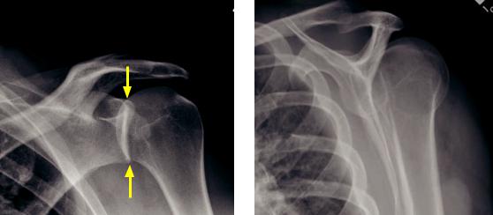
•The Hill–Sachs lesion is a compression fracture of the posterolateral aspect of the humeral head (Hill–Sachs and humerus both start with H). On radiography, the HillSachs lesion is best demonstrated on an AP radiograph with the arm in internal rotation.
•The Bankart lesion is an injury of the anterior-inferior rim of the glenoid. It usually involves the cartilaginous labrum only. A bony Bankart is a fracture of the bony glenoid.
Posterior glenohumeral dislocation
Posterior shoulder dislocation: Grashey (40 degree oblique; left image) radiograph shows overlap of the humeral head and the glenoid (arrows); normally, the glenohumeral joint space should be clear on this view. Posterior displacement of the humerus with respect to the glenoid is confirmed on the transscapular Y view (right image).
•Posterior dislocation represents only 2–3% of dislocations and is usually due to severe muscle spasm, such as from seizure or electrocution. The humeral head is dislocated posteriorly in these situations since the posterior pulling muscles of the back are generally stronger than the anterior pushing muscles.
•Diagnosisofaposteriordislocationismuchmorechallengingcomparedtoananterior dislocation,asthestandardfrontalradiographoftheshouldermaylookclosetonormal.
•Transscapular Y view or axillary view will best show the posterior dislocation.
When possible, diagnosis is usually straightforward on an axillary view; however, many patients with posterior shoulder dislocation will not be able to abduct the arm.
•In a normal shoulder, a Grashey (40 degree oblique) view should show clear space between the glenoid and the humeral head; however, in case of posterior dislocation there will be overlap of the medially displaced humeral head with the glenoid.
•The lightbulb sign describes the appearance of the humeral head due to the fixed internal rotation of the arm often seen in posterior dislocation.
•The trough sign describes a compression fracture of the anteromedial aspect of the humeral head, also known as a reverse Hill–Sachs, from impaction of the humeral head on the posterior glenoid rim upon recoil.
Inferior glenohumeral dislocation (luxatio erecta)
•Inferior dislocation, also called luxatio erecta, is rare (occurring in approximately 1% of dislocations) and is usually caused by direct force to a fully abducted arm.
•Inferiordislocationisoftenassociatedwithrotatorcufftearandgreatertuberosityfracture.
•Inferior dislocation may cause injury to the axillary nerve or artery.
439
Dislocations and associated sequela
normal glenoid alignment
|
bicipital groove |
|
|
humeral |
anterior |
|
labrum |
|
|
head |
|
|
|
|
|
posterior |
glenoid |
|
|
|
|
labrum |
|
anterior dislocation |
interval relocation |
|
Bankart
anterior glenoid
Hill−Sachs posterolateral humeral head
Hill−Sachs: Axial T1-weighted MR arthrogram with fat suppression shows a wedge-shaped compression fracture of the posterolateral humeral head (arrow).
Brigham and Women’s Hospital.
posterior dislocation |
interval relocation |
trough sign

 (reverse Hill−Sachs) anteromedial humeral head
reverse Bankart
posterior glenoid
440
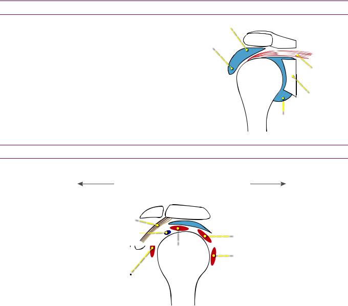
Shoulder bursae
The subacromial and subdeltoid bursae normally communicate with each other. This combined subacromial/subdeltoid bursa is not normally in communication with the glenohumeral joint.
Fluid injected into the glenohumeral joint that extends into the subacromial/subdeltoid bursa is seenwithacompleterotatorcuff tear.Agapintherotatorcuffwill almostalwaysbeevident.
subacromial bursa
subdeltoid bursa
|
|
|
conjoined |
|
|
|
|
humerus |
|
|
rotator cu |
|
|
|
|
|
|
|
glenoid |
|
|
||
|
|
|
|
|
|
||
|
|
|
|
|
glenohumeral joint |
||
Coracoacromial arch and impingement syndrome
sagittal schematic of the coracoacromial arch
anterior |
posterior |
clavicle
coracoacromial ligament 
 biceps tendon
coracoid 

subscapularis

 subacromial bursa
|
|
|
|
|
|
|
infraspinatus |
|
|
|
|
|
|
|
|
|
|
|
|
|
|
|
|
|
|
|
|
|
|
|
|
|
|
|
|
|
|
|
|
|
|
|
|
|
|
|
|
supraspinatus |
|
|
|
teres minor |
|||
|
humerus |
|
|
|
|||
|
|
|
|
|
|||
mpingement syndrome is a clinical diagnosis that can have a variety of causes and
MRI findings. Shoulder impingement was originally described as chronic compression/ irritation of the structures that pass through the coracoacromial arch, including the supraspinatus tendon, biceps tendon, and subacromial bursa.
Rotator cuff tears are often associated with chronic impingement, although there is controversy whether the cuff tears are a result of chronic mechanical impingement or whether degeneration leads to cuff tears, resulting in impingement syndrome.
Extrinsic impingement is caused by structural abnormalities external to the joint and is divided into primary and subcoracoid impingement.
Primaryexternalimpingement iscommonandisduetoavariationintheanatomyofthecoracoacromial arch(e.g.,subacromialenthesophyte,hookedacromion,ACjointosteophytes,osacromiale,orthickened coracoacromialligament).Primaryexternalimpingementistypicallyseeninyoungathletesinvolved withthrowingorrepetitiveoverheadmovement.Chronicprimaryexternalimpingementmaycause thesupraspinatustendonand/ortheproximalaspectofthelongheadofthebicepstendontobecome degenerated,partiallytorn,orevencompletelytorn.Thesubacromialbursa,immediatelyinferiortothe acromion,maybecomeinflamed,leadingtosubacromial/subdeltoidbursitis.
Subcoracoid impingement is less common and occurs when the coracohumeral distance narrows.
Intrinsicimpingement isrelatedtoglenohumeralinstabilityandiscausedbyabnormalities oftherotatorcuffandjointcapsule,withoutabnormalityofthecoracoacromialarch.
441
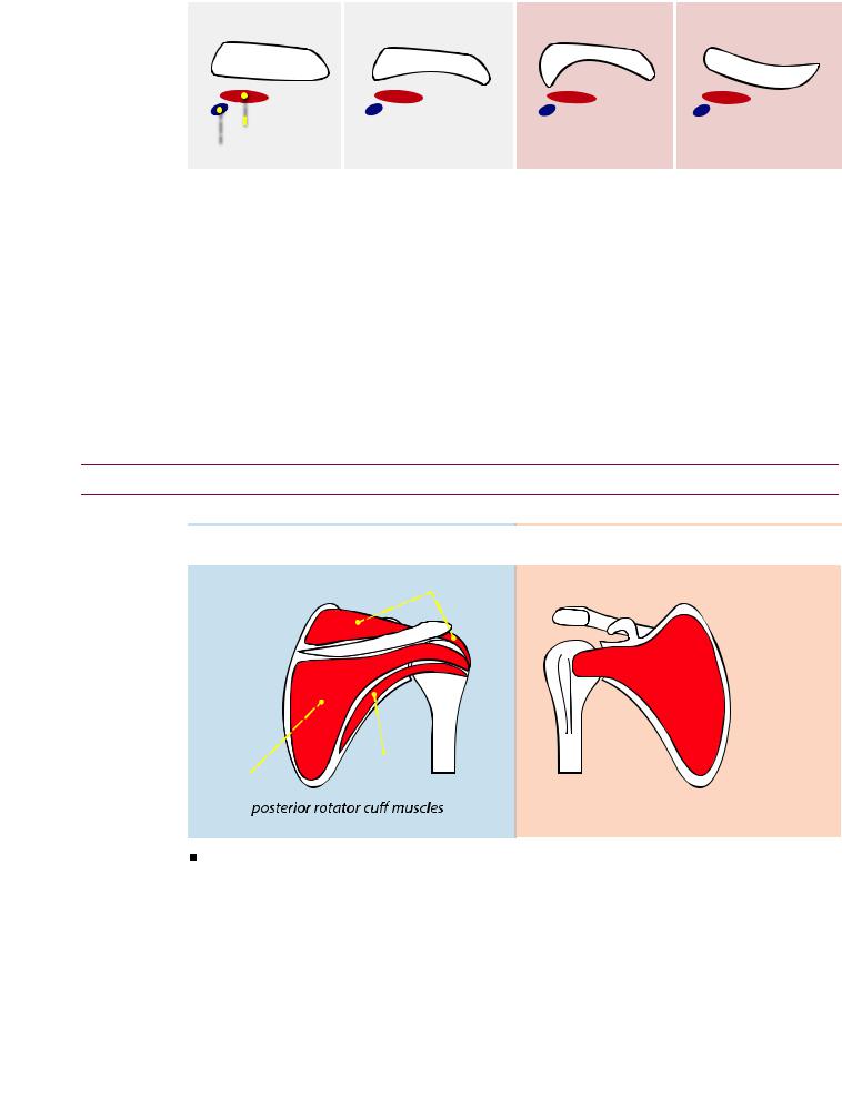
Acromial shape and orientation |
|
|
|
||
|
|
type I |
type II |
type III |
type IV |
|
acromion |
|
|
|
|
|
|
|
|
|
|
|
|
|
|
|
|
|
|
|
|
|
|
|
|
|
|
|
|
|
|
|
|
|
|
|
|
|
|
|
|
 supraspinatus biceps tendon
The impact of acromial shape on impingement syndrome is controversial. An acromion with a hooked undersurface (type III) or convex undersurface (type IV) may contribute to impingement (primary external type) and lead to subsequent rotator cuff tears.
Friction of the acromion against the immediately underlying supraspinatus and biceps tendons may predispose to injury from impingement.
Os acromiale
An os acromiale is a persistent accessory ossification center of the acromion seen in
up to 15% of patients. It is best evaluated on the axial images.
Although the presence of an os acromiale may be an asymptomatic normal variant, the presence of marrow edema suggests that the os may be a cause of pain.
Treatment is resection or fusion.
Rotator cuff
Anatomy of the rotator cuff
posterior |
anterior |
|
|
supraspinatus
subscapularis
|
|
teres minor |
|
infraspinatus |
|
subscapularis inserts |
|
|
|
||
|
|
|
|
insert on greater tuberosity |
on lesser tuberosity |
||
The rotator cuff is an essential active stabilizer of the glenohumeral joint and is formed by the four rotator cuff muscles (supraspinatus, infraspinatus, teres minor, and subscapularis) and their tendons.
The supraspinatus, infraspinatus, and teres minor are located posterior to the body of the scapula and insert on the greater tuberosity. The subscapularis is anterior to the scapula and inserts on the lesser tuberosity.
442
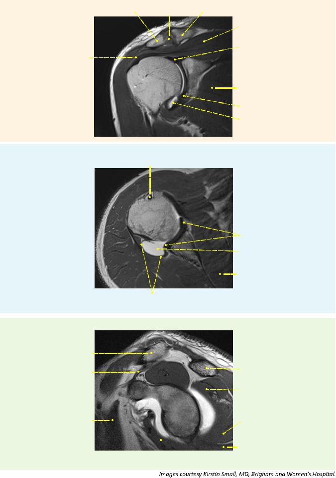
coronal
axial
acromion |
AC joint |
clavicle |
|
|
|
||
|
|
|
|
|
|
|
supraspinatus |
|
|
|
|
|
|
|
|
|
|
|
|
|
|
|
|
|
|
|
|
|
|
|
muscle |
|
|
|
|
|
|||
supraspinatus |
|
|
|
|
|
|
superior labrum |
|
|
|
|
|
|
|
|
tendon |
|
|
glenoid |
|
|
|
|
humeral |
|
|
|
||||
|
|
|
|
|
|||
|
head |
|
|
|
|
subscapularis |
|
|
|
|
|
||||
|
|
|
|
|
|
|
muscle |
deltoid |
|
|
|
|
|
|
inferior labrum |
muscle |
|
|
|
|
|
|
glenohumeral joint |
|
|
|
|
|
|
|
|
long head of biceps tendon
|
|
|
|
|
|
|
|
|
|
|
|
|
|
|
|
|
|
humeral |
|
|
|
|
|
head |
|
|
|
|
|
|
|
|
|
|
labrum |
deltoid |
|
|
|
glenohumeral joint |
|
muscle |
|
|
|
(distended with contrast) |
|
|
|
|
|
|
subscapularis |
|
|
|
|
||
|
|
|
|
|
muscle |
posterior joint capsule |
|
|
|
|
|
sagittal
clavicle |
|
|
|
|
|
|
|
|
|
|
|
|
acromion |
|
|
|
|
|
|
|
|
|
|
|
|
||
coracoclavicular |
|
|
|
|
|
|
supraspinatus |
|
|
|
|||
|
|
|
|
|
|
|
|
|
|
||||
ligament |
|
|
|
|
|
|
|
|
|
|
|||
|
|
|
|
|
|
|
|
|
|
|
|
|
|
|
|
|
|
coracoid |
|
|
|
infraspinatus muscle |
|||||
pectoralis minor |
|
|
|
|
|
|
|
|
glenoid |
|
|
|
|
|
|
|
|
|
|
|
|
|
|
|
|
teres minor muscle |
|
|
|
|
|
|
|
|
|
|
|
|
|
||
|
|
|
|
|
|
|
|
|
|
|
|
|
teres major muscle |
|
|
|
|
|
|
|
|
|
|
|
|
|
|
|
|
|
|
|
|
|
|
|
|
||||
|
|
|
|
|
|
|
|
|
|
||||
|
|
|
|
|
|
|
|
|
|
|
|
||
|
|
|
|
|
|
|
|
|
|
|
|
|
|
|
|
|
|
|
|
|
|
|
|
|
|
|
|
|
|
|
|
|
|
subscapularis |
|
|
|
|
|||
443
Tendinosis/tendinopathy
•Tendinosis (also called tendinopathy) is mucoid degeneration of the tendon, without active inflammation. Tendinosis manifests on imaging as diffuse or focal thickening and intermediate signal intensity on T1and T2-weighted images. On T2-weighted images, the tendinosis does not appear as hyperintense as fluid.
•The magic-angle phenomenon may simulate tendinosis or even a partial tear. The magic-angle effect is due to the anisotropy of the tendon’s collagen fibers and occurs when the collagen fibers are oriented at 55 degrees relative to the magnetic field (B0) with short TE times (for instance T1-weighted images, proton density, and GRE).
Overview of rotator cuff tears
•Tears of the rotator cuff can be partial or complete.
•Complete rotator cuff tears allow communication between the subacromial/ subdeltoid bursa and the glenohumeral joint.
•The footprint is the attachment of the tendons at the greater tuberosity. The critical zone is a potentially undervascularized portion of the distal supraspinatus tendon approximately 1 cm proximal to the insertion on the footprint. Relatively decreased vascular supply may predispose to rupture in this location.
Partial-thickness rotator cuff tear
Partial articular surface supraspinatus tear:
Coronal T1-weighted MR arthrogram with fat suppression demonstrates discontinuity of the articular surface of the distal fibers of the supraspinatus tendon (arrow).
Case courtesy Kirstin M. Small, MD, Brigham and Women’s Hospital.
•The most commonly injured rotator cuff muscle is the supraspinatus, followed by the infraspinatus. The teres minor is the least commonly injured rotator cuff muscle.
•A partial-thickness tear features abnormal signal in a portion of the muscle or tendon but not extending through its entire thickness. MR arthrography will not show communication between the injected glenohumeral joint and the subacromial/ subdeltoid bursa.
Partial-thickness tears can be challenging to diagnose without MR arthrography. Reparative granulation tissue can obscure the abnormal signal that would otherwise indicate a tear.
•Partial tears can be divided into bursal-surface, articular surface, or intrasubstance tears. Articular-surface tears are by far the most common type of partial tear.
•A rim rent tear is the most common type of partial tear. Also called the PASTA lesion (partial thickness articular supraspinatus tendon avulsion), the rim rent tear is an avulsion-type partial tear of the articular surface of the supraspinatus as it inserts on the greater tuberosity.
Less commonly, an analogous lesion can be seen in the infraspinatus.
•The PAINT (partial articular tear with intratendinous extension) lesion is a partial rotator cuff tear at the articular surface that also extends into the tendon.
444
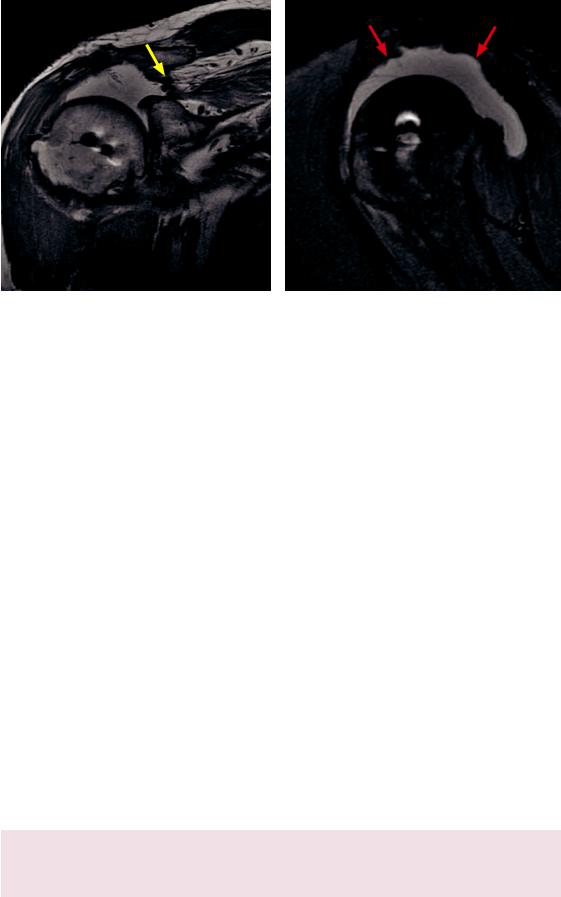
Full-thickness rotator cuff tear
Full-thickness rotator cuff tear: Coronal proton-density (left image) and sagittal fat-saturated protondensity (right image) conventional MRI demonstrates complete discontinuity of the supraspinatus tendon with the tendon retracted proximally (yellow arrow). There is alarge effusion, demonstrating continuity of the glenohumeral joint and the subacromial/subdeltoid bursa (red arrows). Susceptibility artifact is present in the humeral head, consistent with prior rotator cuff repair.
Case courtesy Kirstin M. Small, MD, Brigham and Women’s Hospital.
•Afull-thicknessrotatorcufftearisseenonMRarthrographywhenfluidinjectedinto theglenohumeraljointextendsintothesubacromial/subdeltoidbursa.Intheabsence ofinjectedgadolinium,afull-thicknesstearcanbediagnosedifabnormalsignalextends throughthefibersofthetendon.Agapinthetendonisalmostalwayspresent.
•Similar to partial tears, the supraspinatus is most commonly torn.
•Approximately 30–40% of supraspinatus tears are also associated with a tear of the infraspinatus.
The infraspinatus is rarely torn in isolation; however, isolated infraspinatus tears can be seen in the setting of posterior instability. The posterior capsule and labrum should be carefully evaluated in such cases.
•A chronic full-thickness rotator cuff tear due to rheumatoid arthritis has a classic imaging appearance. The humeral head migrates superiorly and may articulate with the acromion in severe cases.
•Full-thickness rotator cuff tears are classified by the length of the affected tendon/ muscle, ranging from small (<1 cm) to massive (>5 cm).
Rotator cuff atrophy
•Atrophyoftherotatorcuffismostcommonlyduetochronicrotatorcufftear.Fatty degenerationmayoccurwithin4weeksofinjuryandisusuallyirreversible.Theextent ofmuscledegenerationcorrelateswithoutcomefollowingsurgicalrepair.Thedegreeof fattyinfiltrationoftherotatorcuffmusclesshouldberoutinelyassessedinthesagittal plane.ThedegreeoffattyreplacementcanbegradedwiththeGoutallierclassification:
Grade 0: Normal muscle, no fat. |
Grade 3:Fattyinfiltration,equalmuscleandfat. |
Grade 1: Muscle contains a few fatty streaks. |
Grade 4: Fatty infiltration, more fat than |
Grade 2: Fatty infiltration, more muscle than fat. |
muscle. |
|
•Lesscommonly,rotatorcuffatrophymaybesecondarytodenervationatrophy, subsequentlydiscussed.Denervationatrophyisusuallyassociatedwithaparalabralcyst.
445
Adhesive capsulitis
Adhesivecapsulitis,clinicallyknownas frozen shoulder,isthickeningandcontractionof theglenohumeraljointcapsuleduetoinflammationofthejointcapsuleandsynovium.
Adhesive capsulitis is primarily a clinical diagnosis, but MRI findings include thickening
of the joint capsule and synovium >4 mm, assessed at the level of the axillary pouch.
Shoulder ligaments and rotator interval
Anatomy of the shoulder ligaments
|
acromion |
|
coracohumeral |
|
|
ligament (CHL) |
|
|
attaches to |
|
|
greater |
SGHL |
|
tuber |
||
|
MGHL |
|
long head |
IGHL |
|
biceps |
||
|
||
tendon |
|
|
(LHBT) |
|
coracoid  process
 rotator interval contains CHL, LHBT, and SGHL
superior glenohumeral ligament (SGHL) and middle glenohumeral ligament (MGHL) connect the
lesser tuberosity to the supraglenoid tubercle of the scapula.
the inferior glenohumeral ligament (IGHL) connects the inferior glenoid labrum with the anatomic neck of the humerus.
The rotator interval is a triangular region that allows rotational motion around the coracoid process. It is located between the supraspinatus and subscapularis tendons.
The rotator interval contains the coracohumeral ligament (CHL), the long head of the biceps tendon (LHBT), and the superior glenohumeral ligament (SGHL).
The CHL connects the lateral aspect of the coracoid process to the greater tubercle of the humerus, where the CHL blends with the supraspinatus tendon. The CHL helps to stabilize the intraarticular biceps tendon together with the superior glenohumeral igament as the biceps pulley The CHL is located external to the joint capsule.
The three glenohumeral ligaments are derived from the labrum and form a portion of
the joint capsule
The superior glenohumeral ligament (SGHL) connects the lesser tuberosity to the
supraglenoid tubercle of the scapula.
The middle glenohumeral ligament (MGHL) also connects the lesser tuberosity to the supraglenoid tubercle of the scapula. The MGHL is congenitally absent in up to one third of patients. Tears of the MGHL are associated with superior labral tears.
The inferior glenohumeral ligament (IGHL) connects the anatomic neck of the humerus to the inferior glenoid labrum. The IGHL is by far the most important component of the capsulolabral complex for maintaining stability in abduction and external rotation. The IGHL contains three components (the anterior band, the axillary pouch, and the posterior band), all of which are all well-seen on sagittal MR:
The anterior band of the IGHL provides anterior stability. There are several lesions of the anterior band of the IGHL that are associated with anterior instability, discussed on the following page.
The IGHL forms the axillary pouch of the glenohumeral joint capsule.
The posterior band contributes to posterior stability. The IGHL is the only glenohumeral ligament to have a posterior component.
The long head of the biceps tendon (LHBT) attaches to the anterosuperior glenoid rim
along with the SGHL and MGHL
446
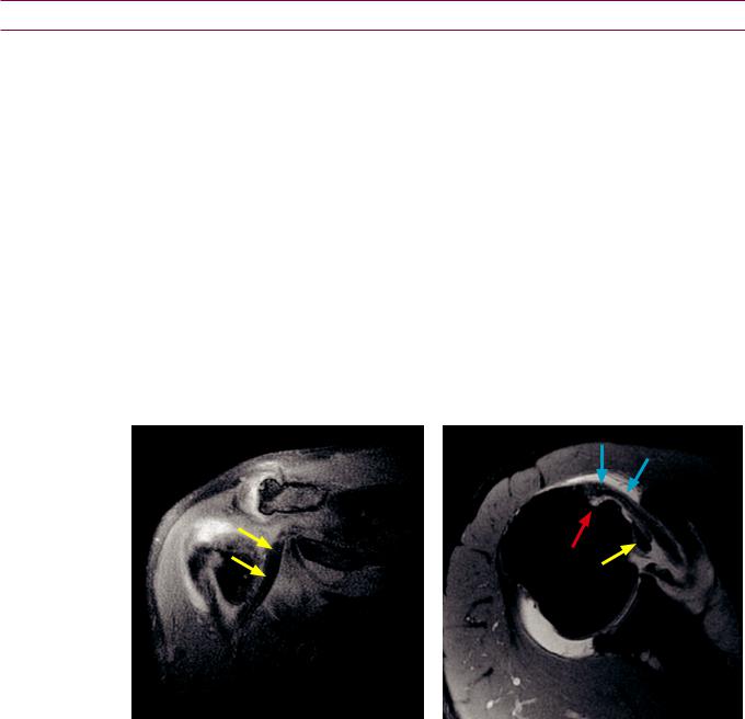
Biceps tendon
Anatomy of the biceps tendon
•The long head of the biceps tendon originates at the superior labrum and courses in the intertubercular groove. The biceps tendon sheath communicates with the glenohumeral joint. Fluid in the tendon sheath may be normal.
•The biceps pulley is a capsuloligamentous complex that stabilizes the biceps tendon in a sling within the bicipital groove. The biceps pulley is formed by the superior glenohumeral ligament, coracohumeral ligament, and the distal fibers of the subscapularis tendon.
Biceps tendon tear
•Approximately 33% of supraspinatus tendon tears are associated with biceps tendon injury (complete tear, partial tear, or degeneration).
•The biceps tendon is at risk for impingement due to its location partially underneath the supraspinatus tendon in the coracoacromial arch.
•In cases of an acute tear, the biceps muscle may retract distally. An empty intertubercular groove may signify a complete tear with retraction or a biceps tendon dislocation.
Biceps tendon subluxation
Biceps tendon subluxation: Coronal (left image) and axial T1-weighted MR arthrogram with fat suppression shows medial intra-articular subluxation of the biceps tendon (yellow arrows). The bicipital groove is empty, best seen on the axial image (red arrow). The blue arrows denote the subscapularis tendon, seen on the axial image.
Case courtesy Kirstin M. Small, MD, Brigham and Women’s Hospital.
•Biceps tendon subluxation is defined as a tendon displaced from, but remaining in contact with, the bicipital groove.
•Biceps tendon subluxation and dislocation are associated with injury to the transverse ligament. The transverse humeral ligament attaches to the greater and lesser tuberosities and normally holds the biceps tendon in place.
447
Biceps tendon dislocation
Biceps tendon dislocation:
Axial T1-weighted image with fat suppression from an MR arthrogram shows medial dislocation of the biceps tendon (yellow arrow) into the subscapularis. An interstitial tear of the subscapularis tendon is present (blue arrow).
Case courtesy Kirstin M. Small, MD, Brigham and Women’s Hospital.
•Biceps tendon dislocation is often seen in conjunction with injury of both the transverse ligament and the biceps pulley.
•Medial dislocation of the biceps tendon is associated with tear of the subscapularis tendon (in contrast to biceps tears, which are more frequently associated with supraspinatus injury).
•Axial MRI best demonstrates biceps tendon dislocation. The bicipital groove is empty and the tendon can usually be seen medial to the bicipital groove.
Instability: Overview and anatomy
Overview of instability
•Shoulder instability is abnormal motion of the humeral head with respect to the glenoid during movement, producing pain, clicking, or the feeling of an unstable joint.
•Instability can manifest as dislocation, subluxation, or microinstability (repetitive motion).
•The stability of the glenohumeral joint is mostly provided by dynamic and static softtissue stabilizers, as there is minimal inherent osseous stability.
•The dynamic stabilizers of the joint include the rotator cuff and biceps tendons.
•The static stabilizer of the joint is the capsulo-labro-ligamentous complex, which is composed of the bony glenoid fossa, labrum, coracohumeral ligament, and superior, middle, and inferior glenohumeral ligaments.
•Thereareseverallabral,ligamentous,andosseousinjuriesassociatedwithinstability,butit isunclearwhetherthestructuralabnormalitiesaresecondarytoinstabilityoracauseofit.
•There are various classifications of instability incorporating clinical, etiological, and imaging findings.
•Instability can also be classified as traumatic or atraumatic.
Traumatic instability usually causes instability in one direction only, is associated with a Bankart lesion, and is treated surgically. Traumatic instability is referred to as TUBS (traumatic, unidirectional, Bankart, surgical).
Atraumatic instability can be divided into two types. The AIOS (acquired, instability, overstress, surgery) pattern is usually seen in athletes with repetitive movements causing microinstability. AMBRI (atraumatic, multidirectional, bilateral, rehabilitation, inferior capsule shift) lesions are due to congenital joint laxity.
•Radiologists usually classify instability based on the direction of joint subluxation (anterior, posterior, multidirectional, or superior). Similar to traumatic shoulder dislocations, anterior instability accounts for the vast majority (95%) of cases.
448
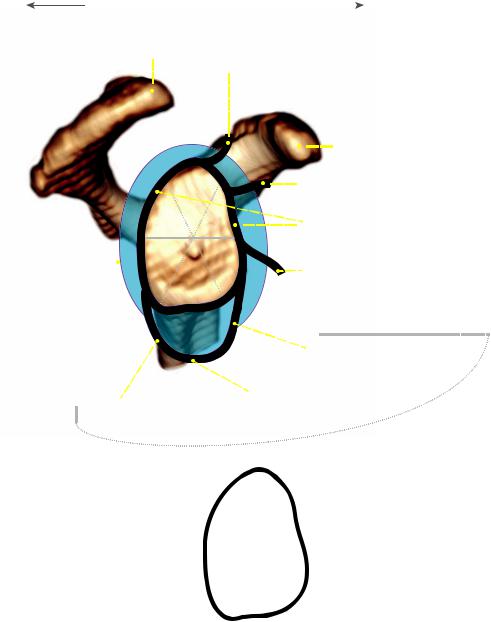
Anatomy of the glenoid labrum
The glenoid labrum is a fibrocartilaginous ring-like structure that rests on the glenoid hyaline cartilage and surrounds the glenoid. The labrum increases the surface area and depth of the glenoid cavity. It also provides a suction effect, adhering the humeral head to the glenoid with motion.
sagittal schematic of the glenoid fossa, glenoid labrum, and glenohumeral ligaments
posterior |
|
anterior |
|
|
|
|
|
||||
acromion |
long head of biceps tendon |
||||
|
|
(LHBT) |
|||
|
|
||||
|
|
||||
|
|
|
|
|
|
|
|
|
|
|
|
|
|
|
|
|
|
|
|
|
|
|
|
|
|
|
|
|
|
|
|
|
|
|
|
|
|
|
|
|
|
|
coracoid |
|
|
|
|
|
|
|
|
|
|
|
|
|
|
|
|
|
|
||
|
|
|
|
|
|
sup |
|
|
|
|
|
|
|
|
|
|
superior glenohumeral ligament |
||
|
|
|
|
|
|
|
|
|
|
|
|
|
|
|
|||||
|
|
|
|
|
|
|
|
|
|
|
|
|
|
|
|
|
(SGHL) |
||
|
|
|
|
|
|
post |
ant |
|
|
labrum |
|||||||||
|
|
|
|
|
|
sup |
sup |
|
|
|
|
|
|||||||
|
|
|
|
|
|
|
|
|
|
||||||||||
glenohumeral |
|
|
|
|
|
post |
ant |
|
|
|
|
|
|||||||
|
|
|
|
|
inf |
inf |
|
|
middle glenohumeral ligament |
||||||||||
|
|
|
|
|
|||||||||||||||
joint capsule |
|
|
|
|
|
|
|
|
|
|
|
|
|||||||
|
|
|
|
|
|
|
|
|
|
|
|
(MGHL) |
|||||||
|
|
|
|
|
|
inf |
|
|
|
|
|
|
|
|
|
|
|||
|
|
|
|
|
|
|
|
|
|
|
|
|
|
|
|
|
|
|
|
inferior
|
|
|
l |
|
|
ra |
|
|
me |
|
|
ohu |
|
|
|
glen |
|
|
|
|
|
|
|
|
|
|
|
|
|
|
|
|
x |
|
|
|
|
|
|
|
|
|
|
|
|
le |
|
|
|
|
|
|
|
|
|
|
|
|
p |
|
|
|
|
|
|
|
|
|
|
|
|
m |
|
|
|
|
|
|
|
|
|
|
|
|
o |
|
|
|
|
|
|
|
|
|
|
|
|
c |
|
|
|
|
|
|
|
|
|
|
|
|
) |
|
|
|
|
|
|
|
|
|
|
|
|
L |
|
|
|
|
|
|
|
|
|
|
|
|
H |
|
|
|
|
|
|
|
|
|
|
|
|
G |
|
|
|
|
|
|
|
|
|
|
|
|
(I |
|
|
|
|
|
|
|
|
|
|
|
|
t |
|
|
|
|
|
|
|
|
|
|
|
|
en |
|
|
|
|
|
|
|
|
|
|
|
|
am |
|
|
|
|
|
|
|
|
|
|
|
|
|
lig |
|
|
|
|
|
|
|
|
|
|
|
|
|
the location of labral |
|
12:00 |
injury can be described as |
|
|
the sextants outlined above, |
|
|
or can also be described |
9:00 |
3:00 |
on a clock face, with 12 o’clock superior and
3 o’clock anterior
6:00
Other important stabilizers of the joint attach directly to the labrum, including the glenohumeral ligaments, the biceps tendon, and the joint capsule
Labral normal variants
A sublabral foramen (seen in 10% of patients) is a normal variant where the anterosuperior segment is not attached to the bony glenoid.
A Buford complex is a less common (seen in up to 6.5%) normal variant where a cord-like middle glenohumeral ligament is seen in combination with an absent anterosuperior labrum.
449
Instability: Associated lesions
•Most of the alphabet-soup instability lesions are located at the anterior-inferior aspect of the glenohumeral joint and are associated with the anterior band of the inferior glenohumeral ligament.
Bankart lesion
•The Bankart lesion is an injury of the anteroinferior labrum due to anterior glenohumeral dislocation. Specifically, a Bankart is detachment of the anteroinferior labrum from the glenoid, with stripping of the scapular periosteum.
•The labrum may migrate superiorly and appear as a balled-up mass-like object, producing the glenoid labrum ovoid mass (GLOM) sign. The differential diagnosis for a black intra-articular mass on MRI includes:
GLOM sign (if a Bankart lesion is present inferiorly).
Dislocated biceps tendon.
Air bubble (if an MR arthrogram was performed).
•An osseous Bankart (or “bony Bankart”) is a fracture of the anterior-inferior glenoid rim, which predisposes to recurrent dislocation due to glenoid insufficiency.
Hill–Sachs
•Hill–Sachs is an impaction fracture of the posterolateral humeral head caused by anterior dislocation. The Hill–Sachs lesion can be diagnosed by scrolling through sequential axial images: With standard 5 mm slices, the normal humeral head should appear round in three consecutive slices starting superiorly. If a Hill–Sachs lesion is present, there will be a posterolateral notch in the humeral head.
•Subtle Hill–Sachs can also be detected as bone marrow edema in this location, without fracture.
Anterior labro-ligamentous periosteal sleeve avulsion (ALPSA)
•An anterior labro-ligamentous periosteal sleeve avulsion (ALPSA) is a variant of the Bankart lesion and also represents an anterior-inferior labral injury. In contrast to the Bankart lesion, the scapular periosteum is intact with an ALPSA.
•Similar to the GLOM sign discussed above, the avulsed anterior-inferior labrum is balled up. However, since the labrum remains attached to the periosteum in an ALPSA lesion, the labrum is displaced inferomedially relative to the glenoid (as opposed to superiorly with the GLOM sign).
Perthes lesion
•The Perthes lesion is an avulsion of the anterior-inferior labrum, where the labrum remains attached to the scapular periosteum.
•Because the labrum remains attached to periosteum and may remain in its anatomic position, the Perthes lesion can be very difficult to visualize. MR arthrography with ABER (abduction–external rotation) positioning is often necessary for diagnosis.
Humeral avulsion of the inferior glenohumeral ligament (HAGL)
•Humeral avulsion of the inferior glenohumeral ligament (HAGL) is avulsion of the humeral attachment of the IGHL. This lesion occurs on the opposite (humeral) site of the IGHL compared to the Bankart/ALPSA/Perthes lesions.
•HAGL is associated with subscapularis tendon tears.
Bony HAGL (BHAGL)
•A BHAGL is a HAGL lesion with an additional bony avulsion of the anatomic neck of the humerus, caused by avulsion of the IGHL.
450
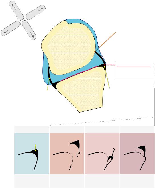
Summary of anterior-inferior instability lesions
lateral |
anterior |
posterior |
|
medial |
|
oblique axial schematic
IGHL humeral attachment
HAGL
BHAGL
Floating AIGHL (both)
humeral head

 IGHL - anterior band
IGHL - posterior band
normal |
Bankart |
anteriorinferior labrum
glenoid
labrum detached from glenoid with stripping of the scapular periosteum
ALPSA
anterior labro-ligamentous periosteal sleeve avulsion
Bankart variant with balled-up labrum and intact scapular periosteum
Perthes
avulsion of labrum, which remains attached to scapular periosteum
451
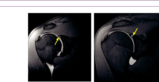
Posterior instability
•Posterior instability is uncommon, accounting for less than 5% of instability.
•The injuries associated with posterior instability are highly variable and include several posterior variations of anterior lesions such as posterior HAGL (PHAGL), reverse Bankart (posterior glenoid fracture), and reverse Hill–Sachs/trough sign.
•Tears of the infraspinatus and teres minor are associated with posterior instability, as these rotator cuff muscles insert on the posterior aspect of the greater tuberosity.
•Hypoplastic posterior glenoid may be a congenital cause of posterior instability.
•The Bennett lesion, also known as thrower’s exostosis, is extra-articular posterior ossification associated with posterior labral injury. The Bennett lesion is often seen in baseball pitchers and is thought to be the result of avulsion of the posterior band of the IGHL. Ossification can be confirmed with an axillary shoulder radiograph.
Miscellaneous lesions not associated with instability
Superior labrum anterior posterior (SLAP) tear
SLAP tear (type II): Coronal T1-weighted MR arthrogram with fat suppression shows high signal within the superior labrum (yellow arrows) extending from anterior to posterior.
Case courtesy Kirstin M. Small, MD, Brigham and Women’s Hospital.
•Asuperiorlabrumanteriorposterior(SLAP)tearisananterior–posteriororientedtearof thesuperiorlabrumcenteredattheattachmentofthebicepstendon.Thebicepstendon insertsontotheanterior-superiorlabrum.TherewereoriginallyfourtypesofSLAPlesions described;however,nowthereareupto10differentsubcategoriesofSLAPlesions.
•Type I SLAP is isolated fraying of the superior portion of the labrum without a frank tear. The biceps tendon is intact.
•Type II SLAP is the most frequent type of SLAP and features labral fraying in conjunction with stripping of the superior labrum. Type II SLAP lesions are most commonly associated with repetitive microtrauma.
•Type III SLAP is a bucket-handle tear of the superior labrum, analogous to a buckethandle tear of the knee meniscus.
•Type IV SLAP is also a bucket-handle tear of the superior labrum, with additional extension into the biceps tendon.
452
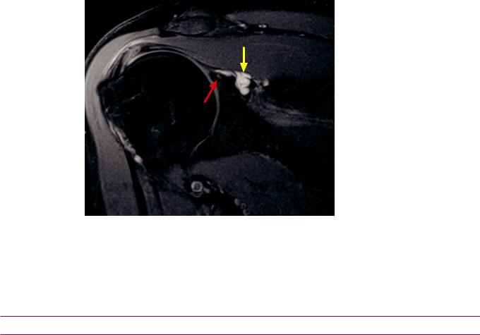
Glenoid labral articular disruption (GLAD)
•Glenoid labral articular disruption (GLAD) is a superficial tear of the anterior-inferior labrum, with adjacent glenoid articular cartilage injury.
•Although the GLAD lesion occurs at the anterior-inferior labrum, the same site as the Bankart, ALPSA, and Perthes lesions, GLAD is usually not caused by or associated with instability.
•GLAD lesions may lead to post-traumatic arthritis and intra-articular bodies.
Paralabral cysts
Paralabral cyst and labral tear:
Coronal T2-weighted MRI with fat suppression shows a lobulated paralabral cyst (yellow arrow) medial to the glenoid.
There is globular intermediate signal within the labrum (red arrow), highly suggestive of labral tear especially in the presence of an adjacent paralabral cyst.
Case courtesy Kirstin M. Small, MD, Brigham and Women’s Hospital.
•Similar to the formation of parameniscal cysts adjacent to meniscal tears, paralabral cysts form adjacent to labral tears. Paralabral cysts are usually seen in the soft tissues adjacent to the labrum but may extend into bone.
•The presence of a paralabral cyst is a very specific finding for a labral tear even when abnormal labral signal is not seen.
•A paralabral cyst may compress a nerve and cause an entrapment neuropathy.
Entrapment neuropathies
Quadrilateral space syndrome
•The quadrilateral space is located at the posterior aspect of the axilla. It is formed by the humerus (laterally), the long head of the triceps muscle (medially), teres minor (superiorly), and teres major (inferiorly).
•The axillary nerve and posterior humeral circumflex artery travel through the quadrilateral space. The axillary nerve provides motor fibers to the teres minor and deltoid.
•Entrapment of the axillary nerve in the quadrilateral space may cause teres minor paresthesias and eventual atrophy, with or without deltoid involvement.
Suprascapular nerve entrapment at the suprascapular notch
•The proximal suprascapular nerve provides motor innervation to the supraspinatus and infraspinatus muscles. Entrapment of the suprascapular nerve at the suprascapular notch, most commonly from a paralabral cyst associated with a superior labral tear, causes atrophy of both the supraspinatus and infraspinatus.
453
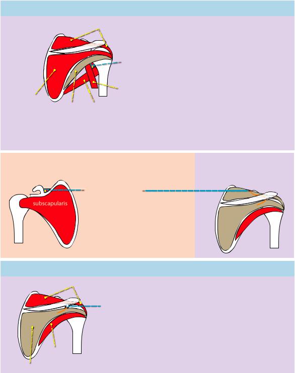
Suprascapular nerve entrapment at the spinoglenoid notch
The distal suprascapular nerve provides motor innervation to the infraspinatus muscle only. Entrapment of the distal suprascapular nerve at the spinoglenoid notch will cause isolated atrophy of the infraspinatus.
Summary of entrapment neuropathies
quadrilateral space
supraspinatus |
posterior view |
|
 quadrilateral
 space contains
 axillary nerve and
posterior circum ex humeral artery
triceps long head
infraspinatus |
teres minor teres major |
|
compression of axillary nerve causes teres minor (±deltoid, not pictured) weakness/atrophy
the borders of the quadrilateral space: humerus (lateral)
teres minor (superior) triceps long head (medial) teres major (inferior)
|
|
|
|
suprascapular notch |
|
|
||
|
|
|
|
|
|
|
|
|
|
|
|
|
|
|
anterior view |
|
posterior view |
|
|
|
|
|
|
|
|
|
|
|
|
|
suprascapular notch |
|
|
|
|
|
|
|
|
|
|
|
||
subscapularis |
|
contains suprascapular nerve and |
|
|
||||
|
suprascapular artery |
|
|
|||||
compression of the suprascapular nerve causes atrophy/weakness of both the infraspinatus and supraspinatus
spinoglenoid notch
supraspinatus
posterior view
spinoglenoid notch
contains distal branch of suprascapular nerve
compression of the distal branch of the suprascapular nerve
at the spinoglenoid notch causes isolated atrophy of the infraspinatus
teres minor
infraspinatus
Parsonage–Turner
Parsonage–Turner syndrome is idiopathic brachial neuropathy, which may cause rotator cuff atrophy. It is a diagnosis of exclusion and a structural cause for atrophy
(e.g., a mass or cyst compressing a nerve) must be ruled out.
454
Elbow and forearm
Elbow and forearm trauma
Elbow dislocation
•The most common elbow dislocation is posterior dislocation of both the radius and ulna with respect to the humerus.
•Elbow dislocations are frequently associated with ulnar fractures. These fractures may be more distal than imaged in a typical elbow radiograph, so forearm radiographs should be obtained in the setting of an elbow dislocation.
Radial head fracture
•A radial head fracture is the most common elbow fracture in adults.
•The fat pad sign is elevation of the anterior and/or posterior fat pads due to an elbow effusion. Elevation of the posterior fat pad is considered nearly diagnostic for fracture (most commonly radial head fracture in adults). The sail sign represents elevation of the anterior fat pad only and is less specific for fracture.
•If a fat pad sign is present and no fracture is seen, additional views (typically of the radial head) or CT should be obtained.
Supracondylar fracture
•Supracondylar fracture is the most common elbow fracture in children.
Essex–Lopresti fracture-dislocation
Essex–Lopresti fracture dislocation: Frontal radiograph of the wrist (left image) shows a probable ulnar (medial) dislocation of the ulna at the distal radioulnar joint. Although the dislocation is subtle, the ulna is abnormally rotated as evidenced by the radial displacement of the radial styloid (yellow arrow).
Frontal elbow radiograph shows a nondisplaced radial head fracture (red arrow).
•Essex–Lopresti fracture-dislocation is radial head fracture and tearing of the interosseous membrane with ulnar dislocation at the distal radioulnar joint.
455
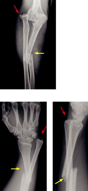
Monteggia fracture-dislocation
Monteggia fracture-dislocation:
Frontal radiograph of the proximal forearm shows a middiaphyseal ulnar fracture (yellow arrow) and radial head dislocation (red arrow).
Case courtesy Stacy Smith, MD, Brigham and Women’s Hospital.
•Monteggia fracture-dislocation is an ulnar fracture and radial dislocation at the elbow.
Galeazzi fracture-dislocation
Galeazzi fracture-dislocation:
Frontal and lateral radiographs of the forearm show a radial diaphyseal fracture of the distal third of the radial diaphysis (yellow arrows) and distal ulnar dislocation (red arrows). The radial shaft fracture has radial and volar angulation and foreshortening.
Case courtesy Stacy Smith, MD, Brigham and Women’s Hospital.
•Galeazzi fracture-dislocation is fracture of the distal third of the radius with ulnar dislocation at the distal radioulnar joint.
Colles fracture
•Colles fracture is a distal radius fracture with dorsal angulation. The fracture is usually intra-articular. Colles fracture is the most common injury to the distal forearm.
•Colles fracture typically results from a fall on an outstretched hand (FOOSH).
Hutchinson (chauffeur’s) fracture
•Hutchinson (chauffeur’s) fracture is a fracture of the radial (lateral) aspect of the distal radius extending into the radial styloid and the radiocarpal joint.
456
Wrist and hand
Common hand and wrist fractures
mallett nger
injury of extensor tendon
boutonniere deformity injury of extensor tendon
volar plate fracture associated with dislocation
gamekeeper’s thumb ulnar collateral ligament injury injury
Bennet fracture
Rolando fracture (comminuted) trapezium fracture
scaphoid fracture
scapholunate ligament injury
triquetral fracture best seen on lateral
Kienboch disease
AVN of the lunate
457
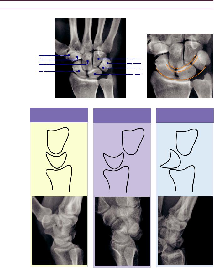
Wrist
Carpal anatomy and alignment
Arcs of Gilula should be smooth and continuous. Disruption may be a sign of igamentous injury or fracture.
trapezium |
hook of |
hamate |
scaphoid
s |
|
c |
|
a |
|
p |
|
h |
|
oi |
|
d |
|
fossa |
luna |
|
radius
te
fossa
hamate pisiform triquetrum
lunate
ulnar styloid
ulna
Lunate and perilunate dislocation
normal alignment |
|
perilunate dislocation |
|
|
|
capitate |
|
capitate |
lunate |
lunate |
radius |
radius |
lunate dislocation
capitate
lunate
radius
n a perilunate dislocation, the lunate is aligned with the radius, but capitate is not aligned with the lunate. Dislocation is next to the lunate (peri- unate).
n a lunate dislocation, the lunate is dislocated volarly, but the capitate is still relatively aligned with the radius. The lunate itself is dislocated (lunate dislocation)
458
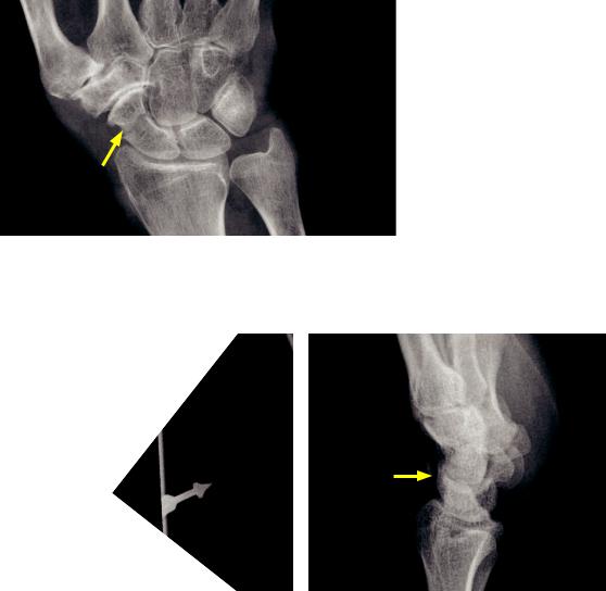
Scapholunate ligament injury
•Scapholunate ligament injury is one of the many patterns of injury caused by a fall on an outstretched arm.
•The Terry Thomas signrepresentsincreaseddistanceofscapholunateinterval,thought toresemblethegapbetweenthefrontteethofthefamousBritishactorfromthe60s.
•Scapholunate advanced collapse (SLAC) wrist is a sequela of chronic scapholunate ligament injury, which may be secondary to osteoarthritis, CPPD arthropathy, or chronic untreated scapholunate dissociation.
•The capitate migrates proximally to fill the abnormal gap between the scaphoid and lunate.
Scaphoid fracture
Scaphoid fracture: Radiograph of the wrist shows a minimally displaced fracture of the scaphoid (arrow).
•Blood supply to the scaphoid is interosseous, from distal to proximal. The proximal pole of the scaphoid has a tenuous blood supply and proximal fractures have the highest risk of avascular necrosis.
Triquetral fracture
Triquetral fracture: Frontal radiograph (left image) of the wrist is normal. Lateral radiograph shows an avulsion fragment dorsal to the triquetrum (arrow).
•Fracture of the triquetrum can be subtle to detect on the frontal radiograph. Triquetral fractures are best seen on the lateral view of the wrist as an avulsion fragment dorsal to the triquetrum.
Kienbock disease
•Kienbock disease is avascular necrosis of the lunate. It is associated with negative ulnar variance (i.e., ulna shorter than the radius), and thought to be caused by increased load on the lunate.
459
Fingers and hand trauma
Extension tendon injuries
normal anatomy |
mallet nger |
boutonniere deformity |
||
distal phalanx |
|
|
|
|
lateral extensor slips |
|
mallet nger |
|
|
converge on base of distal phalanx |
|
disruption of extensor tendon |
||
|
at distal phalanx |
|
|
|
|
|
|
|
|
middle phalanx |
|
|
|
|
central band of extensor tendon |
|
|
boutonniere deformity |
|
|
|
|||
inserts on base of middle phalanx |
|
|
|
disruption of extensor tendon |
|
|
|
|
at middle phalanx |
|
|
|
|
|
Malletfinger Malletfingerisdisruptionoftheextensortendonatthedistalphalanx.It iscausedbydirectimpactonthetipofthefinger.Malletfingermaybeassociatedwith anavulsionfragmentofthedistalphalanx,whichisclinicallyanimportantdistinction.
On physical exam, the distal interphalangeal (DIP) joint cannot be straightened.
Boutonniere deformity is injury to the medial slip of the extensor tendon. It is seen most commonly in the setting of rheumatoid arthritis, but it may be post-traumatic.
The proximal interphalangeal (PIP) joint herniates through the lateral slips of the common extensor tendon and becomes entrapped, causing fixed flexion of the PIP and extension of the DIP.
Gamekeeper’s thumb (skier’s thumb)
Gamekeeper’s thumb with associated nondisplaced avulsion fragment:
Radiograph of the thumb demonstrates a nondisplaced avulsion fragment at the base of the thumb proximal phalanx (arrow).
Gamekeeper’s (also known as skier’s) thumb is injury to the ulnar collateral ligament (UCL) at the base of the thumb proximal phalanx. It is caused by forced abduction of the thumb. Gamekeeper’s thumb may be associated with an avulsion fragment of the base of the proximal phalanx.
ncomplete UCL tears can be treated conservatively; however, complete UCL tears must be treated surgically.
The Stener lesion is a complication of UCL injury and represents complete UCL disruption with interposition of the adductor aponeurosis. The Stener lesion must be treated surgically.
460
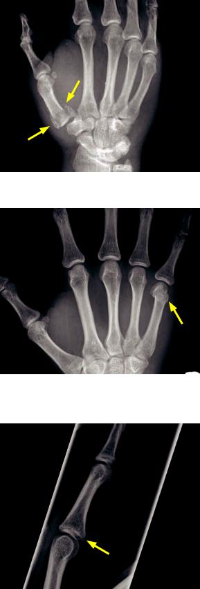
Bennett fracture
•A Bennett fracture is an intra-articular fracture of the base of the thumb metacarpal.
Rolando fracture
Rolando fracture: PA radiograph of the hand demonstrates a comminuted
intra-articular fracture at the base of the thumb metacarpal (arrows).
• A Rolando fracture is a comminuted Bennett fracture.
Boxer’s fracture
Boxer’s fracture: PA radiograph of the hand shows a fracture of the 5th metacarpal neck (arrow).
•A Boxer’s fracture is a metacarpal neck fracture, most commonly occurring in the 5th metacarpal.
Volar plate fracture
Volar plate fracture: Lateral radiograph shows a subtle tiny avulsion fragment (arrow) of the volar aspect of the middle phalanx at the PIP joint.
•A volar plate fracture is an avulsion fracture of the volar aspect of the proximal middle phalanx, caused by hyperextension.
461

References, resources, and further reading
General References:
Brower A.C. Arthritis in Black and White. (2nd ed.). Saunders. (1997).
Chew, F.S. & Roberts, C.C. Musculoskeletal Imaging: A Teaching File (2nd ed.). Lippincott Williams & Wilkins. (2005).
Greenspan, A. Orthopedic Imaging: A Practical Approach (5th ed.). Lippincott Williams & Wilkins. (2010).
Helms, C.A. et al. Musculoskeletal MRI (2nd ed.). Saunders. (2008).
Manaster, B., May, D. & Disler, D.G. Musculoskeletal Imaging: The Requisites (3rd ed.). Mosby. (2006).
Weissman, B. Imaging of Arthritis and Metabolic Bone Disease (1st ed.). Mosby. (2009).
Arthritis:
Bennett D., Ohashi K., Elkhoury G. Spondyloarthropathies: ankylosing spondylitis and psoriatic arthritis. Radiologic Clinics of North America, 42(1), 121-34(2004).
Jacobson J.A. et al. Radiographic Evaluation of Arthritis: Inflammatory Conditions. Radiology, 248(2), 378-89(2008).
JacobsonJ.A.etal.RadiographicEvaluationofArthritis:DegenerativeJointDiseaseandVariations.Radiology,248(3),737-47(2008).
Steinbach L.S. Calcium pyrophosphate dihydrate and calcium hydroxyapatite crystal deposition diseases: imaging perspectives. Radiologic Clinics of North America, 42(1), 185-205, vii(2004).
Neoplasms:
Miller T.T. Bone tumors and tumorlike conditions: analysis with conventional radiography. Radiology, 246(3), 662-74(2008).
Nomikos G.C. et al. Primary bone tumors of the lower extremities. Radiologic Clinics of North America, 40, 971-90(2002).
Infection:
Bancroft, L.W. MRI imaging of infectious processes of the knee. Radiologic clinics of North America, 45(6), 931–41, v(2007).
Donovan, A. & Schweitzer, M.E. Current concepts in imaging diabetic pedal osteomyelitis. Radiologic clinics of North America, 46(6), 1105–24, vii(2008).
Pineda, C., Vargas, A. & Rodríguez, A.V. Imaging of osteomyelitis: current concepts. Infectious disease clinics of North America, 20(4), 789–825(2006).
Foot and Ankle:
Choplin, R.H. et al. CT with 3D rendering of the tendons of the foot and ankle: technique, normal anatomy, and disease. Radiographics, 24(2), 343-56(2004).
Kong, A., Cassumbhoy, R. & Subramaniam, R.M. Magnetic resonance imaging of ankle tendons and ligaments: part I - anatomy. Australasian radiology, 51(4), 315-23(2007).
Leffler, S. & Disler, D.G. MRI imaging of tendon, ligament , and osseous abnormalities of the ankle and hindfoot. Radiologic Clinics of North America, 40, 1147-70(2002).
Rosenberg, Z.S., Beltran, J. & Bencardino, J.T. From the RSNA Refresher Courses MRI Imaging of the Ankle and Foot. Radiographics, (20), 153-79(2000).
Knee:
Barr,M.S.&Anderson,M.W.Theknee:bonemarrowabnormalities.RadiologicClinicsofNorthAmerica,40,1109-20(2002).
Fox, M.G. MRI imaging of the meniscus: review, current trends, and clinical implications. Radiologic clinics of North America, 45(6), 1033-53, vii(2007).
Frick, M.a., Wenger, D.E. & Adkins, M. MRI imaging of synovial disorders of the knee: an update. Radiologic clinics of North America, 45(6), 1017-31, vii(2007).
Mccauley, T.R. MRI imaging of chondral and osteochondral injuries of the knee. Radiologic Clinics of North America, 40, 1095-107(2002).
462

Phillips, M. & Pomeranz, S. Imaging of Osteochondritis Dissecans of the Knee. Operative Techniques in Sports Medicine, 16(2), 52-64(2008).
Hip:
Anderson, S.E., Siebenrock, K.A. & Tannast, M. Femoroacetabular impingement: evidence of an established hip abnormality. Radiology, 257(1), 8-13(2010).
Hong, R.J., Hughes, T.H., Gentili, A. & Chung, C.B. Magnetic resonance imaging of the hip. Journal of Magnetic Resonance Imaging: JMRI, 27(3), 435-45(2008).
Ikemura, S. et al. MRI evaluation of collapsed femoral heads in patients 60 years old or older: Differentiation of subchondral insufficiency fracture from osteonecrosis of the femoral head. AJR. American journal of roentgenology, 195(1), W63–8(2010).
Kwek, E.B.K. et al. An emerging pattern of subtrochanteric stress fractures: a long-term complication of alendronate therapy? Injury, 39(2), 224-31(2008).
Palmer, W.E. Femoroacetabular impingement: caution is warranted in making imaging-based assumptions and diagnoses. Radiology, 257(1), 4-7(2010).
Ragab, Y., Emad, Y. & Abou-Zeid, A. Bone marrow edema syndromes of the hip: MRI features in different hip disorders. Clinical rheumatology, 27(4), 475-82(2008).
Sweeney, J.P., Helms, C.A., Minagi, H. & Louie, K. The Widened Teardrop Distance: A Plain Film indicator of Hip Joint Effusion in Adults. Radiology, 149, 117-19(1987).
Spine:
Bagley, L.J. Imaging of spinal trauma. Radiologic Clinics of North America, 44(1), 1-12, vii(2006).
Greenbaum, J., Walters, N. & Levy, P.D. An evidenced-based approach to radiographic assessment of cervical spine injuries in the emergency department. The Journal of Emergency Medicine, 36(1), 64-71.(2009).
Phal, P.M. & Anderson, J.C. Imaging in spinal trauma. Seminars in roentgenology, 41(3), 190-5(2006).
Shoulder:
Chang, D., Mohana-Borges, A., Borso, M. & Chung, C.B. SLAP lesions: anatomy, clinical presentation, MRI imaging diagnosis and characterization. European journal of radiology, 68(1), 72-87(2008).
Morag, Y. et al. MRI Imaging of Rotator Cuff Injury: What the Clinician Needs to Know. Radiographics, 26, 1045-66(2006).
Omoumi, P., Teixeira, P., Lecouvet, F. & Chung, C.B. Glenohumeral joint instability. Journal of magnetic resonance imaging: JMRI, 33(1), 2-16(2011).
Opsha, O. et al. MRI of the rotator cuff and internal derangement. European journal of radiology, 68(1), 36-56(2008).
Elbow, Forearm, Wrist, and Hand:
Chen, C. et al. Scapholunate advanced collapse: a common wrist abnormality in calcium pyrophosphate dihydrate crystal deposition disease. Radiology, 177(2), 459(1990).
Hauger, O. et al. Pulley system in the fingers: normal anatomy and simulated lesions in cadavers at MRI imaging, CT, and US with and without contrast material distention of the tendon sheath. Radiology, 217(1), 201-12(2000).
Sof, C.M. & Potter, H.G. Imaging of elbow injuries in the child and adult athlete. Radiologic Clinics of North America, 40, 251-65(2002).
Staebler, A., Heuck, A. & Reiser, M. Imaging of the hand: degeneration, impingement and overuse. European journal of radiology, 25(2), 118-28(1997).
463