


C H A P T E R 9
SPLEEN
Cases 9.1–9.6 Inflammatory Diseases
Cases 9.7–9.21 Masses
Benign
Malignant
Cases 9.22–9.29 |
Miscellaneous |

This page intentionally left blank
9. SPLEEN 755
CASE 9.1
Findings |
Discussion |
|
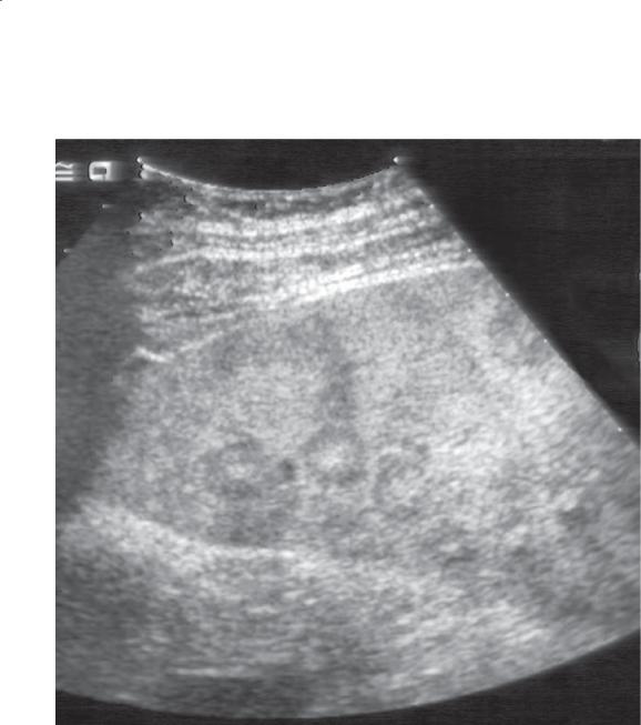
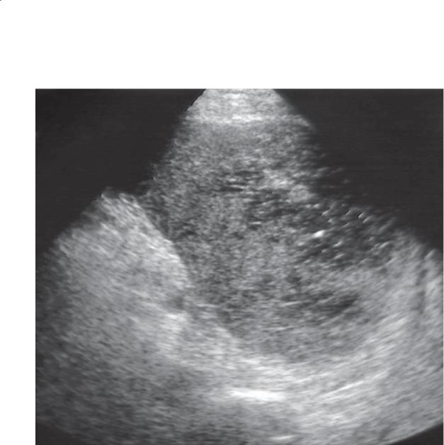
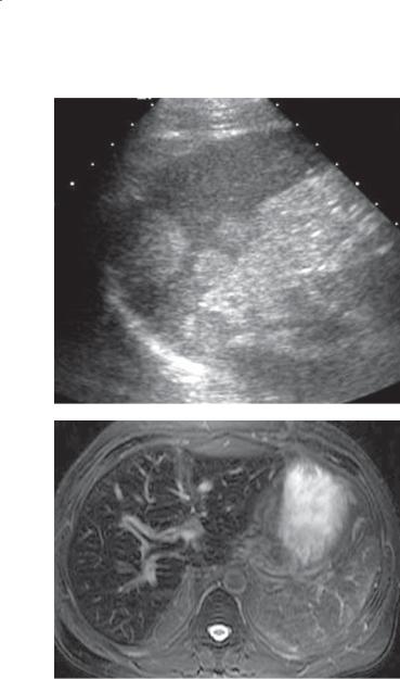
Sonogram of the spleen. Multiple target lesions are |
Fungal infections involving the liver and spleen usually |
|
present throughout the spleen. |
develop in immunocompromised persons receiving |
|
|
|
chemotherapy. These infections can be difficult to |
Di erential Diagnosis |
detect with imaging studies because of their small size. |
|
1. |
Metastases |
In 50% of blood cultures, results are negative. In these |
2. |
Lymphoma |
cases, biopsy and histologic confirmation of infection |
3. |
Candida microabscesses |
may be necessary. |
4. |
Mycobacterial infection |
Th e typical target or bull’s-eye lesion at sonography |
|
|
is an echogenic center surrounded by a hypoechoic |
Diagnosis |
band. As the lesions heal, they often become |
|
Candida microabscesses |
homogeneously hypoechoic. Most lesions are 5 to |
|
|
|
10 mm in diameter. The finding of similar-appearing |
|
|
lesions in the liver or kidneys is helpful to suggest |
|
|
Candida infection. |
Disease type: Inflammatory Diseases
756 MAYO CLINIC GASTROINTESTINAL IMAGING REVIEW
CASE 9.2
Findings
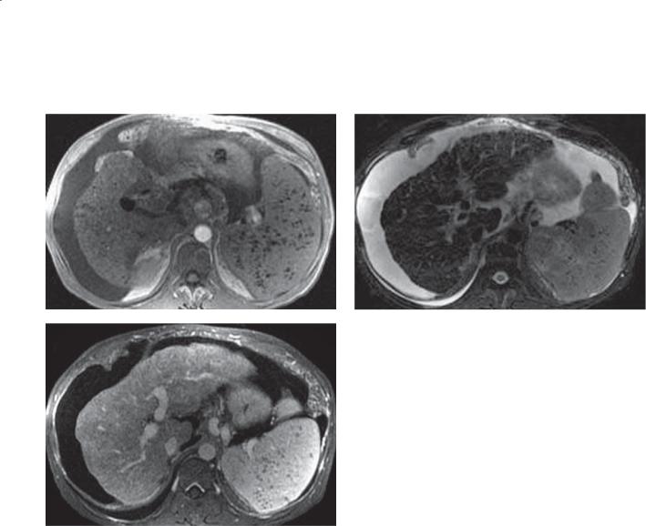
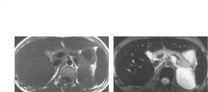
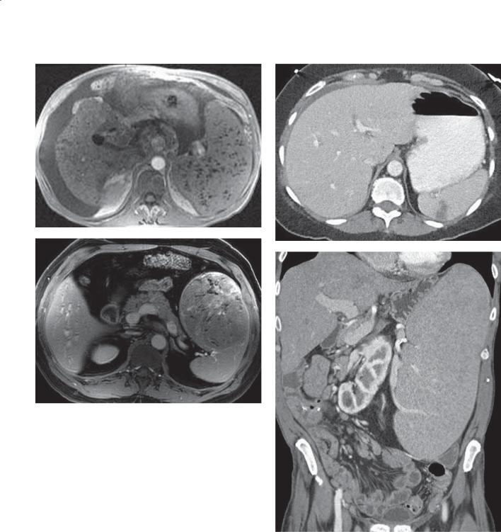
T2-weighted unenhanced MRI. The spleen is of abnormally low signal intensity. There are multiple tiny high-intensity masses throughout.
Di erential Diagnosis
1.Metastases
2.Lymphoma
3.Candida microabscesses
4.Mycobacterial infection
Diagnosis
Candida microabscesses and hemosiderosis
Discussion
In patients with suspected Candida microabscesses and a negative abdominal sonogram, MRI can be helpful to assess for these tiny abscesses. This patient had received multiple transfusions for a hematopoietic malignancy, and hemosiderosis (secondary hemochromatosis)
had developed. Excess iron from transfusions is accumulated in the reticuloendothelial system and lowers the splenic signal. Suppression of the normal splenic signal increases contrast and visibility of the high-signal microabscesses.
Disease type: Inflammatory Diseases
9. SPLEEN 757
CASE 9.3 |
CASE 9.4 |
A |
|
B
Findings
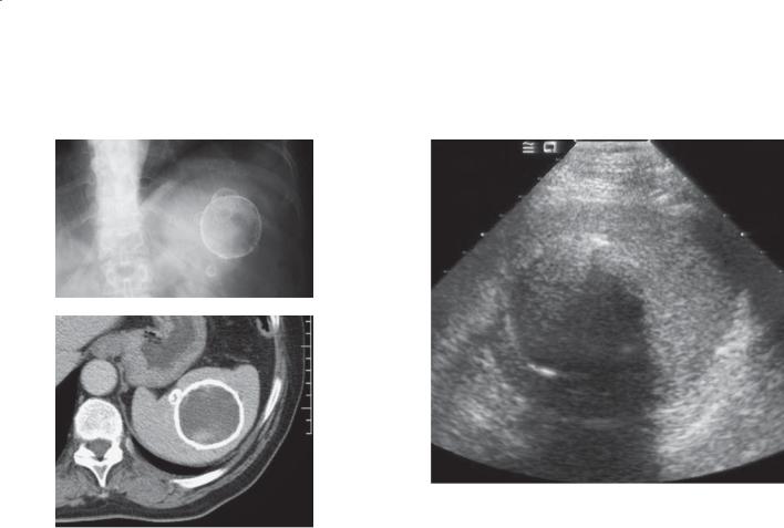
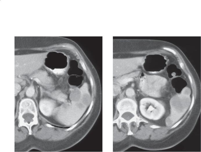
CASE 9.3. A. Abdominal radiograph. Several peripherally calcified masses of variable size are present in the spleen.
B. Contrast-enhanced CT. The peripherally calcified masses contain water-attenuation fluid.
CASE 9.4. Splenic sonogram. A large mass is present in the spleen. The rim of the mass is hyperechoic, and there is a strong acoustic shadow from the rim indicating calcification.
Di erential Diagnosis
1.Echinococcal cysts
2.Traumatic splenic cyst
3.Intrasplenic aneurysm
4.Metastases
Diagnosis
Echinococcal cysts
Discussion
Splenic involvement in patients with echinococcal disease is unusual—occurring in less than 2%
of patients with this disease. Liver, lung, bone, and brain are more commonly involved. Splenic
involvement usually is due to systemic dissemination or intraperitoneal spread. Echinococcus granulosus
is the most common form to affect the spleen, with sharply defined cysts and daughter cysts. Echinococcus multilocularis presents with tiny cystic masses that have the appearance of an invasive malignancy. Liver cysts usually are present if splenic cysts exist.
Sonographic findings include anechoic splenic cysts with or without daughter cysts. Calcification of the wall (as in this case) is helpful. Occasionally the abnormality presents as a solid mass. CT is helpful for showing a cyst with subtle wall calcification. The interior attenuation of the cyst may be higher than water because of the presence of hydatid sand and inflammatory cells.
Positive radiographic findings, clinical suspicion in patients from an endemic area, and positive serologic tests are most helpful for making the diagnosis.
Disease type: Inflammatory Diseases
758 MAYO CLINIC GASTROINTESTINAL IMAGING REVIEW
CASE 9.5
A B
Findings
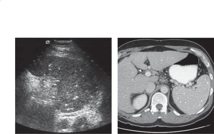
A.Abdominal sonogram. Multiple tiny hyperechoic masses are present throughout the spleen.
B.Contrast-enhanced CT. Multiple tiny calcifications are present in the spleen.
Di erential Diagnosis
1.Healed granulomatous infection
2.Candida microabscesses
3.Extrapulmonary Pneumocystis infection
4.Treated lymphoma
5.Treated metastases
Diagnosis
Healed granulomatous infection, histoplasmosis
Discussion
Multiple calcified granulomas in the spleen are common, and they are usually the sequelae of healed infection from prior histoplasmosis. Radiographically similar findings can be found in patients with healed tuberculosis, Mycobacterium avium-intracellulare, and sarcoidosis and in patients who have had infection with Pneumocystis carinii. Multiple punctate calcifications could develop in patients who have had successful treatment for lymphoma and metastases with splenic involvement.
Disease type: Inflammatory Diseases
9. SPLEEN 759
CASE 9.6
Findings |
Discussion |
|
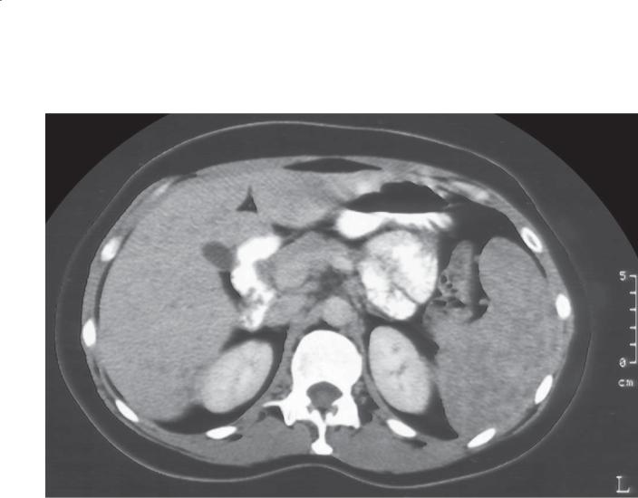
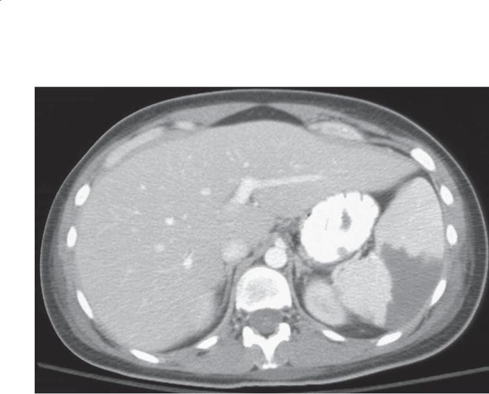
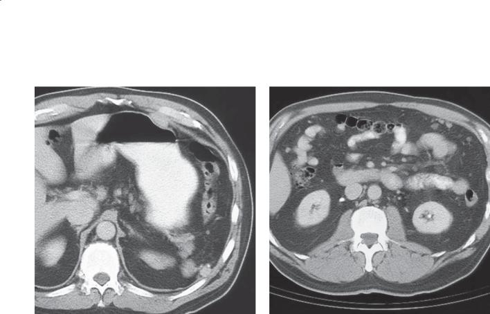
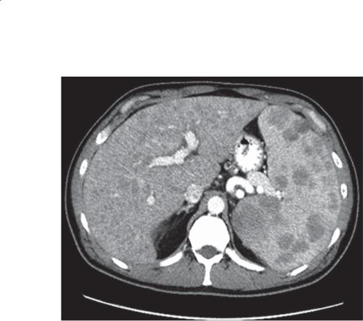
Contrast-enhanced CT (delayed). Multiple |
Sarcoidosis is a disease of unknown cause that |
|
hypoattenuating masses are present throughout the |
usually affects the lung and mediastinum. Abdominal |
|
spleen and liver. |
sarcoidosis is unusual; when it occurs, it can affect |
|
|
|
the solid organs of the abdomen and lymph nodes. |
Di erential Diagnosis |
Although pulmonary involvement is often present |
|
1. |
Metastases |
concurrently, it is not required. Sarcoid can present in |
2. |
Lymphoma |
various ways, with hepatosplenomegaly, multiple low- |
3. |
Candidiasis |
attenuation filling defects within the solid organs (as in |
4. |
Mycobacterial infection |
this case), diffuse calcifications, or lymphadenopathy. |
5. |
Granulomatous infection |
|
Diagnosis
Granulomatous infection, sarcoidosis
Disease type: Inflammatory Diseases

760 MAYO CLINIC GASTROINTESTINAL IMAGING REVIEW
TABLE 9.1
Inflammatory Diseases of the Spleen |
CASE |
|
|
|
|
Candidiasis |
Multiple tiny low-attenuation splenic lesions. Target appearance on |
9.1 and 9.2 |
|
sonography. Liver also usually involved. Immunosuppressed patients |
|
|
|
|
Echinococcal cysts |
Peripherally calcified cyst and daughter cysts. Liver disease usually |
9.3 and 9.4 |
|
present when spleen is involved. Usually from endemic area |
|
|
|
|
Healed granulomatous |
Multiple tiny calcified splenic granulomas. Calcified granulomas in the |
9.5 |
infection, histoplasmosis |
lungs and calcified hilar and mediastinal nodes often present |
|
|
|
|
Sarcoidosis |
Multiple tiny hypoattenuating splenic lesions. Chronic changes can |
9.6 |
|
result in calcified splenic granulomas. Adenopathy may be present |
|
|
|
|
Disease type: Inflammatory Diseases
9. SPLEEN 761
CASE 9.7 |
CASE 9.8 |
A |
|
B
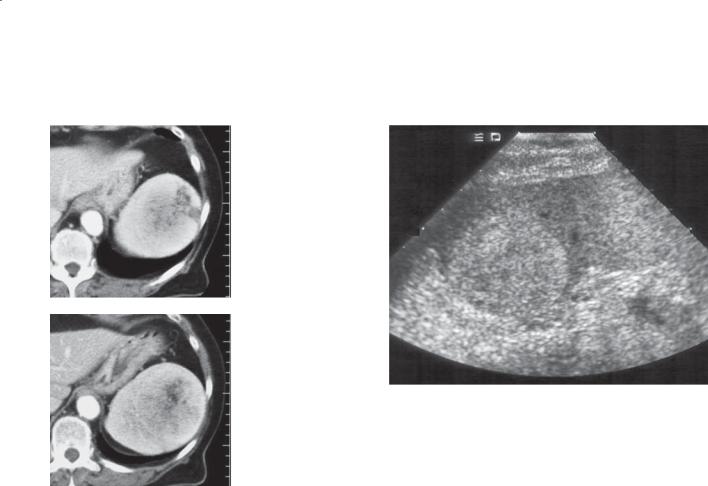
Findings
CASE 9.7. Contrast-enhanced CT. A and B. A large, welldefined, solitary hyperenhancing mass is present in the spleen. Several hypoattenuating regions are present within the mass.
CASE 9.8. Abdominal sonogram. A solitary, moderately heterogeneous echogenic mass is present within the spleen.
Di erential Diagnosis
1.Metastases
2.Lymphoma
3.Hemangioma
4.Hamartoma
Diagnosis
Hemangioma
Discussion
Hemangiomas of the spleen have many of the same characteristics as hemangiomas within the liver. They are the most common benign tumor to affect the spleen. They are usually incidental findings. Pathologically, splenic hemangiomas are similar, with endothelium-lined vascular spaces of various sizes. Kasabach-Merritt syndrome may occur with large hemangiomas—with findings of anemia, infarction, high-output congestive heart failure, thrombocytopenia, and coagulopathy.
Although hemangiomas may enhance after intravenous contrast material in a pattern similar to that of hepatic hemangiomas, our experience is that this occurs in a minority of cases. In most instances, a heterogeneously enhancing mass is discovered which has nonspecific imaging characteristics. In these cases, comparison with prior films or follow-up imaging
to assess for growth can be helpful. The sonographic appearance of hemangiomas is also similar to that found in the liver. These lesions are usually echogenic, but they may appear more complex. If necessary, percutaneous biopsy can be performed safely.
Disease type: Masses
762 MAYO CLINIC GASTROINTESTINAL IMAGING REVIEW
CASE 9.9
A B
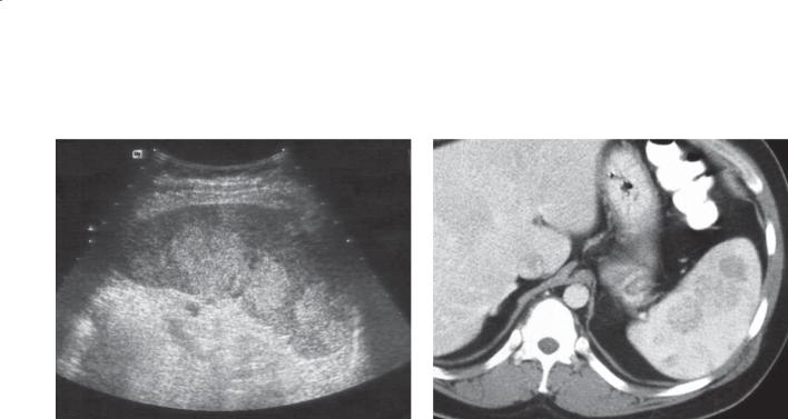
Findings
A.Abdominal sonogram. Multiple homogeneous echogenic masses are present throughout the spleen.
B.Contrast-enhanced CT. Several masses are present in the spleen. Some of these masses contain globular enhancement.
Di erential Diagnosis
1.Hemangiomas
2.Metastases
3.Lymphoma
Diagnosis
Multiple hemangiomas
Discussion
Hemangiomas are usually solitary but can be multiple. Splenic hemangiomas can be part of generalized angiomatosis (Klippel-Trénaunay-Weber syndrome) or associated with the Kasabach-Merritt syndrome. They also have been reported to be associated with portal venous hypertension.
Disease type: Masses
CASE 9.10
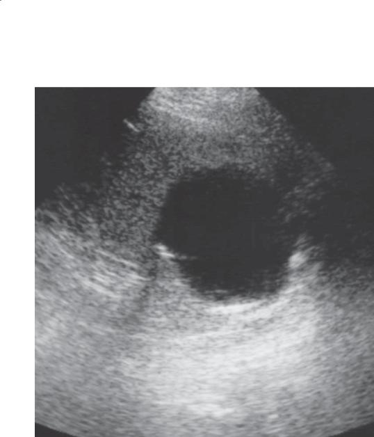
Findings
Abdominal sonogram. A well-defined anechoic mass with posterior acoustic enhancement is present within the spleen. The mass contains a dependent septation.
Di erential Diagnosis
Benign splenic cyst
Diagnosis
Benign splenic cyst
9. SPLEEN 763
Discussion
Splenic cysts are usually anechoic structures with welldefined walls and increased through-transmission at sonography. Intracystic debris due to hemorrhage can be seen within these lesions. Septations (trabeculation) are seen in up to one-third of cysts.
Disease type: Masses
764 MAYO CLINIC GASTROINTESTINAL IMAGING REVIEW
CASE 9.11
Findings |
Discussion |
Contrast-enhanced CT. Solitary, septated cyst is |
Splenic cysts are divided pathologically into either |
present within the spleen. |
true (with a cellular lining) or false (without a cellular |
|
lining) cysts. True cysts that are not parasitic in origin |
Di erential Diagnosis |
often are referred to as epidermoid cysts. They are |
1. Epidermoid cyst |
believed to arise from an embryonic inclusion of the |
2. Parasitic cyst |
surface mesothelium in the developing spleen. False |
3. Secondary cyst |
cysts have a smooth fibrous lining and are believed |
|
to arise from prior splenic trauma, infarction, or |
Diagnosis |
infection. It can be difficult to distinguish the two types |
Epidermoid cysts |
of cysts radiologically. Peripheral calcification can be |
|
seen with either type of cyst. Parasitic cysts usually are |
|
echinococcal in origin. |
|
CT findings of a well-defined, water-attenuation |
|
mass without enhancement are typical. Peripheral |
|
septations can occur and sometimes are referred to as |
|
trabeculation. Some authorities believe that these are |
|
more commonly found in true cysts. |
Disease type: Masses

9. SPLEEN 765
CASE 9.12
A B
C
Findings
A.T1-weighted MRI. Large mass of heterogeneous signal intensity is within the anterior aspect of the spleen.
B.T2-weighted MRI. The mass is well circumscribed and of heterogeneous signal intensity.
C.Contrast-enhanced MRI. The mass enhances heterogeneously.
Di erential Diagnosis
1.Metastasis
2.Hemangioma
3.Hamartoma
Diagnosis
Hamartoma
Discussion
Hamartomas of the spleen are benign tumors composed of normal cellular elements in abnormal quantity, degree of differentiation, cellular arrangement, or a combination of all 3 features. Pathologically, they are classified into 3 types: red pulp (vascular), white pulp (lymphomatous), or mixed. They are usually solid tumors without encapsulation. Clinically they may be asymptomatic, or they
may be associated with hypersplenism (anemia, thrombocytopenia, pancytopenia).
At sonography they usually appear homogeneously echogenic, but they may contain cystic spaces. The appearance at CT is variable—usually presenting as a hypoattenuating or isoattenuating mass. Large lesions can have scars, cystic regions, and calcification.
Disease type: Masses
766 MAYO CLINIC GASTROINTESTINAL IMAGING REVIEW
CASE 9.13
A B
Findings
A.Contrast-enhanced CT. A well-circumscribed, heterogeneously enhancing mass is present in the spleen.
B.Abdominal sonogram. The mass is of heterogeneous echotexture and its borders are well defined.
Di erential Diagnosis
1.Lymphoma
2.Metastases
3.Hemangioma
4.Infl ammatory pseudotumor
5.Hamartoma
Diagnosis
Inflammatory pseudotumor
Discussion
Inflammatory pseudotumor is a rare entity composed of a localized area of inflammatory and fibroblastic cells surrounding regions of hemorrhage and necrosis. These lesions usually are discovered incidentally. The radiographic appearance is nonspecific, except that the mass is usually well defined and heterogeneous internally. Calcification may be present. Occasionally, a central stellate scar is present.
Disease type: Masses
9. SPLEEN 767
CASE 9.14
A B
C
Findings
A.T1-weighted MRI.
B.T2-weighted MRI.
C.Contrast-enhanced MRI. Multiple nodules of very low signal intensity are throughout the spleen. Ascites and cirrhotic liver are seen.
Di erential Diagnosis
Siderotic nodules (Gamna-Gandy bodies)
Diagnosis
Siderotic nodules (Gamna-Gandy bodies)
Discussion
In patients with cirrhosis and portal venous hypertension, small nodules (3-8 mm in diameter) can develop within the spleen from prior organized hemorrhage. The deposited hemosiderin explains the findings that are best appreciated on gradient echo pulse sequences. Approximately 10% of patients with portal venous hypertension have these nodules. They can occur within a normal or enlarged spleen. The siderotic nodules have no clinical significance themselves, except to indicate the presence of portal venous hypertension.
Disease type: Masses
768 MAYO CLINIC GASTROINTESTINAL IMAGING REVIEW
CASE 9.15
Findings
Contrast-enhanced CT. There is a wedge-shaped lowattenuation defect within the spleen.
Di erential Diagnosis
Splenic infarction
Diagnosis
Splenic infarction
Discussion
Splenic infarction usually is caused by emboli (most commonly in older persons) or thrombi (due to an underlying hematologic disorder). Patients usually present with sudden pain in the left upper quadrant. Infarction results in a weakened area of the spleen which can rupture and tissue that is prone to infection. Close observation and follow-up for one of these complications usually are recommended.
Although this case illustrates the most classic findings of splenic infarction with a wedge-shaped peripheral defect, infarction can present as a heterogeneous mass that may be indistinguishable from a neoplasm. If the clinical history is not revealing, a biopsy may be necessary.
Disease type: Masses
9. SPLEEN 769
CASE 9.16
Findings |
Discussion |
|
Abdominal sonogram. Several hypoechoic regions are |
Th e appearance of a splenic infarct at sonography |
|
present within the spleen. |
depends on its age. Acutely, splenic infarction appears |
|
|
|
as a poorly delineated hypoechoic region(s). A wedge- |
Di erential Diagnosis |
shaped abnormality may be present. Aging infarcts |
|
1. |
Lymphoma |
become better marginated and echogenic, with focal |
2. |
Metastases |
atrophy and scarring. |
3. |
Infarction |
Th is case illustrates several hypoechoic regions, |
4. |
Abscess |
some with poor and some with good delineation. The |
|
|
larger region contains some central hyperechoic islands |
Diagnosis |
within it—likely representing evolution of a healing |
|
Splenic infarction |
infarct. |
|
Disease type: Masses
770 MAYO CLINIC GASTROINTESTINAL IMAGING REVIEW
CASE 9.17
A B
Findings
Contrast-enhanced CT. A. Arterial phase of contrast enhancement shows multiple regions of hypoattenuation throughout the spleen. B. Portal
venous phase of contrast enhancement shows a normal homogeneous spleen.
Di erential Diagnosis
Splenic pseudomass
Diagnosis
Splenic pseudomass
Discussion
Enhancement of the spleen is often heterogeneous during the early phases of contrast enhancement because of variable rates of flow through the red pulp. This finding should not be considered an abnormality unless it persists into the later portal venous phase of enhancement. Generally, a homogeneous splenic blush should be seen within 2 minutes after bolus injection of contrast material.
Disease type: Masses
9. SPLEEN 771
CASE 9.18
A B
Findings
Contrast-enhanced CT. A and B. Several well-defined hypoattenuating masses are present within the spleen.
Di erential Diagnosis
1.Metastases
2.Lymphoma
3.Multiple hemangiomas
4.Multiple benign cysts
Diagnosis
Lymphoma
Discussion
Lymphoma is the commonest malignant tumor to affect the spleen. Splenic involvement occurs in approximately one-third of patients with lymphoma (either Hodgkin or non-Hodgkin). Current imaging techniques are not optimal for detecting the disease. Splenomegaly is one of the most common findings
in patients with lymphoma. However, the presence of an enlarged spleen does not necessarily indicate lymphomatous involvement, especially in patients with bone marrow involvement and splenomegaly due to extramedullary hematopoiesis. As a result, splenectomy is often necessary to detect splenic involvement. Splenomegaly also is unreliable in Hodgkin disease because one-third of patients with splenic involvement have a normal-sized spleen, and one-third of patients with splenomegaly do not have splenic involvement.
Splenic lymphoma occurs in 4 major patterns: homogeneous enlargement, miliary involvement, multiple masses, and a solitary large mass.
The accuracy of CT for detecting lymphoma is approximately 60%. Regional and systemic adenopathy can be helpful for suggesting lymphoma. Splenic infarction can be seen in patients with splenic lymphoma, with the characteristic peripheral, wedgeshaped regions of hypoattenuation.
Disease type: Masses
772 MAYO CLINIC GASTROINTESTINAL IMAGING REVIEW
CASE 9.19
Findings
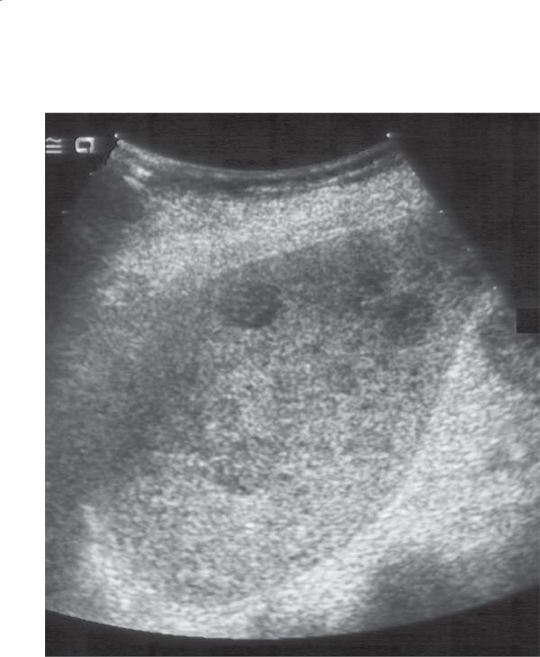
Abdominal sonogram. The spleen is enlarged and contains multiple hypoechoic masses.
Di erential Diagnosis
1.Metastases
2.Lymphoma
Diagnosis
Lymphoma
Discussion
Lymphoma usually presents as a diffuse or focal (small nodular lesions or large lesions) hypoechoic pattern. Low-grade disease usually presents with the diffuse or small nodular pattern, whereas high-grade disease usually presents with large masses. Detection of adenopathy elsewhere in the abdomen also can be helpful for suggesting the diagnosis.
Disease type: Masses
CASE 9.20
Findings
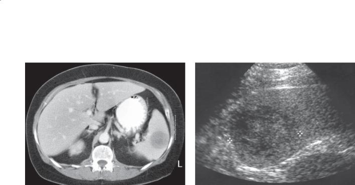
CASE 9.20. Contrast-enhanced CT. A well-defined focal mass is present within the spleen.
CASE 9.21. Abdominal sonogram. A solid mass in the spleen contains heterogeneous echotexture (slightly hypoechoic compared with the remainder of the spleen).
Di erential Diagnosis
1.Metastases
2.Lymphoma
3.Hamartoma
Diagnosis
Metastases (melanoma primary)
9. SPLEEN 773
CASE 9.21
Discussion
Metastasis to the spleen usually occurs by hematogenous arterial seeding. Venous seeding can occur in patients with portal venous hypertension. The spleen is a relatively uncommon site for metastasis, occurring in less than 10% of patients at autopsy. The commonest primary tumors to metastasize to the spleen include melanoma, breast, ovary, and lung.
Metastases can have a widely variable appearance, ranging from cystic to heterogeneous to solid masses. Some will be well defined and others infiltrative.
The keys for detection are to identify regions of hypoattenuation at CT (or hypoechoic or hyperechoic regions at sonography) and to correlate these findings with the clinical history of a malignancy. Patients with melanoma metastases can have lesions with cystic necrosis and fluid visualized centrally or a sonographic halo around the circumference of the mass.
Disease type: Masses

774 MAYO CLINIC GASTROINTESTINAL IMAGING REVIEW
TABLE 9.2
Masses of the Spleen |
|
CASE |
|
|
|
Benign |
|
|
Hemangioma |
Most common benign tumor of spleen. May have peripheral |
9.7–9.9 |
|
globular enhancement and hyperechoic appearance, as in liver. |
|
|
Frequently atypical in appearance |
|
Splenic cyst |
True cysts (epidermoid cysts) have a cellular lining. False cysts |
9.10 and 9.11 |
|
(due to trauma, infection, or infarction) have only a fibrous lining. |
|
|
Anechoic with posterior acoustic enhancement. Thin peripheral |
|
|
calcification in both true and false splenic cysts |
|
Hamartoma |
Benign tumor with disorganized normal cellular elements. |
9.12 |
|
Solid tumor with variable echogenicity and enhancement |
|
Inflammatory pseudotumor
Siderotic nodules (Gamna-Gandy bodies)
Infarct
Pseudomass
Candidiasis
Rare, well-defined mass surrounding hemorrhage and necrosis. |
9.13 |
Occasional central stellate scar |
|
Tiny, benign, iron-containing nodules in the spleen in patients |
9.14 |
with portal hypertension. Multiple low-intensity splenic nodules |
|
on gradient echo MRI |
|
Classically wedge-shaped, peripheral, low-attenuation, |
9.15 and 9.16 |
hypoechoic lesion. Can appear masslike |
|
Heterogeneous enhancement of spleen on early phases. |
9.17 |
Findings should resolve on portal venous phase |
|
Multiple tiny low-attenuation splenic lesions. Target appearance |
9.1 and 9.2 |
on sonography. Liver also usually involved. Immunosuppressed |
|
patients |
|
Echinococcal cysts |
Peripherally calcified cyst and daughter cysts. Liver disease usually |
9.3 and 9.4 |
present when spleen is involved. Usually from endemic area |
|
|
|
|
|
Sarcoidosis |
Multiple tiny hypoattenuating splenic lesions. Adenopathy may |
9.6 |
be present |
|
|
|
|
|
|
|
|
Malignant |
|
|
Lymphoma |
Commonest malignant tumor of spleen. Solitary or multiple |
9.18 and 9.19 |
|
masses. Adenopathy common |
|
Metastases |
Uncommon site for metastases. Most commonly melanoma, |
9.20 and 9.21 |
|
breast, ovary, and lung |
|
|
|
|
Disease type: Masses
9. SPLEEN 775
CASE 9.22
Findings
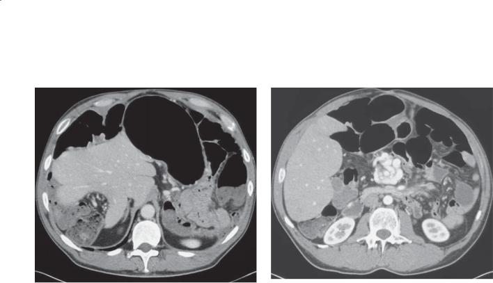
Contrast-enhanced CT. A mixed-attenuation mass is present within a subcapsular location about the lateral aspect of the spleen.
Di erential Diagnosis
Subcapsular splenic hematoma
Diagnosis
Subcapsular splenic hematoma
Discussion
Th e spleen is the most commonly injured organ after blunt abdominal trauma, constituting 25% of solid-organ injuries. Physical complaints and
findings of splenic injury after blunt trauma include left upper quadrant pain, referred left shoulder pain, hypotension, and shock. Diagnostic peritoneal lavage may be used in the emergency department to assess for hemoperitoneum in the unstable patient. The presence
of hemoperitoneum has a high association with splenic injury. In stable patients, contrast-enhanced CT is the preferred method to assess for splenic injuries.
Splenic injuries can be classified at CT as
1)subcapsular or intrasplenic hematomas,
2)lacerations, 3) fractures, or 4) vascular pedicle injuries. Intrasplenic hematomas usually appear as a hypoattenuating focal mass. Aging hematomas at unenhanced CT may appear isoattenuating. Subcapsular hematomas (as in this case) appear as a crescent of fluid or mixed attenuation collections. Small subcapsular collections can be difficult to differentiate from perisplenic fluid. Lacerations are
low-attenuation linear abnormalities that do not cross the spleen entirely. Fractures extend entirely through the spleen and may result in devascularized fragments. They are found most commonly in the splenic poles. Vascular pedicle injuries usually result in severe bleeding and hemodynamic instability. Patients usually are taken to the operating room for emergency repair.
Disease type: Miscellaneous
776 MAYO CLINIC GASTROINTESTINAL IMAGING REVIEW
CASE 9.23
Findings
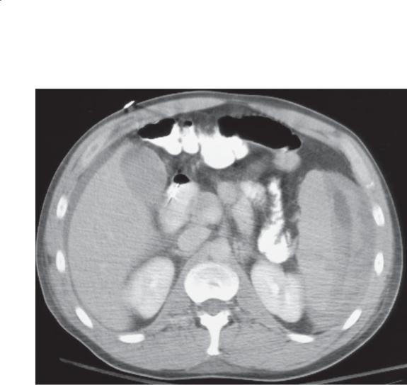
Contrast-enhanced CT. Multiple linear abnormalities divide the spleen into multiple small pieces. There is fluid about the spleen and a left pleural effusion.
Di erential Diagnosis
Multiple splenic lacerations
Diagnosis
Multiple splenic lacerations (shattered spleen)
Discussion
A shattered spleen is a severe splenic injury with a high incidence of vascular pedicle injuries. Because of the dual blood supply to the spleen by way of the splenic artery and the short gastric arteries, this condition may result in nonenhancement of the lower polar spleen with upper pole perfusion by way of the short gastric arteries. In some cases, lacerations can be subtle findings. The findings of perisplenic fluid (clot) and thickening of the anterior pararenal fascia and the lateral conal fascia can be useful indicators of a splenic injury.
Several grading systems exist for classifying splenic injuries; none have been adopted for standard care and reporting.
Disease type: Miscellaneous
CASE 9.24
A
Findings
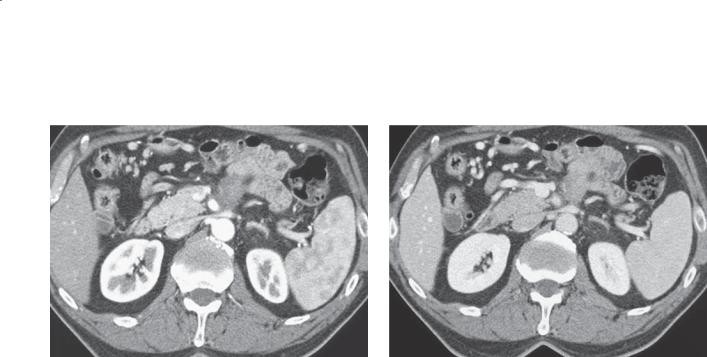
Contrast-enhanced CT. A and B. Several soft tissue attenuation nodules are present in the splenic bed. Soft tissue nodules also are present in the region of the omentum.
Di erential Diagnosis
1.Metastases
2.Splenosis
Diagnosis
Splenosis
9. SPLEEN 777
B
Discussion
Splenosis refers to autotransplantation of splenic tissue. Splenic trauma is the most common cause, with seeding of splenic tissue throughout the peritoneal cavity, where it can grow if blood supply is established. Splenosis also has been reported within the pleural space, retroperitoneum, pericardium, lung, and subcutaneous tissues. One-fourth to two-thirds of splenic injuries result in splenosis. The condition usually is asymptomatic, but it can be confused with other peritoneal conditions, including metastases, mesothelioma, or endometriosis. A history of splenic trauma is often key for suggesting the diagnosis.
Splenules discovered at CT usually are round or oval with homogeneous enhancement after intravenous contrast administration. Nuclear scintigraphy is usually the best test for making this diagnosis.
Disease type: Miscellaneous
778 MAYO CLINIC GASTROINTESTINAL IMAGING REVIEW
CASE 9.25
A B
Findings
MRI of the abdomen. A. T1-weighted. B. T2-weighted. The liver, spleen, and bone marrow are of abnormally low signal intensity. The pancreas is of normal signal.
Di erential Diagnosis
1.Secondary hemochromatosis
2.Primary hemochromatosis
Diagnosis
Secondary hemochromatosis
Discussion
Secondary hemochromatosis is a condition of excess iron deposition within the reticulo-endothelial system, usually due to blood transfusions or rhabdomyolysis. It should be distinguished from
primary hemochromatosis, in which there is abnormal iron absorption and deposition and subsequent cellular damage within the liver, heart, and pancreas. Secondary hemochromatosis has little clinical significance, whereas primary hemochromatosis requires treatment to prevent organ damage.
Secondary hemochromatosis is most easily recognized at MRI on gradient echo or T2-weighted pulse sequences. There is abnormally low signal within the liver, spleen, and bone marrow (as in this case). The pancreas is of normal signal. At CT, the liver and spleen are hyperattenuating (usually Hounsfield units are in excess of 100). Primary hemochromatosis has iron deposition (and low MRI signal) in the liver, pancreas, and heart. Involvement of the pancreas is the easiest key finding for differentiating primary from secondary hemochromatosis.
Disease type: Miscellaneous
CASE 9.26
Findings
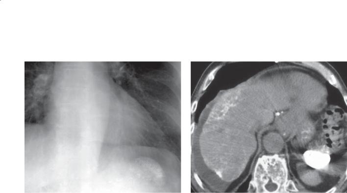
CASE 9.26. Abdominal radiograph. The spleen is abnormally dense, and there is a suggestion of hyperdense nodules in the upper abdomen.
CASE 9.27. Unenhanced CT. The spleen is hyperattenuating. There are hyperattenuating lymph nodes in the gastrohepatic ligament. The liver is of heterogeneous attenuation.
Di erential Diagnosis
1.Th orium dioxide exposure
2.Autosplenectomy from sickle cell disease
Diagnosis
Th orium dioxide exposure
9. SPLEEN 779
CASE 9.27
Discussion
Both of these patients had prior thorium dioxide administration. Thorium dioxide was previously used as an intravascular contrast agent. Thorium 232 is an alpha emitter with a very long half-life which accumulates within the reticuloendothelial system. Uptake of the agent occurs in the spleen, liver, and lymph nodes. Splenic changes include fibrosis and atrophy. The radiation associated with this agent predisposes patients to angiosarcoma, hepatocellular carcinoma, and cholangiocarcinomas.
At CT, the most common finding is hyperattenuating spleen and liver. Filling defects or heterogeneous attenuation within the liver or spleen should prompt investigation for a complicating neoplasm. Patients with sickle cell disease would not be expected to have changes in the liver or lymph nodes.
Disease type: Miscellaneous
780 MAYO CLINIC GASTROINTESTINAL IMAGING REVIEW
CASE 9.28
A B
Disease type: Miscellaneous
9. SPLEEN 781
C
Findings
Contrast-enhanced CT. A through C. The spleen is not present in the left upper quadrant, but it is visible in the pelvis. The mesenteric vasculature has a whorled appearance.
Di erential Diagnosis
Wandering spleen
Diagnosis
Wandering spleen
Discussion
A wandering spleen is the result of splenic ligamentous laxity, likely due to abnormal embryologic fusion of
the posterior mesogastrium or to lack of formation of the normal splenorenal and gastrosplenic ligaments. Because of its hypermobility, the spleen can move
to abnormal locations within the peritoneal cavity. Usually a mass is discovered in an otherwise asymptomatic patient. Occasionally, vascular torsion of the splenic pedicle can result in splenic congestion or infarction. Torsion can be chronic and intermittent.
Radiographic findings at CT show an abnormal location or change in splenic shape. Patients with infarction have the typical features of an infarct(s) with a portion of or the entire spleen hypoperfused. Treatment in symptomatic patients usually is operative splenopexy. Splenectomy is reserved only for patients with infarction.
Disease type: Miscellaneous
782 MAYO CLINIC GASTROINTESTINAL IMAGING REVIEW
CASE 9.29
Findings
Contrast-enhanced CT. The spleen is shrunken and calcified.
Di erential Diagnosis
1.Autosplenectomy from sickle cell disease
2.Th orium dioxide exposure
Diagnosis
Autosplenectomy from sickle cell disease
Discussion
Sickle cell anemia (disease) results from homozygous inheritance of the hemoglobin 5-producing gene and affects 1 in 500 African Americans. Sickle cell trait (heterozygous form) is found in 1 in 10 African
Americans. Abnormal hemoglobin 5 can result in rigid,
crescentic, or sickle-shaped erythrocytes. This can lead to hemolysis and vascular occlusion(s). In patients with homozygous sickle cell disease, splenic infarctions can lead to loss of function, gradual decrease in size, and ultimately fibrosis and calcification (autosplenectomy). Loss of splenic function results in an increased susceptibility to aggressive infections.
Autosplenectomy in sickle cell disease appears as a small, densely calcified spleen on CT or plain radiography. The calcified spleen may be as small as 1 cm. Technetium Tc 99m liver-spleen scans show no splenic uptake. Thorium dioxide exposure can
result in a similar appearance of the spleen. However, liver and lymph node calcification also is often present with thorium dioxide exposure. Clinical history usually easily differentiates these two disease processes.
Disease type: Miscellaneous

|
|
9. SPLEEN 783 |
|
|
|
|
TABLE 9.3 |
|
Miscellaneous Splenic Conditions |
CASE |
|
|
|
|
Subcapsular hematoma |
Crescent-shaped, nonenhancing fluid collection in a |
9.22 |
|
subcapsular location |
|
|
|
|
Shattered spleen |
Multiple fracture lines traverse spleen. Associated perisplenic |
9.23 |
|
stranding and fluid |
|
|
|
|
Splenosis |
Solitary or multiple nodules, usually left upper quadrant, variable |
9.24 |
|
size. Enhancement mimics spleen |
|
|
|
|
Hemochromatosis |
Primary, may not affect spleen. Secondary, spleen always affected |
9.25 |
|
Secondary, low MRI signal diffusely in spleen, marrow, and |
|
|
liver. Pancreas usually spared |
|
|
|
|
Thorium dioxide exposure |
Dense, atrophic spleen, regional high-density lymph nodes. Liver |
9.26 and 9.27 |
|
also usually affected |
|
|
|
|
Wandering spleen |
Soft tissue mass with enhancement similar to spleen. Spleen not |
9.28 |
|
present in usual fossa. Can undergo torsion |
|
|
|
|
Autosplenectomy |
Dense, atrophic spleen. History of sickle cell disease |
9.29 |
|
|
|
Disease type: Miscellaneous

784 MAYO CLINIC GASTROINTESTINAL IMAGING REVIEW
Diff erential Diagnoses
Solid Masses
BENIGN
Hemangioma
Hamartoma
Inflammatory pseudotumor Splenic infarct
MALIGNANT
Lymphoma
Metastasis
Angiosarcoma
Cystic Masses
True cyst (epidermoid cyst) False cyst (posttraumatic cyst) Echinococcal cyst
Bacterial abscess Cystic metastasis
TABLE 9.4
Multiple Small (<1 cm) Lesions
Fungal microabscesses (Candida, histoplasmosis) Multiple bacterial abscesses
Lymphoma
Sarcoidosis Gamna-Gandy bodies Metastases
Pseudotumors (early phase of contrast enhancement)
9. SPLEEN 785
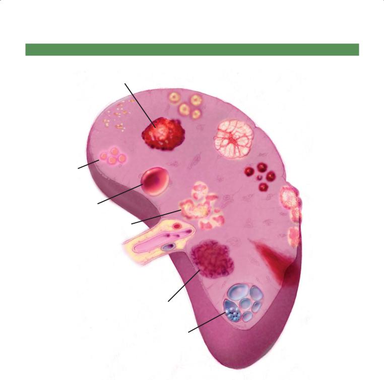
SPLENIC MASSES
Hemangioma Candidiasis
Healed granulomatous
infection
Hamartoma
Siderotic nodules
Sarcoidosis
Cyst
Metastases
Lymphoma
D.F.
© MAYO 2013
Infarct
Inflammatory pseudotumor
Echinococcal
cyst
Figure 9.30 (Used with permission of Mayo Foundation for Medical Education and Research.)
786 MAYO CLINIC GASTROINTESTINAL IMAGING REVIEW
QUESTIONS
Multiple Choice (choose the best answer)
9.1.Which of the following is the most likely diagnosis for the findings in the figure?
a.Hemangioma
b.Epidermoid cyst
c.Laceration
d.Infarction
e.Lymphoma
9. SPLEEN 787
9.2.In this case of an immunocomprised patient, which of the following is most often associated with the condition shown in the figure?
a.Fever
b.Intestinal obstruction
c.Peritonitis
d.Increased level of carcinoembryonic antigen
e.Increased level of α-fetoprotein
9.3.For the case shown in Question 9.2, how can the diagnosis best be confirmed?
a.Operation
b.Urine culture
c.Ultrasound-guided biopsy
d.Nasal swab
e.MRI
788 MAYO CLINIC GASTROINTESTINAL IMAGING REVIEW
A B
C
D
Match images A through F with the responses listed below.
9.4. |
______ Atrial fibrillation |
9.6. |
______ Myelofibrosis |
9.5. |
______ Alcoholism |
9.7. |
______ Nodular enhancement |
9. SPLEEN 789
E
F
790 MAYO CLINIC GASTROINTESTINAL IMAGING REVIEW
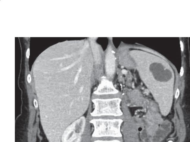
9.8.Which of the following is the most likely diagnosis for the findings in the figure?
a.Candidiasis
b.Echinococcal disease
c.Sarcoidosis
d.Metastases
e.Infl ammatory pseudotumor

9. SPLEEN 791
ANSWERS
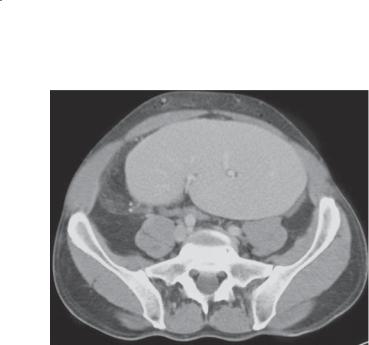
9.1.Answer c.
Th e spleen has several linear low-attenuation changes within it, and there is free intraperitoneal fluid. This patient had had abdominal trauma. The findings are consistent with a splenic laceration and hemoperitoneum.
Because the changes in the spleen do not look masslike, hemangioma, epidermoid cyst, and lymphoma can be excluded. Infarction usually presents with a wedgeshaped low-attenuation region, without ascites. The history in this case is very helpful for making the diagnosis.
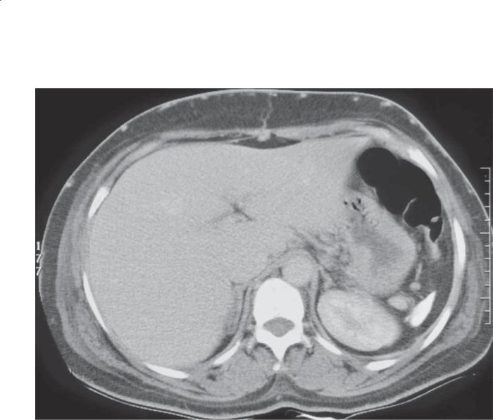
9.2.Answer a.
Th is patient has multiple filling defects within the liver and spleen. In an immunocomprised host, candidiasis is the most likely diagnosis.
Intestinal obstruction and peritonitis are not associated with Candida infection.
Metastases from some tumors could be associated with an increased level of carcinoembryonic antigen. There is no clue of a primary tumor on this image, and the question indicates that the patient is immunocompromised.
Multicentric hepatocellular carcinoma is associated with an increased level of α-fetoprotein. There is no evidence of cirrhosis or portal venous hypertentsion on this image.
9.3.Answer c.
Percutaneous ultrasound-guided biopsy of one of these lesions with examination for hyphae is the best way to confirm the diagnosis, unless the patient has had positive results for Candida infection on blood cultures.
Operation is not necessary or curative.
Urine culture results may be negative because the abscess cavities are located in the liver and spleen.
Nasal swab results could be negative. The patient could have concurrent oral thrush, but this finding would not confirm that the changes in the liver or spleen are due to Candida infection.
MRI shows changes similar to those on CT and is not diagnostic.
9.4.Answer b.
In figure b, a wedge-shaped area of low attenuation in the spleen is characteristic of an infarct. Infarct can have several causes, including atrial fibrillation.
9.5.Answer a.
Alcoholism leads to cirrhosis and portal hypertension. These conditions can lead to Gamna-Gandy bodies in the spleen (as in figure a) due to local hemorrhage. The hemosiderin in the healed areas of hemorrhage are of low signal intensity at MRI.
9.6.Answer d.
Splenomegaly develops in patients with myelofibrosis as a result of extramedullary hematopoiesis.
9.7.Answer e.
In figure e, multiple hyperechoic masses are present in the spleen as a result of multiple hemangiomas. Images of these tumors can be similar to those in the liver, and enhancement characteristics at CT may also be similar.
Figure c shows a splenic hamartoma. There is a large, focal, heterogeneous intensity mass within the spleen. The imaging characteristics are not specific.
Figure f shows hemosiderosis. The spleen, liver, and
marrow on this T2-weighted MRI are of low signal intensity as a result of excessive iron deposition. This finding is most common in patients requiring multiple transfusions. No end-organ damage occurs in hemosiderosis. Differentiation from primary hemochromatosis is by identification of low signal in the pancreas and liver—usually with normal signal in the spleen and marrow.
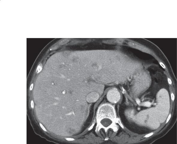
9.8.Answer d.
Multiple hypoattenuating masses are present within the liver and spleen. The most likely diagnosis is metastases or lymphoma. Candidiasis presents with smaller lesions, usually in an immunocompromised host. Echinococcal disease presents with multiple cysts and daughter cysts and the patient has a travel history to an endemic area. Sarcoidosis could present with these findings; usually, adenopathy and other findings on chest radiography are present. Inflammatory pseudotumors are rare; they often present as a solitary, heterogeneously enhancing mass.
This page intentionally left blank