

C H A P T E R 7
BILE DUCTS AND GALLBLADDER
BILE DUCTS
Cases 7.1–7.13 Inflammatory Diseases
Cases 7.14–7.37 Masses and Filling Defects
Benign Tumors
Malignant Tumors
Cases 7.38–7.45 |
Miscellaneous |
GALLBLADDER
Cases 7.46–7.64 Inflammatory Diseases
Cases 7.65–7.77 Masses and Filling Defects
Benign Tumors
Malignant Tumors
Cases 7.78–7.81 |
Miscellaneous |

This page intentionally left blank
7. BILE DUCTS AND GALLBLADDER 563
CASE 7.1
Findings
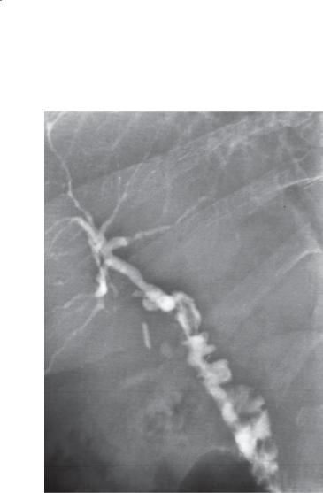
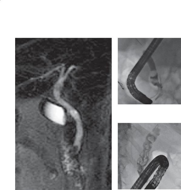
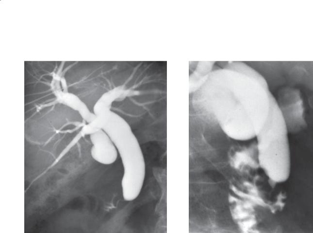
ERCP. The intrahepatic and extrahepatic bile ducts are irregularly narrowed. Bandlike strictures affect the extrahepatic duct. Alternating areas of normal and narrowed duct affect the intrahepatic bile ducts.
Di erential Diagnosis
Primary sclerosing cholangitis
Diagnosis
Primary sclerosing cholangitis
Discussion
Primary sclerosing cholangitis (PSC) is a cholestatic disease of unknown origin most often affecting young (<45 years) males who have ulcerative colitis. The natural history of PSC is one of chronic progression; the median duration of survival is approximately 12 years from diagnosis without a transplant. Pathologically, fibrosing inflammation is the key finding. The disease
usually follows a progression from initial cholangitis to periportal hepatitis, to septal fibrosis, to bridging necrosis, and finally to cirrhosis. The disease most commonly affects both the intrahepatic and the extrahepatic ducts; however, intrahepatic involvement alone is visible at cholangiography in about 20% of patients. Direct gallbladder involvement is present in about 15% of patients.
Cholangiographic features of PSC include the following:
1.Band strictures (as in this case) leading to a beaded appearance
2.Nonuniform, segmental strictures 1 to 2 cm long
3.Pruned-tree appearance of intrahepatic ducts (due to obliteration of ducts)
4.Diverticular outpouchings
5.Mural irregularity—this can vary from fine to shaggy contour changes
6.Combinations of the above
Bile Ducts: Inflammatory Diseases
564 MAYO CLINIC GASTROINTESTINAL IMAGING REVIEW
CASE 7.2
A B
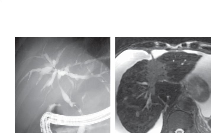
Findings
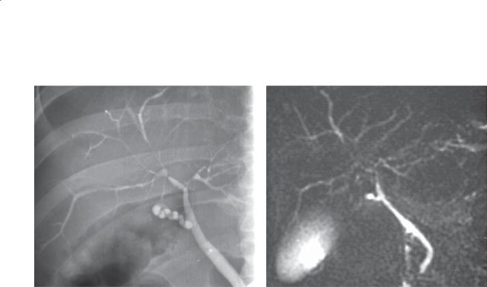
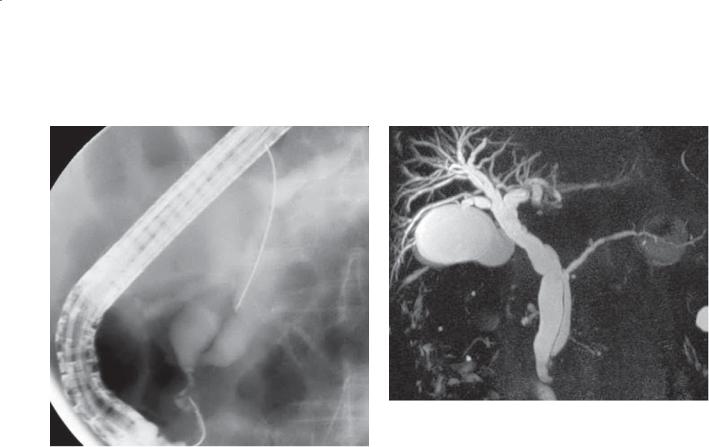
A. ERCP. B. MRCP. Diffuse long strictures are present throughout the intrahepatic ducts. The extrahepatic ductal system is normal in appearance.
Di erential Diagnosis
Primary sclerosing cholangitis
Diagnosis
Primary sclerosing cholangitis
Discussion
Th is case illustrates long strictures that occasionally occur in primary sclerosing cholangitis (PSC). The case also shows the normal appearance of the extrahepatic ducts, a finding in up to 20% of patients with PSC. Even with a normal radiographic appearance of the extrahepatic ducts, the vast majority of patients with PSC have histologic changes of the disease in the extrahepatic ducts.
Bile Ducts: Inflammatory Diseases
CASE 7.3
A
Findings
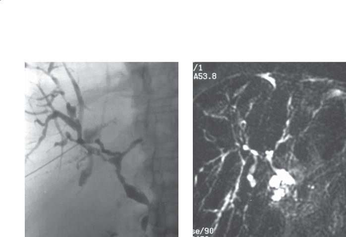
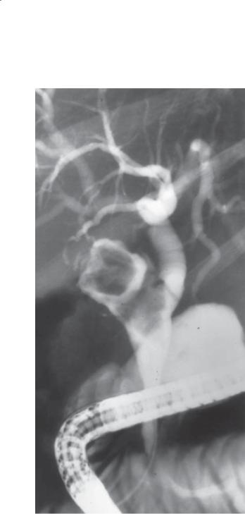
A.Percutaneous cholangiogram. Multiple strictures of varying length involve both the intrahepatic and the extrahepatic biliary system. The left intrahepatic ductal system is not opacified.
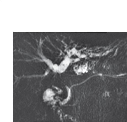
B.MRCP. Multiple biliary strictures correlate with those seen on the cholangiogram. The left intrahepatic ducts are visible and have an appearance similar to that of the right ducts. There is a high-grade stricture of the proximal left ductal system near the bifurcation of the common hepatic duct.
Di erential Diagnosis
Primary sclerosing cholangitis
Diagnosis
Primary sclerosing cholangitis
7. BILE DUCTS AND GALLBLADDER 565
B
Discussion
Th is case is a good example of random segmental strictures (1-2 cm in length) commonly seen in primary sclerosing cholangitis. In this case, nonfilling of the left ductal system is present on the percutaneous cholangiogram. Because MRCP does not depend
on opacification of the bile ducts, the ductal system beyond an obstruction can be visualized. The dominant stricture, in this case in the main left bile duct, should raise concern that a complicating cholangiocarcinoma may be present.
Bile Ducts: Inflammatory Diseases
566 MAYO CLINIC GASTROINTESTINAL IMAGING REVIEW
CASE 7.4
A B
Findings
A.ERCP. The intrahepatic and extrahepatic bile ducts are diffusely abnormal and have regions of narrowing and mural irregularity.
B.MRCP. Changes are similar to those on the ERCP; however, multiple intrahepatic ducts are better visualized.
Di erential Diagnosis
1.Primary sclerosing cholangitis
2.Ascending cholangitis
Diagnosis
Primary sclerosing cholangitis
Discussion
Th is case illustrates the mural irregularity that often occurs in primary sclerosing cholangitis. Although the same changes can be seen on both ERCP and MRCP, the spatial resolution at MRI is inferior to that at ERCP. Fine changes of mural irregularity could be overlooked at MRCP. Therefore, if clinical suspicion of the disease is high, ERCP is indicated despite a normal MRCP. MRCP is superior to ERCP for identifying bile ducts that are isolated from the main ductal system. These dilated, obstructed ducts may be a source of infection. MRCP can be a helpful study before a biliary drainage procedure to evaluate for an isolated duct(s). ERCP allows biopsy and brushings of dominant strictures to assess for a complicating cholangiocarcinoma.
Bile Ducts: Inflammatory Diseases
CASE 7.5
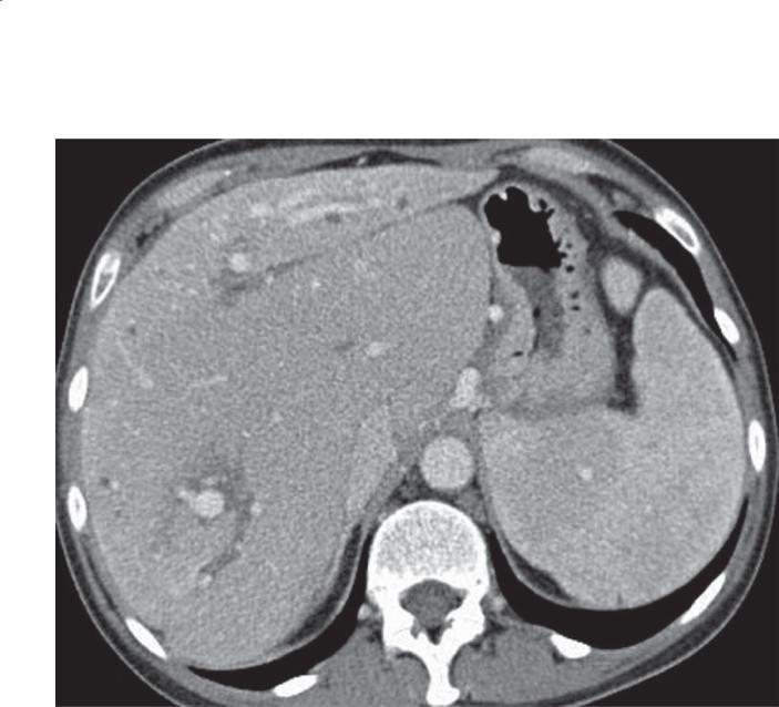
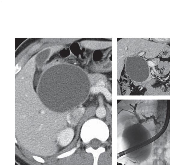
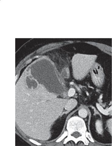
Findings
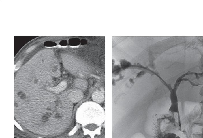
Contrast-enhanced CT. The caudate lobe is massively enlarged, and the left lobe relatively small. There is segmental bile duct dilatation. Splenomegaly is present.
Di erential Diagnosis
1.Cirrhosis, cause uncertain
2.Primary sclerosing cholangitis, with secondary cirrhosis
Diagnosis
Primary sclerosing cholangitis, with secondary cirrhosis
7. BILE DUCTS AND GALLBLADDER 567
Discussion
Changes of primary sclerosing cholangitis can be detected at CT and MRI with the identification of segmental bile duct dilatation and strictures.
Occasionally, uniform ductal wall thickening can be visualized. Chronic biliary obstruction can lead to cirrhosis. The segmental nature of the obstruction can lead to bizarre lobar changes in the liver; some segments are markedly enlarged (the caudate lobe in
this case), and other lobes are small as a result of biliary obstruction (the left lobe in this case).
Bile Ducts: Inflammatory Diseases
568 MAYO CLINIC GASTROINTESTINAL IMAGING REVIEW
CASE 7.6
A B
Findings
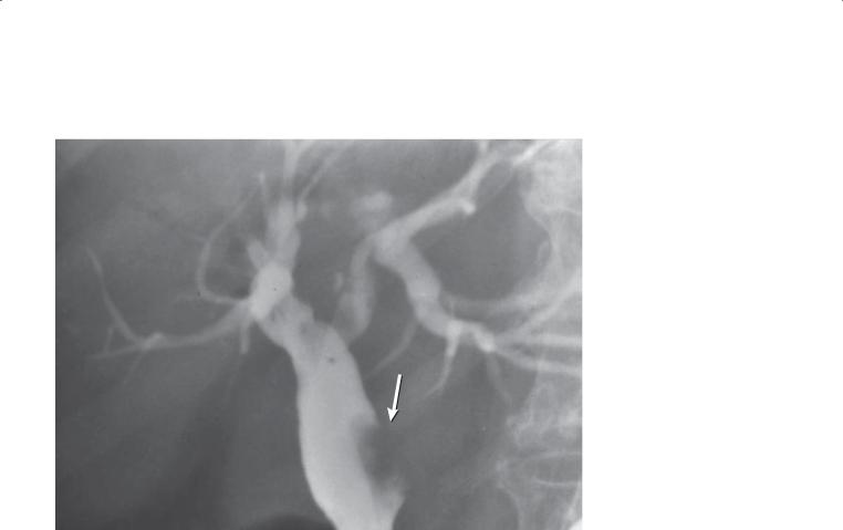
A.ERCP. The bile ducts are diffusely irregular and have areas of stricture and dilatation. A dominant stricture involves the proximal common hepatic duct.
B.T2-weighted MRI. A large, hyperintense mass arises at the junction of the medial and lateral hepatic segments and extends into the porta hepatis.
Di erential Diagnosis
Cholangiocarcinoma complicating primary sclerosing cholangitis
Diagnosis
Cholangiocarcinoma complicating primary sclerosing cholangitis
Discussion
Cholangiocarcinoma is a dreaded complication of primary sclerosing cholangitis (PSC). Because of the diffuse and unpredictable ductal changes in patients with PSC, distinguishing benign strictures from cholangiocarcinoma often is difficult. Dominant strictures, those strictures that are most severe
and critical in their location, should be considered suspicious for cholangiocarcinoma. Comparison of the cholangiogram with prior studies, ERCP-guided brushings for histologic analysis, and MRI can be helpful to assess the nature of a new stricture.
Bile Ducts: Inflammatory Diseases
7. BILE DUCTS AND GALLBLADDER 569
CASE 7.7 |
CASE 7.8 |
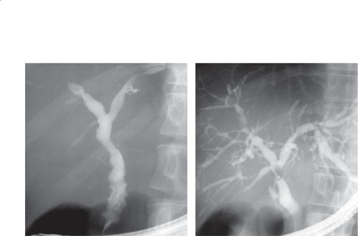
Findings
CASE 7.7. T-tube cholangiogram. The intrahepatic bile ducts appear crowded and pruned, and there is some tortuosity and deformity of the peripheral ducts.
CASE 7.8. Unenhanced CT. The liver has a diffusely nodular appearance without bile duct dilatation.
Di erential Diagnosis
1.Hepatic cirrhosis
2.Small-duct primary sclerosing cholangitis
3.Primary biliary cirrhosis
Diagnosis
Primary biliary cirrhosis
Discussion
Primary biliary cirrhosis (PBC) is a cholestatic disease of unknown origin. T-cell–mediated immune response plays an important role in bile duct injury. Other autoimmune diseases often are associated
(rheumatoid arthritis, Sjögren syndrome, Hashimoto thyroiditis, and others). Serologic markers, including antimitochondrial antibody, often are present. The disease predominantly affects females. Small duct destruction with an inflammatory cellular infiltrate is characteristic of PBC histologically.
Early in the disease, cholangiograms are normal. Later, the cholangiographic features are those of cirrhosis with crowding, tortuosity, and deformity of the bile ducts (as in this case). Enlarged benign reactive lymph nodes in the hepatic portal are common and can cause mass effect on the extrahepatic ducts. Although primary sclerosing cholangitis (PSC) affecting the large ducts (cases 7.1 through 7.4) has many features that are different from those of PBC, small-duct PSC may be indistinguishable. Further, other causes of hepatic cirrhosis also may result in a similar appearance. Correlation of the imaging studies with the clinical and laboratory findings is critical to a correct diagnosis. In many cases, a liver biopsy is necessary.
Bile Ducts: Inflammatory Diseases
570 MAYO CLINIC GASTROINTESTINAL IMAGING REVIEW
CASE 7.9
A B
Findings
ERCP. A and B. The extrahepatic bile duct is mildly dilated, and the duct is diffusely irregular. The intrahepatic ducts are mildly dilated and have multiple segmental strictures. Endoscopic balloon in the common bile duct (B).
Di erential Diagnosis
1.Primary sclerosing cholangitis
2.Ascending cholangitis
3.Acquired immunodeficiency syndrome cholangiopathy
Diagnosis
Acquired immunodeficiency syndrome cholangiopathy
Discussion
AIDS is due to the human immunodeficiency virus, which destroys CD4 (T helper-inducer) lymphocytes. Affected persons have progressive loss of cellular immunity and become susceptible to opportunistic infection and tumors. Infectious organisms found
in the gastrointestinal tract which can be clinically
important in these patients include Candida albicans, cytomegalovirus, herpes simplex virus,
Cryptosporidium, Mycobacterium tuberculosis, and Mycobacterium avium-intracellulare. Common tumors in these patients include Kaposi sarcoma and nonHodgkin lymphoma.
Biliary tract abnormalities in patients with AIDS are related to infection with either Cryptosporidium or cytomegalovirus. Patients present with right upper quadrant abdominal pain, fever, leukocytosis, and abnormal liver function tests. The characteristic cholangiographic features of AIDS cholangiopathy include segmental strictures of the biliary system which are identical to those in patients with primary sclerosing cholangitis. In addition, some patients have papillary stenosis alone or in combination with the bile duct strictures. The combination of intrahepatic ductal strictures and papillary stenosis is unique to AIDS-related cholangitis. Acalculous cholecystitis also has been reported. Treatment of this condition is based on eradication of infectious agents. Endoscopic papillotomy can provide relief of symptoms and improve results of liver function tests.
Bile Ducts: Inflammatory Diseases
CASE 7.10
A
Findings
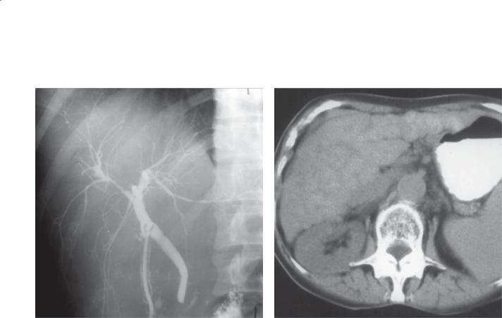
A.Contrast-enhanced CT. Multiple water-attenuation masses within the liver appear to communicate with the biliary system.
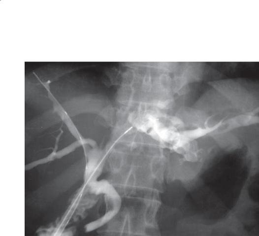
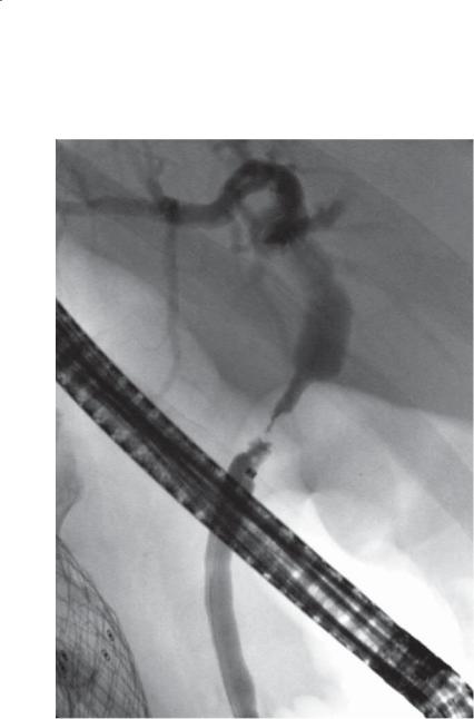
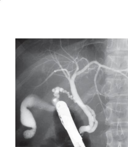
B.Transhepatic cholangiogram. Multiple saccular cavities communicate with the right hepatic biliary system. The right-sided bile ducts are mildly dilated and irregular in contour. A stricture within the right hepatic duct is bridged by a transhepatic biliary stent. There is a freely functioning choledochojejunostomy.
Di erential Diagnosis
1.Ascending cholangitis with communicating hepatic abscess
2.Caroli disease
Diagnosis
Ascending cholangitis with communicating hepatic abscess
7. BILE DUCTS AND GALLBLADDER 571
B
Discussion
Ascending cholangitis is usually due to obstruction of the biliary tree with bacterial infection of the bile. Increased pressure within the biliary tree can drive bacteria into the bile canniliculi, hepatic veins, and lymphatics, a process leading to a pyogenic hepatic abscess. Abscess cavities can communicate with the
biliary system (as in this case). Bacteremia can occur in 25% to 40% of patients, and it may be fatal if untreated. Transhepatic cholangiography was performed in this case because of the choledochojejunostomy and the likely difficulty of obtaining biliary access at ERCP.
Bile Ducts: Inflammatory Diseases
572 MAYO CLINIC GASTROINTESTINAL IMAGING REVIEW
CASE 7.11
A B
C
Bile Ducts: Inflammatory Diseases

Findings
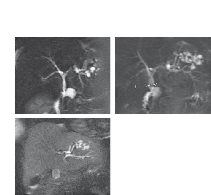
MRCP. A through C. The extrahepatic bile duct is mildly dilated to the level of a choledochoenterostomy. Several saccular fluid collections communicating with the biliary system are present in the lateral segment of the left hepatic lobe. Small filling defects are present within these fluid collections.
Di erential Diagnosis
1.Caroli disease
2.Liver abscesses
Diagnosis
Liver abscesses due to bacterial cholangitis
7. BILE DUCTS AND GALLBLADDER 573
Discussion
Abscesses can develop in the liver as a result of bacterial cholangitis, usually due to bile stasis or obstruction. Patients usually present with signs and symptoms of an infection and jaundice. Escherichia coli is the most common organism. The abscesses typically communicate with the biliary system. Treatment is focused on relieving the obstruction and administering antibiotics. Chronic cholangitis can result in bile duct strictures, attenuation of the peripheral bile ducts at cholangiography, and biliary cirrhosis.
Bile Ducts: Inflammatory Diseases
574 MAYO CLINIC GASTROINTESTINAL IMAGING REVIEW
CASE 7.12
A B
C
Bile Ducts: Inflammatory Diseases
7. BILE DUCTS AND GALLBLADDER 575
CASE 7.13
Findings
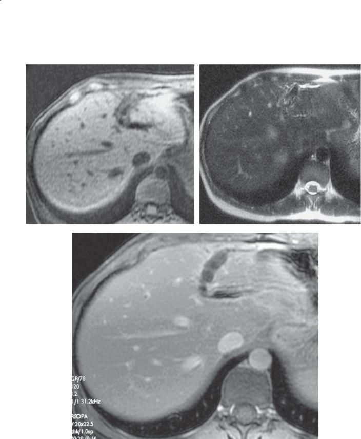
CASE 7.12. A. T1-weighted MRI. There are 2 low-in- tensity tubular structures in the left lobe of the liver. B. T2-weighted MRI. Low-intensity T2 structures appear to be present in 2 dilated left lobe bile ducts. C. Gadolinium-enhanced MRI. Very low-intensity stones are visible within the dilated bile ducts.
CASE 7.13. Percutaneous cholangiogram. The intrahepatic bile ducts within the left lobe of the liver are dilated and contain several filling defects. There is a stricture of the distal left hepatic ducts. (Adapted from MacCarty RL. Inflammatory disorders of the biliary tract. In: Gore RM, Levine MS, editors. Textbook of gastrointestinal radiology. Vol 2. 2nd ed. Philadelphia [PA]: WB Saunders Company; c2000. p. 1375-94. Used with permission.)
Di erential Diagnosis
1.Cholangiocarcinoma
2.Oriental cholangiohepatitis
3.Benign biliary stricture—origin unknown
Diagnosis
Oriental cholangiohepatitis
Discussion
Oriental cholangiohepatitis is most common in Southeast Asia. The cause of the disease is not known, but associations with clonorchiasis, ascariasis,
and nutritional deficiency have been suggested. Dilatation of intrahepatic and extrahepatic ducts containing pigmented stones and infected fluid is seen pathologically. The infection results in biliary strictures. Patients usually present with recurrent attacks of abdominal pain, fever, and jaundice. Localized intrahepatic segmental ductal stenosis with upstream dilatation may occur, especially in the
lateral segment of the left lobe or posterior segment of the right hepatic lobe. Sonographic and CT findings include intrahepatic or extrahepatic duct stones, dilatation of the intrahepatic or extrahepatic ducts, increased periportal echogenicity, segmental lobar atrophy, and gallstones. Cholangiographic findings include bile duct stones, ductal dilatation and focal strictures, acute peripheral tapering, and decreased arborization of the intrahepatic bile ducts.
Bile Ducts: Inflammatory Diseases
576 MAYO CLINIC GASTROINTESTINAL IMAGING REVIEW
TABLE 7.1
Inflammatory Diseases of the Bile Ducts |
|
CASE |
|
|
|
Primary sclerosing cholangitis (PSC) |
Band strictures (beaded appearance), segmental |
7.1–7.6 |
|
strictures, pruned-tree appearance, mural |
|
|
irregularity, and diverticular outpouchings. |
|
|
Increased risk of cholangiocarcinoma |
|
|
|
|
Primary biliary cirrhosis |
Autoimmune cholestatic disease involving |
7.7 and 7.8 |
|
small bile ducts. Cirrhosis results in crowding |
|
|
and deformity of bile ducts |
|
|
|
|
Acquired immunodeficiency syndrome |
Indistinguishable from PSC. PSC changes plus |
7.9 |
cholangiopathy |
papillary stenosis highly suspicious |
|
|
|
|
Bacterial cholangitis |
Ductal irregularity, strictures, small abscess |
7.10 and 7.11 |
|
cavities that communicate with bile ducts |
|
|
|
|
Oriental cholangiohepatitis |
Focal strictures, intraductal stones, intrahepatic |
7.12 and 7.13 |
|
and extrahepatic duct dilatation, straight rigid |
|
|
intrahepatic ducts |
|
|
|
|
Bile Ducts: Inflammatory Diseases
7. BILE DUCTS AND GALLBLADDER 577
CASE 7.14 |
CASE 7.15 |
CASE 7.16
Findings
CASE 7.14. MRCP. The intrahepatic and extrahepatic bile ducts are dilated to the level of the distal common bile duct. There is a filling defect within the distal common bile duct.
CASE 7.15. ERCP. Two filling defects are visible within the dilated common bile duct.
CASE 7.16. ERCP. There are multiple common bile duct filling defects.
Di erential Diagnosis
Choledocholithiasis
Diagnosis
Choledocholithiasis
Discussion
Choledocholithiasis can develop de novo within the bile ducts or as calculi that have passed through the cystic duct from the gallbladder. Choledocholithiasis is found in 12% to 15% of patients undergoing cholecystectomy, but this rate increases with age, duct size, and chronicity of symptoms. Typical symptoms include biliary colic, jaundice, cholangitis, and pancreatitis. The correct imaging test to begin the evaluation depends on the clinical suspicion of choledocholithiasis. If the suspicion is high, ERCP usually is indicated so the calculus can be removed at the same time. MRCP is an excellent noninvasive
approach for imaging the bile ducts and is indicated in patients less likely to require intervention.
Bile Ducts: Masses and Filling Defects
578 MAYO CLINIC GASTROINTESTINAL IMAGING REVIEW
CASE 7.17 |
CASE 7.18 |
A |
|
B
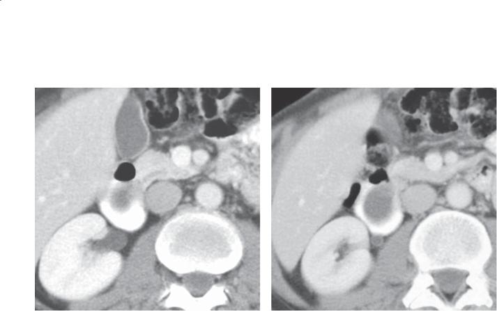
Findings
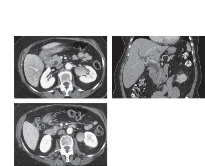
CASE 7.17. Contrast-enhanced CT. A. The intrapancreatic portion of the common bile duct is dilated. B. The distal common bile duct contains a filling defect. A rim of fluid attenuation is seen around the filling defect.
CASE 7.18. Coronal contrast-enhanced CT. Intrahepatic and extrahepatic bile ducts are dilated. A filling defect is visible in the distal common bile duct.
Di erential Diagnosis
Choledocholithiasis
Diagnosis
Choledocholithiasis
Discussion
Th e composition of bile duct calculi can vary in the percentage of calcium bilirubinate, cholesterol, and pigment. Stones containing calcium are the easiest to recognize because of their high attenuation. Pure cholesterol stones are of low attenuation and very difficult to detect at CT. Most calculi are of mixed composition, and the vast majority can be recognized at CT, especially with proper CT technique. Two helpful findings have been described to represent the appearance of a calculus within the bile duct imaged in cross-section. The commonest finding is referred to as the target sign with a ring of water attenuation
around the stone (as in case 7.17B). The crescent sign is a variant of the target sign with only a crescent of water attenuation surrounding the calculus. Coronal images (case 7.18) can also be very helpful.
Bile Ducts: Masses and Filling Defects
CASE 7.19
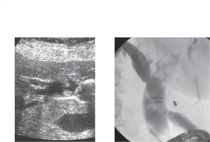
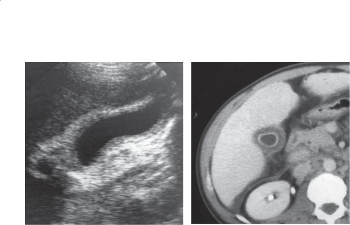
Findings
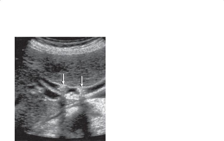
Sonogram. Longitudinal view of the common bile duct has 2 echogenic structures (arrows) in a dilated duct which have posterior acoustic shadowing.
Di erential Diagnosis
Choledocholithiasis
Diagnosis
Choledocholithiasis
7. BILE DUCTS AND GALLBLADDER 579
Discussion
Because ultrasonography is noninvasive, inexpensive, and readily available, it is usually the test of choice when patients present with biliary-related symptoms. The best sensitivities reported for detection of common bile duct stones by sonography are approximately 75%. A nondilated duct, nonshadowing stones, bowel gas, arterial calcifications, and cholecystectomy clips often make this diagnosis difficult. The dilated bile duct, posterior shadowing of the stones, and location of the stones in the mid common bile duct make the diagnosis easy in this case. When clinical suspicion of choledocholithiasis is high but no stones are seen with sonography, MRCP or ERCP is often recommended for further evaluation.
Bile Ducts: Masses and Filling Defects
580 MAYO CLINIC GASTROINTESTINAL IMAGING REVIEW
CASE 7.20
A B
Findings
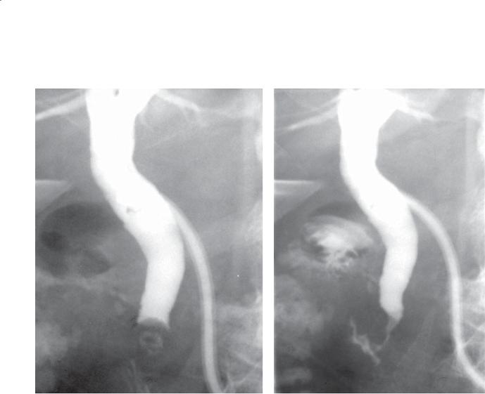
T-tube cholangiogram. A. A meniscus is formed from a filling defect in the distal common bile duct. B. The filling defect is no longer seen.
Di erential Diagnosis
1.Passage of a common bile duct stone
2.Ampullary carcinoma
3.Pseudocalculus
Diagnosis
Pseudocalculus
Discussion
A pseudocalculus is an apparent filling defect within the ampullary segment of the common bile duct due to spasm of the sphincter of Oddi. With relaxation of the sphincter (which can be assisted with the use of intravenous glucagon or by waiting 10–15 minutes), the filling defect disappears and contrast material flows into the duodenum normally. A key finding distinguishing a real stone from a pseudocalculus is the presence of contrast outlining the inferior aspect of the filling defect of a true calculus. In this case, only the superior meniscus is present. It is important to be aware of this entity so that an interventional procedure is not performed needlessly.
Bile Ducts: Masses and Filling Defects
CASE 7.21
A
Findings
Transhepatic cholangiogram. A and B. There is moderate dilatation of the intrahepatic and
extrahepatic bile ducts to the level of the papilla. There is a short segment of narrowing at the level of the papilla.
Di erential Diagnosis
1.Spasm of the sphincter of Oddi
2.Papillary stenosis
3.Postcholecystectomy syndrome
Diagnosis
Papillary stenosis
7. BILE DUCTS AND GALLBLADDER 581
B
Discussion
Papillary stenosis is an anatomical abnormality of the sphincter of Oddi with partial or complete
narrowing of this segment of the duct. The finding is due to inflammation or fibrosis, usually caused by pancreatitis, choledocholithiasis, or surgical trauma. Papillary stenosis also is found in patients with acquired immunodeficiency syndrome-related cholangiopathy. Treatment usually is papillotomy performed at ERCP.
Spasm of the sphincter of Oddi (sphincter of Oddi dyskinesia) is a functional abnormality of the sphincter, which does not relax normally (case 7.20). The sphincter normally has alternating low and high muscular tone several times per minute. Patients with this disorder have increased baseline pressures of the sphincter or an increase in the amplitude
and frequency of the phasic contractions. There is also a paradoxic increase in muscular tone to cholecystokinin. This diagnosis is usually made at
manometry. Postcholecystectomy syndrome refers to spasm of the sphincter of Oddi in patients who have had a cholecystectomy and biliary-type pain.
Bile Ducts: Masses and Filling Defects
582 MAYO CLINIC GASTROINTESTINAL IMAGING REVIEW
CASE 7.22
A B
Findings
A.MRCP. Both the biliary and the pancreatic ducts are dilated to the level of the ampulla. There is focal narrowing of the ampullary portion of the common bile duct.
B.ERCP. The common bile duct and pancreatic duct are dilated. Focal narrowing is present in the ampullary region.
Di erential Diagnosis
1.Spasm of the sphincter of Oddi
2.Papillary stenosis
3.Pseudocalculus
4.Ampullary carcinoma
Diagnosis
Ampullary carcinoma
Discussion
Fixed narrowing of the ampulla is confirmed with the finding of upstream dilatation of both the common bile and the pancreatic ducts. The appearance is unchanged between MRCP and ERCP. Both pseudocalculous and spasm of the sphincter of Oddi would be expected
to vary in appearance and not be associated with significant upstream dilatation. No meniscus is seen, and thus the diagnosis of pseudocalculous is less likely. Papillary stenosis is a fixed area of narrowing that can cause obstruction. ERCP with biopsy and brushings would be needed to distinguish papillary stenosis from ampullary carcinoma. Patients with familial polyposis syndrome (as in this patient) and hereditary nonpolyposis syndrome have a 100to 200-fold increase in the incidence of ampullary carcinoma. Surveillance endoscopy is recommended in these patients. Patients who have familial polyposis syndrome also have an increased risk for development of premalignant adenomatous duodenal polyps.
Bile Ducts: Masses and Filling Defects
7. BILE DUCTS AND GALLBLADDER 583
CASE 7.23
Findings |
Discussion |
ERCP. There is a large filling defect within the cystic |
Mirizzi syndrome is an inflammatory stricture of |
duct and secondary narrowing of the common |
the common hepatic or common bile duct due to |
hepatic duct. |
an impacted stone in the cystic duct or neck of the |
|
gallbladder. The biliary stricture can lead to stasis, |
Di erential Diagnosis |
obstruction, cholangitis, and jaundice. In chronic |
Mirizzi syndrome |
cases, secondary biliary cirrhosis can develop. ERCP is |
|
the best procedure for making this diagnosis, although |
Diagnosis |
findings at CT and sonography also can be diagnostic. |
Mirizzi syndrome |
|
Bile Ducts: Masses and Filling Defects
584 MAYO CLINIC GASTROINTESTINAL IMAGING REVIEW
CASE 7.24
Findings
MRCP. The intrahepatic bile ducts are dilated and contain numerous small filling defects. The common hepatic duct is narrowed for a long segment, and the common bile duct and pancreatic duct are of normal caliber.
Di erential Diagnosis
1.Extrinsic compression of the common hepatic duct by adenopathy
2.Mirizzi syndrome
3.Primary sclerosing cholangitis
4.Cholangiocarcinoma
Diagnosis
Extrinsic compression of the common hepatic duct by adenopathy (with intrahepatic bile duct stones)
Discussion
Th e differential possibilities for narrowing of the biliary system can be shortened by considering the location of the narrowing. Hilar lymphadenopathy is the commonest cause for narrowing of the common hepatic duct. Mirizzi syndrome should be considered if there are clinical symptoms of acute cholecystitis. Primary sclerosing cholangitis does not result in mass effect, but rather bile duct narrowing. Although cholangiocarcinoma can occur anywhere within the ducts, the confluence of the right and left ducts is a much more common location.
Bile Ducts: Masses and Filling Defects
7. BILE DUCTS AND GALLBLADDER 585
CASE 7.25
A B
Findings
A.Sonogram. Several polypoid masses are present within the dilated extrahepatic duct.
B.ERCP. Multiple small polypoid filling defects are present in the extrahepatic and visualized intrahepatic bile ducts.
Di erential Diagnosis
1.Intraductal mucous plugs
2.Intraductal calculi or sludge balls
3.Intraductal blood clots
4.Intraductal cholangiocarcinoma
5.Biliary papillomatosis
Diagnosis
Biliary papillomatosis
Discussion
Biliary papillomatosis is an extremely rare condition; fewer than 100 cases have been described in the English literature. It is characterized by multiple and
recurrent papillary adenomas in the biliary tract. The extrahepatic ducts and large intrahepatic ducts are most commonly involved. Patients usually present in their sixth or seventh decade with cholangitis or obstructive jaundice. Sonographic findings usually include multiple solid filling defects attached to the wall of a dilated extrahepatic duct. Differentiating
biliary papillomatosis from other endoluminal masses (nonshadowing stones, sludge balls, mucous plugs, blood clots, or cholangiocarcinoma) is often extremely difficult. Final diagnosis still relies on histologic results.
Papillary adenomas can secrete mucin, and mucinous discharge from a dilated ampulla is a common finding at ERCP. Some of the elongated filling defects seen on ERCP likely are mucous plugs. Atypical and dysplastic changes often are present
in biliary papillary adenomas, and transition to adenocarcinoma has been reported frequently. Because of this, surgical resection of the affected segment(s) or liver transplantation is the recommended therapy for this disease.
Bile Ducts: Masses and Filling Defects
586 MAYO CLINIC GASTROINTESTINAL IMAGING REVIEW
CASE 7.26
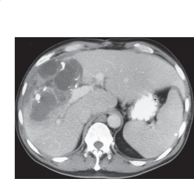
Findings
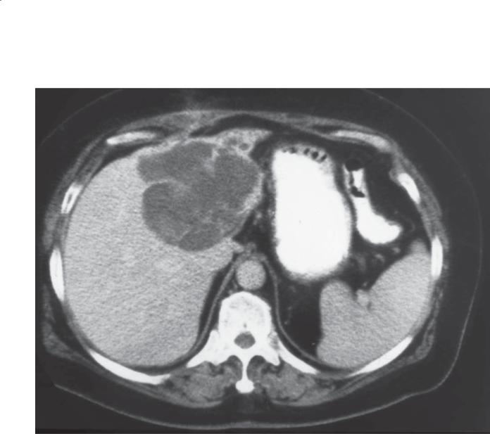
Contrast-enhanced CT. There is a large multilocular cystic mass in the left lobe of the liver.
Di erential Diagnosis
1.Echinococcal cyst
2.Biliary cystadenoma or carcinoma
3.Metastatic mucinous cystadenocarcinoma
4.Hepatic abscess
Diagnosis
Biliary cystadenoma
Discussion
Biliary cystadenomas are rare, premalignant hepatic cystic tumors that originate from biliary duct precursors, lined by mucin-secreting columnar epithelium. These tumors usually are multilocular with a well-defined thick capsule (as in this case). Biliary cystadenomas are most commonly found in middleaged white women (female:male ratio, 4:1). Because
these tumors can be large, patients can present with chronic abdominal pain and a palpable abdominal mass. Many are discovered incidentally.
Several other pathologic entities can have a similar radiographic appearance. Patients with echinococcal disease of the liver (case 6.25) usually have peripheral intracystic cysts and wall calcification with a history of travel to an endemic area. Patients with hepatic abscesses (case 6.23) often have multiple well-defined cystic masses with rim hyperenhancement and present with fevers and
leukocytosis. Metastatic cystadenocarcinoma should be considered; however, the cyst-within-a-cyst appearance is atypical.
No reliable clinical or radiographic criteria differentiate biliary cystadenoma from cystadenocarcinoma, and because of the malignant potential of biliary cystadenomas, these lesions should be removed surgically. The finding of mural nodules or thick, irregular, enhancing septations should raise concern for cystadenocarcinoma.
Bile Ducts: Masses and Filling Defects
7. BILE DUCTS AND GALLBLADDER 587
CASE 7.27
Findings
Contrast-enhanced CT. A complex, multilocular cystic mass is present in the right lobe of the liver with thick, nodular, enhancing septations and coarse calcifications.
Di erential Diagnosis
1.Liver abscess
2.Biliary cystadenoma or cystadenocarcinoma
3.Echinococcal cyst
Diagnosis
Biliary cystadenocarcinoma
Discussion
Biliary cystadenocarcinomas are very rare primary liver neoplasms arising from the biliary epithelium. The thick septae, enhancing nodular components of this mass, and calcifications favor biliary cystadenocarcinoma over cystadenoma (case 7.26). Pathologists have begun to separate
biliary cystadenomas and cystadenocarcinomas into those that contain ovarian stroma (found only in women) and those that do not (men and women). Those containing ovarian stroma tend to have a better prognosis. A liver abscess (case 6.23) would present in a patient with fever and increased leukocyte count, and the associated calcifications would be extremely unusual. Echinococcal cysts (cases 6.26 and 6.27) usually have thin septations, daughter cysts, and delicate peripheral calcifications.
Bile Ducts: Masses and Filling Defects
588 MAYO CLINIC GASTROINTESTINAL IMAGING REVIEW
CASE 7.28
A C
B
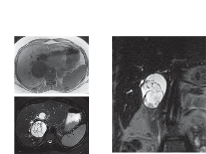
Findings
A.T1-weighted MRI. Mass with low signal intensity is within the posterior segment of the right hepatic lobe.
B.T2-weighted MRI. Mass with high signal intensity contains multiple internal septations.
C.Coronal T2-weighted MRI. Complex mass with high signal intensity contains multiple internal septations. The mass was separate from the gallbladder.
Di erential Diagnosis
1.Echinococcal cyst
2.Biliary cystadenoma or cystadenocarcinoma
3.Complex benign hepatic cyst
4.Cystic metastasis
Diagnosis
Biliary cystadenoma
Discussion
Biliary cystadenomas and cystadenocarcinomas are rare biliary tumors, usually occurring in women in their late 30s. They usually are intrahepatic in origin, are lined by columnar epithelium, and secrete mucin. They are analogous to pancreatic cystadenomas and cystadenocarcinomas. Their cystic appearance is due to their generally being filled with mucin. These tumors are believed to be either premalignant or malignant, and they are usually removed surgically when discovered. Cystadenomas can be unilocular in appearance or contain multiple septations
(as in this case). Imaging findings of nodularity within the mass are worrisome for malignant cystadenocarcinoma. Pathologic findings of ovarian struma in cystadenocarcinomas portend an indolent
course, whereas those without ovarian struma are more aggressive and can lead to death.
Bile Ducts: Masses and Filling Defects
7. BILE DUCTS AND GALLBLADDER 589
CASE 7.29
A B
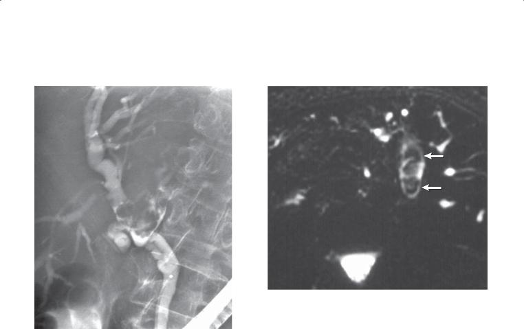
Findings
A.ERCP. There is a polypoid intraluminal filling defect within the common hepatic duct with extension into the left hepatic bile duct, which has an anomalous low insertion. The mass expands the duct. The left ductal system is poorly visualized.
B.Axial MRCP. The left hepatic bile duct is dilated and contains a polypoid filling defect (arrows). Multiple dilated proximal left intrahepatic bile ducts are visible.
Di erential Diagnosis
1.Intraductal cholangiocarcinoma
2.Intraductal blood clot
3.Intraductal mucous plug
4.Intraductal calculi
Diagnosis
Intraductal cholangiocarcinoma
Discussion
Intraductal cholangiocarcinomas usually have a better prognosis than the scirrhous subtypes. These tumors arise within the bile ducts and grow as intraluminal masses. Many are well-differentiated adenocarcinomas and secrete mucin. In some cases, mucin obstructs the duct and obscures the primary tumor. Intraductal cholangiocarcinomas are analogous to intraductal papillary mucinous neoplasm of the pancreas. It is not always possible to differentiate blood clot and mucin from tumor—but either finding should raise suspicion of an underlying neoplasm. Calculi usually have rounded and multifaceted edges.
Bile Ducts: Masses and Filling Defects
590 MAYO CLINIC GASTROINTESTINAL IMAGING REVIEW
CASE 7.30
Findings
ERCP. There is a polypoid filling defect (arrow) in the extrahepatic bile duct. The common hepatic duct is moderately dilated.
Di erential Diagnosis
1.Adenoma
2.Blood clot or mucous plug
3.Adherent debris or calculus
4.Cholangiocarcinoma
Diagnosis
Cholangiocarcinoma
Discussion
Cholangiocarcinoma arising in the common bile duct has the best prognosis. This filling defect is polypoid in appearance, whereas most cholangiocarcinomas are infiltrating. An adenoma could have a similar
appearance. Stones usually are not adherent to the wall, and plugs of mucus are more elongated.
Bile Ducts: Masses and Filling Defects
CASE 7.31
Findings
ERCP. The mid extrahepatic bile duct has annular, irregular narrowing with upstream dilatation.
Di erential Diagnosis
1.Cholangiocarcinoma
2.Pancreatic cancer
3.Ascending cholangitis
4.Periportal adenopathy
5.Primary sclerosing cholangitis
Diagnosis
Cholangiocarcinoma
7. BILE DUCTS AND GALLBLADDER 591
Discussion
Th e irregular, annular, abrupt narrowing indicates a malignant stricture. The stricture is too proximal
for a pancreatic tumor. Its irregular contour suggests a mucosal origin rather than extrinsic adenopathy. Ascending cholangitis would not be focal, and luminal irregularity would be expected of the entire biliary system. Primary sclerosing cholangitis is usually not focal, involving the intrahepatic and extrahepatic bile
ducts with strictures that are characterized as bandlike, alternating narrowing and dilatation, and diverticular outpouchings. Cholangiocarcinomas involving the extrahepatic ducts have a better prognosis than those involving the liver because resection with a wide margin is usually more feasible.
Bile Ducts: Masses and Filling Defects

592 MAYO CLINIC GASTROINTESTINAL IMAGING REVIEW
CASE 7.32
Findings
ERCP. There is high-grade narrowing of the distal right and left hepatic ducts and the proximal common hepatic bile duct. Upstream dilatation of the right and left ducts is seen, above the strictures.
Di erential Diagnosis
1.Cholangiocarcinoma, Klatskin tumor
2.Ischemic stricture, after liver transplant
3.Portal adenopathy
Diagnosis
Cholangiocarcinoma, Klatskin tumor
Discussion
Klatskin tumor refers to a cholangiocarcinoma involving the upper common hepatic duct. The tumor often extends to involve the right or left hepatic duct or both ducts. Because of the proximity of the hepatic artery and portal veins to the bile ducts, involvement of these vital structures is common with Klatskin tumor. Surgical resection is the only effective cure and requires at least 1 hepatic lobe to be tumor-free. ERCP is often used to assess resectability. Tumors that extend beyond the second-order biliary branches bilaterally are usually considered nonresectable.
Bile Ducts: Masses and Filling Defects
CASE 7.33
A
Findings
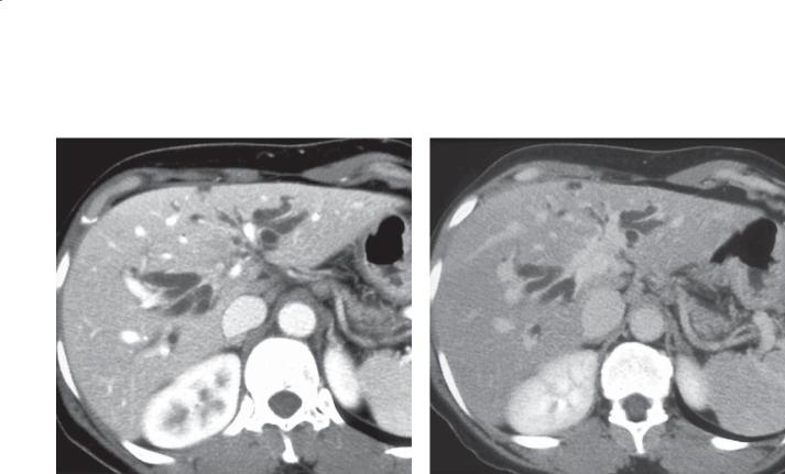
A.Contrast-enhanced CT. The bile ducts are dilated to the central portion of the liver without evidence of an obstructing mass.
B.Delayed enhanced CT. There is an enhancing mass at the level of the bifurcation of the left and right hepatic ducts with secondary obstruction of the bile ducts in both the right lobe and the left lobe of the liver.
Di erential Diagnosis
1.Cholangiocarcinoma
2.Metastasis
Diagnosis
Cholangiocarcinoma (Klatskin type)
7. BILE DUCTS AND GALLBLADDER 593
B
Discussion
Cholangiocarcinomas most commonly arise at the confluence of the right and left hepatic ducts. They are usually scirrhous carcinomas and often are referred to as Klatskin tumors. Most of the tumors are either hypoattenuating or isoattenuating compared with the liver during the arterial and portal venous phases of
contrast enhancement. Scirrhous cholangiocarcinomas incite a desmoplastic reaction. The fibrotic nature of these tumors can be identified as the tumor enhances on delayed imaging (as in this case). The size of these lesions can vary from 2 to 10 cm in diameter. Although most are infiltrating along the course of the bile
ducts, some present as more circumscribed polypoid masses. Tumors that are isoattenuating are the most difficult to identify. Their presence may be suspected by lobar atrophy of a hepatic segment, as a result of chronic biliary obstruction by the tumor. Metastasis to regional lymph nodes occurs in the portacaval and peripancreatic regions. Surgical resection in patients without metastases usually depends on the proximal extent of the tumor. Today, most surgeons do not consider an operation if the tumor extends proximal to the second-order biliary branches.
Bile Ducts: Masses and Filling Defects
594 MAYO CLINIC GASTROINTESTINAL IMAGING REVIEW
CASE 7.34
A B
C
Findings
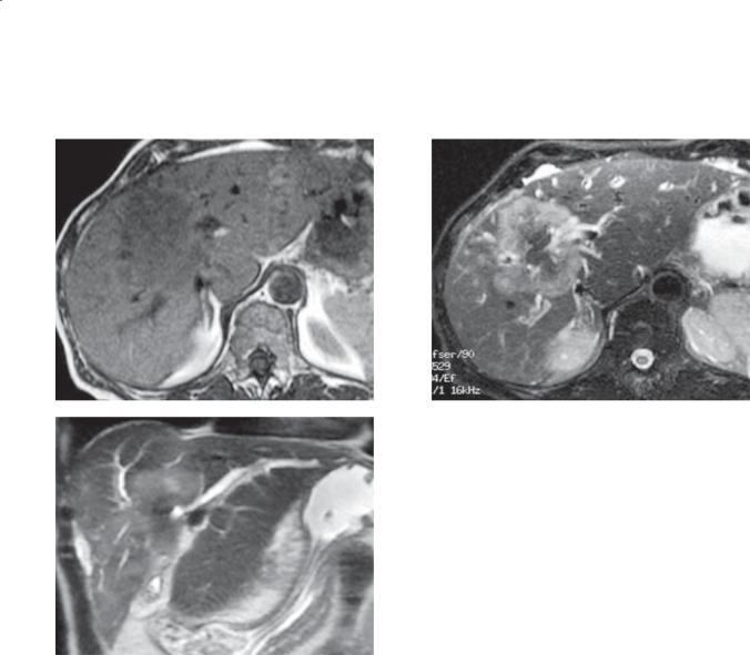
A.T1-weighted MRI. A large hypointense mass is located in the central portion of the liver.
B.T2-weighted MRI. The large mass is heterogeneous in signal intensity and causes obstruction of multiple intrahepatic bile ducts.
C.Coronal MRCP. The mass is slightly hyperintense compared with the liver. Multiple dilated bile ducts are obstructed by the mass.
Di erential Diagnosis
1.Cholangiocarcinoma
2.Metastasis
3.Hepatocellular carcinoma
Diagnosis
Cholangiocarcinoma (Klatskin type)
Discussion
Th is is an example of the expansile form of cholangiocarcinoma occurring at the confluence of the right and left hepatic bile ducts. This large Klatskin tumor is well demarcated on the T2-weighted MRI examination. The excellent contrast resolution available at MRI makes identification of cholangiocarcinoma
superior to either CT or sonography. MRCP is a helpful technique to display the biliary anatomy in a familiar coronal plane. T1-weighted images are particularly helpful to assess for extrahepatic metastases and adenopathy.
Bile Ducts: Masses and Filling Defects

CASE 7.35
A
Findings
A.T1-weighted MRI. There is a linear region of abnormally low signal intensity in the region of the right posterior portal vein.
B.T2-weighted MRI. A linear region of nodular thickening of high signal intensity extends within the distribution of the right posterior portal triad.
Di erential Diagnosis
1.Sclerosing cholangitis
2.Infi ltrative cholangiocarcinoma
3.Ascending cholangitis
Diagnosis
Infiltrative cholangiocarcinoma
7. BILE DUCTS AND GALLBLADDER 595
B
Discussion
Infiltrative cholangiocarcinoma can be very difficult to detect, and assessment of its full extent also can be difficult. MRI is the best imaging examination to
visualize this tumor. Bile duct thickening can be found in patients with sclerosing cholangitis, but it is usually only a few millimeters in thickness. Marked bile duct thickening (as in this case) is nearly always associated with tumor from infiltrating cholangiocarcinoma. This tumor is seen to encase the portal vein and hepatic artery centrally, a feature that renders it unresectable.
Bile Ducts: Masses and Filling Defects
596 MAYO CLINIC GASTROINTESTINAL IMAGING REVIEW
CASE 7.36
A B
C
Findings |
Discussion |
A.T1-weighted MRI. The left lobe of the liver is atrophic, and the signal intensity of this lobe is heterogeneous.
B.T2-weighted MRI. Multiple dilated bile ducts are seen within an atrophic left lobe. They converge to a central point.
C.T2-weighted MRI (different level than in B). The dilated left lobar ducts converge to a central mass of low signal intensity (arrow).
Di erential Diagnosis
Cholangiocarcinoma
Diagnosis
Cholangiocarcinoma with lobar atrophy
Th is case illustrates the typical findings of lobar atrophy in a patient with a slow-growing cholangiocarcinoma obstructing a lobar branch bile duct. The tumor is difficult to see because it is isointense or hypointense compared with the liver. The lesion can be at an advanced stage because its detection is often delayed without clinical symptoms. Surgical resection might be possible if the tumor does not involve the main portal vein and proper hepatic artery.
Bile Ducts: Masses and Filling Defects
CASE 7.37
Findings
Contrast-enhanced CT. A large hypoattenuating mass is present in the left lobe of the liver. Two dilated bile ducts are present in the atrophic left hepatic lobe.
Di erential Diagnosis
1.Metastases
2.Hepatocellular carcinoma
3.Cholangiocarcinoma
4.Biliary cystadenoma
Diagnosis
Intrahepatic cholangiocarcinoma
7. BILE DUCTS AND GALLBLADDER 597
Discussion
Although cholangiocarcinomas are most commonly found at the confluence of the right and left biliary ducts, some arise in the peripheral ductal system. These tumors can become large because they do not always cause symptoms at an early stage. Upstream ductal dilatation usually is present, and, depending on the location of the tumor, there may be lobar atrophy from chronic biliary obstruction.
Hepatocellular carcinomas (cases 6.51 through 6.67) usually are hypervascular tumors, and biliary cystadenomas (case 7.26) predominantly are cystic and septated (although enhancing nodules can be seen with cystadenocarcinomas). A metastasis could be indistinguishable from this lesion.
Bile Ducts: Masses and Filling Defects

598 MAYO CLINIC GASTROINTESTINAL IMAGING REVIEW
TABLE 7.2
Masses and Filling Defects of the Bile Ducts |
CASE |
||
Benigna |
|
|
|
Choledocholithiasis |
Posterior shadowing echogenic foci in a dilated duct on |
7.14–7.19 |
|
|
sonography. Target or meniscus sign on CT. MRCP and ERCP |
|
|
|
are more sensitive |
|
|
Pseudocalculus |
Apparent filling defect due to spasm of sphincter of Oddi. |
7.20 |
|
|
Transient. Absent inferior meniscus |
|
|
Papillary stenosis |
Distal common bile duct stricture |
7.21 |
|
Ampullary carcinoma |
Dilated biliary and pancreatic ducts to narrowed and irregular |
7.22 |
|
|
ampulla |
|
|
Mirizzi syndrome |
Inflammatory stricture of common duct caused by stone |
7.23 |
|
|
impacted in cystic duct |
|
|
Extrinsic compression of |
Smooth narrowing of common hepatic duct |
7.24 |
|
common duct by adenopathy |
|
|
|
Biliary papillomatosis |
Multiple solid filling defects attached to the wall of a dilated |
7.25 |
|
|
duct. High incidence of malignant transformation. Mucous |
|
|
|
filling defects often associated |
|
|
Biliary cystadenoma or |
Unilocular or septated mucin-filled cyst. Nodules suggest |
7.26–7.28 |
|
cystadenocarcinoma |
|||
malignancy |
|
||
|
|
||
|
|
|
|
Malignant |
|
|
|
Cholangiocarcinoma |
Intraductal, polypoid, and distal tumors have the best |
7.29–7.37 |
|
|
prognosis. Scirrhous or infiltrating tumors are the most common |
|
|
type. Confluence of right and left biliary ducts most common site (Klatskin tumor). Lobar atrophy. Large peripheral hypovascular mass
a Other benign biliary filling defects include sludge balls, mucous plugs, and blood clots.
Bile Ducts: Masses and Filling Defects
7. BILE DUCTS AND GALLBLADDER 599
CASE 7.38
Findings |
Discussion |
|
ERCP. There is a low insertion of the cystic duct to the |
Anomalies of the biliary tract are relatively common. |
|
common bile duct. The cholangiogram is otherwise |
The commonest anomaly is an aberrant intrahepatic |
|
unremarkable. |
duct draining into a contralateral main or common |
|
|
hepatic duct. For example, the bile duct draining the |
|
Di erential Diagnosis |
right anterior hepatic lobe can anomalously drain |
|
Low insertion of the cystic duct |
into the left main bile duct or common hepatic |
|
|
duct. Anomalies of the cystic duct also are relatively |
|
Diagnosis |
common (as in this case). Major cystic duct anomalies |
|
Low insertion of the cystic duct |
include the following: |
|
|
1. |
Absence of the cystic duct with direct drainage of |
|
|
the gallbladder into the common hepatic duct |
|
2. |
Low insertion (as in this case) |
|
3. |
Long conjoined segment of the cystic duct with the |
|
|
common duct |
|
4. |
Insertion into the right hepatic duct |
|
5. |
Insertion at the level of bifurcation |
Th ese major cystic duct anomalies are illustrated on page 644, and common intrahepatic duct variations are illustrated on page 645.
Bile Ducts: Miscellaneous
600 MAYO CLINIC GASTROINTESTINAL IMAGING REVIEW
CASE 7.39
A C
B
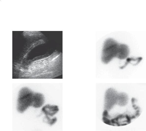
Findings
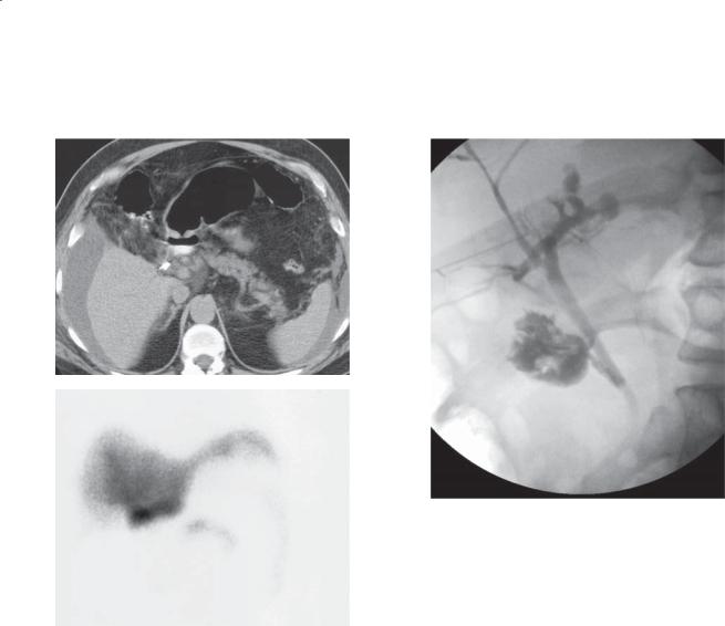
A.Unenhanced CT. Postoperative changes of cholecystectomy are present. There is a moderate amount of ascites in the visualized abdomen.
B.Hepatobiliary scintigram. One-hour hepatoiminodiacetic acid scan shows an abnormal collection of tracer in the post-cholecystectomy bed and additional tracer uptake outlining the peritoneal cavity. No definite tracer is seen to pass into the bowel.
C.Percutaneous transhepatic cholangiogram. There
is leakage of contrast from the cystic duct stump. The common duct remains intact.
Di erential Diagnosis
Biliary leak
Diagnosis
Biliary leak
Discussion
Ninety-five percent of biliary leaks are iatrogenic, and most arise as a result of complications of surgery to the biliary tree. Iatrogenic biliary injuries are more common with laparoscopic cholecystectomy than with open cholecystectomy. Extravasation
of small quantities of bile is fairly common after cholecystectomy and usually is of little clinical significance. The most worrisome leaks are those in which bile ascites is present and no bile is seen to enter the small bowel on hepatoiminodiacetic acid scan. In these cases, prompt drainage is required to decrease morbidity. Percutaneous transhepatic cholangiography or ERCP usually is required to determine the level of the ductal injury and to exclude ligation, laceration, or transection of the bile ducts. These major duct injuries ultimately require choledochojejunostomy or hepaticojejunostomy if clinically feasible.
Bile Ducts: Miscellaneous
7. BILE DUCTS AND GALLBLADDER 601
CASE 7.40
A C
B D
Findings
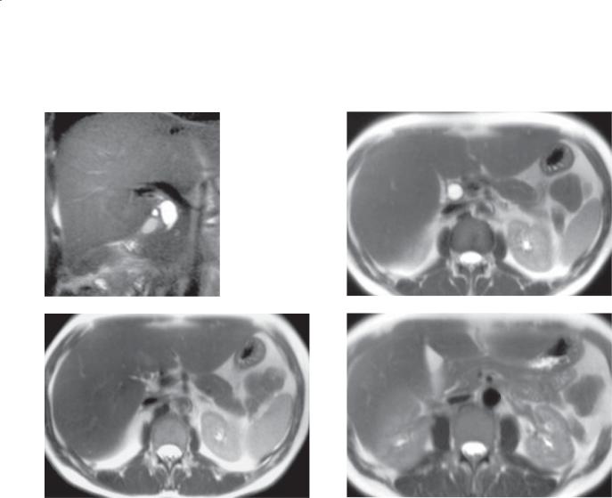
MRCP. A. Coronal image. B through D. Axial images. Fusiform focal dilatation of the common bile duct. The duct above and below this region (B and D) appears normal.
Di erential Diagnosis
1.Choledochal cyst, type I
2.Biliary obstruction, cause undetermined
Diagnosis
Choledochal cyst, type I
Discussion
Choledochal cysts are congenital cystic dilatations of the bile ducts. The cause of choledochal cysts is
not known, but it has been hypothesized that an anomalous connection between the pancreatic duct and the biliary system allows pancreatic secretions to weaken the wall of the duct with subsequent
dilatation. The majority of cases are diagnosed in girls before the age of 10 years. There is an association between choledochal cysts and other biliary tract anomalies (especially biliary tract stenosis and atresia), congenital hepatic fibrosis, and medullary sponge kidney. Complications include rupture, stasis with cholangitis, stone formation, liver abscess, cirrhosis, and cholangiocarcinoma. The classification of choledochal cysts is based on the location of the cyst(s) within the biliary system, as shown in the figure on page 643.
Bile Ducts: Miscellaneous
602 MAYO CLINIC GASTROINTESTINAL IMAGING REVIEW
CASE 7.41
A B
C
Findings
A and B. Contrast-enhanced CT. Large cystic mass is visible in the region of the hepatic porta. The location of the mass is in the expected region of the bile duct (not visualized).
C. ERCP. The cystic mass represents massive dilatation of the extrahepatic bile duct.
Di erential Diagnosis
1.Choledochal cyst, type I
2.Obstructed bile duct (cause not seen)
Diagnosis
Choledochal cyst, type I
Discussion
Choledochal cysts can become very large and can be difficult to diagnose on cross-sectional imaging.
Coronal images can be helpful to identify the tapering ends of the cyst that lead to normal-caliber bile ducts. ERCP is often the reference standard to confirm their biliary origin. (See the figure on page 643.)
Bile Ducts: Miscellaneous
CASE 7.42
A
Findings
Contrast-enhanced CT. A and B. A water-attenuation mass arises from the region of the ampulla and fills most of the duodenal lumen.
Di erential Diagnosis
Choledochocele
Diagnosis
Choledochocele (type III choledochal cyst)
7. BILE DUCTS AND GALLBLADDER 603
B
Discussion
A choledochocele is a protrusion of a dilated intramural segment of the common bile duct into the duodenum. It is considered a type of choledochal cyst (type III). It has been referred to by several terms, including duodenal duplication cyst, diverticulum of the common bile duct, and enterogenous cyst of the duodenum. It is analogous to a ureterocele. Patients with a choledochocele usually complain of nausea, vomiting, and biliary colic. At cholangiography, a saclike dilatation of the common bile duct is seen. Because the dilated ductal segment is filled with bile (it also can contain sludge and stones), it will be water attenuation at CT.
Bile Ducts: Miscellaneous
604 MAYO CLINIC GASTROINTESTINAL IMAGING REVIEW
CASE 7.43
A B
C
Bile Ducts: Miscellaneous

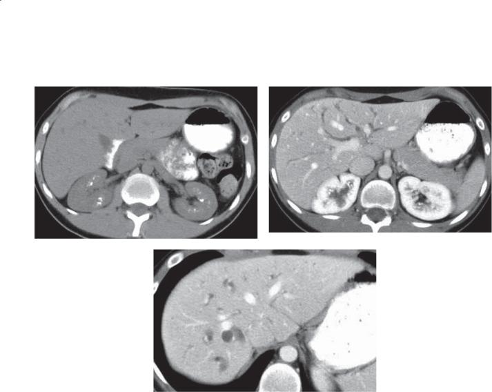
Findings
Unenhanced CT. A. Multiple calcifications within the medullary regions of both kidneys. B. Multiple dilated intrahepatic bile ducts with alternating areas of narrowing and dilatation. Medullary renal calcifications are present. C. Saccular areas of bile duct dilatation are within the right hepatic lobe, and dilatation of left lobar bile duct is only minimal.
Di erential Diagnosis
1.Caroli disease
2.Primary sclerosing cholangitis
3.Biliary obstruction, cause not seen
4.Cholangitis with abscess formation
Diagnosis
Caroli disease
7. BILE DUCTS AND GALLBLADDER 605
Discussion
Caroli disease occurs embryologically (autosomal recessive) with arrest of normal bile duct remodeling and subsequent destructive inflammation and segmental dilatation. Caroli disease exists when
the large ducts are affected. Small interlobular duct involvement results in congenital hepatic fibrosis. Patients can have either segmental or diffuse bile duct dilatation. Usually, alternating regions of narrowing and saccular or fusiform dilatation are seen. Although disease confined to the intrahepatic bile ducts is characteristic, up to half of patients will also have extrahepatic bile duct dilatation.
Various renal diseases can be seen with Caroli syndrome, including medullary sponge kidney (as in this case with multiple medullary calcifications), polycystic renal disease, and medullary cystic disease. The radiologic appearance of primary sclerosing cholangitis is different, because it is rarely saccular in appearance. Cholangitis could present in this way; however, the history would include symptoms and signs of an acute infection. Obstruction would have uniformly dilated and tapering ducts without bile lakes or strictures.
Bile Ducts: Miscellaneous
606 MAYO CLINIC GASTROINTESTINAL IMAGING REVIEW
CASE 7.44
AB
C D
E
Bile Ducts: Miscellaneous

Findings
MRCP. A and B. Marked dilatation of a majority of the intrahepatic biliary system. Some normal-caliber ducts are visible.
C.T1-weighted MRI. Multiple dilated intrahepatic ducts. There is a focal mass within the right lobe of the liver (arrow).
D.T2-weighted MRI. The focal mass is of low signal intensity. The dilated ducts are of high signal intensity.
E.Dynamic contrast-enhanced MRI. The mass within the right lobe of the liver enhances heterogeneously during the early phases of contrast enhancement.
Di erential Diagnosis
Caroli disease with complicating adenocarcinoma
Diagnosis
Caroli disease with complicating adenocarcinoma
7. BILE DUCTS AND GALLBLADDER 607
Discussion
Complications of Caroli disease include stone formation, recurrent cholangitis, liver abscess, and adenocarcinoma (as in this case). There is a 100-fold increase in the risk for development of
adenocarcinoma compared with the risk in the normal population.
Bile Ducts: Miscellaneous
608 MAYO CLINIC GASTROINTESTINAL IMAGING REVIEW
CASE 7.45
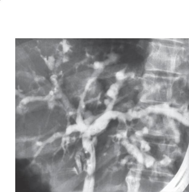
Findings
ERCP. Multiple cystic dilatations of the intrahepatic bile ducts are seen throughout the liver.
Di erential Diagnosis
1.Caroli disease
2.Multiple biliary abscesses
Diagnosis
Caroli disease
Discussion
Caroli disease is a form of choledochal cystic disease with segmental saccular or fusiform dilatation of the intrahepatic ducts and a normal-caliber extrahepatic duct (type V). Patients with Caroli disease usually present during adulthood with symptoms of cholangitis. There is an association between Caroli disease and renal tubular ectasia.
Bile Ducts: Miscellaneous

|
|
7. BILE DUCTS AND GALLBLADDER 609 |
|
|
|
|
|
|
TABLE 7.3 |
|
|
Miscellaneous Bile Duct Conditions |
CASE |
||
|
|
|
|
Cystic duct |
Anomalies |
7.38 |
|
|
Many congenital variants |
(see page 644) |
|
|
|
|
|
Bile leak |
Loculated or free fluid |
7.39 |
|
|
Usually requires cholangiography |
||
|
to define site of leak |
|
|
|
|
|
|
Choledochal cysts |
|
|
|
Type I |
Fusiform |
7.40 and 7.41 |
|
Type II |
Diverticulum |
|
|
Type III |
Choledochocele |
7.42 |
|
Type IV |
Intrahepatic and extrahepatic |
|
|
Type V |
Caroli (intrahepatic) |
7.43–7.45 |
|
|
|
(see page 643) |
|
|
|
|
|
Bile Ducts: Miscellaneous
610 MAYO CLINIC GASTROINTESTINAL IMAGING REVIEW
CASE 7.46 |
CASE 7.47 |
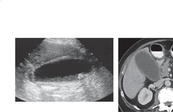
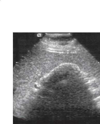
Findings
CASE 7.46. Sonogram. The gallbladder wall is thickened, and sludge and a stone within its lumen cast an acoustic shadow. There is a thin hypoechoic zone about the gallbladder.
CASE 7.47. Contrast-enhanced CT. Gallbladder is diffusely thick-walled and distended.
Di erential Diagnosis
Acute cholecystitis and cholelithiasis
Diagnosis
Acute cholecystitis and cholelithiasis
Discussion
Th ere are many sonographic signs of acute cholecystitis, including cholelithiasis or sludge, intramural thickening with zones of lucency, distention of the gallbladder, sonographic Murphy sign, perihepatitis, and pericholecystic fluid. The most accurate sign is the combination of cholelithiasis, intramural edema, and maximal pain over the gallbladder (sonographic Murphy sign). Perihepatitis manifests as a zone of decreased hepatic echogenicity about the gallbladder (as in case 7.46).
Gallbladder: Inflammatory Diseases
7. BILE DUCTS AND GALLBLADDER 611
CASE 7.48
A B
C D
Findings
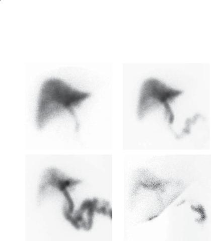
Cholescintigram. A. 10 minutes after injection. Nonvisualization of the gallbladder. B. 20 minutes after injection. Nonvisualization of the gallbladder.
C.60 minutes after injection. Nonvisualization of the gallbladder. Excretion into the small bowel is excellent.
D.4 hours after injection. Nonvisualization of the gallbladder with increased uptake within the liver about the gallbladder.
Di erential Diagnosis
1.Acute cholecystitis
2.Chronic cholecystitis
3.Pancreatitis
4.Hyperalimentation
Diagnosis
Acute cholecystitis
Discussion
Cholescintigraphy usually is performed with technetium Tc 99m-labeled iminodiacetic acid analogues. Today, diisopropyl-iminodiacetic acid (DISIDA) and mebrofenin are the most popular agents because they can be used in patients with hyperbilirubinemia (up to a bilirubin value of 20 mg/ dL). The agent is taken up by the liver and excreted without conjugation. Normally, the gallbladder contains tracer uptake within 30 minutes after administration. Nonvisualization of the gallbladder is suspicious for cystic duct obstruction; however, some gallbladders are slow to fill (distended gallbladders and those with bile stasis), and delayed imaging is needed. False-positive studies are rare, usually due to intermittent obstruction of the cystic duct by a moving stone. False-negative results can be caused by any of the conditions in the differential list above.
In the case shown here, despite delayed imaging, the gallbladder never fills. The 4-hour image shows increased uptake about the gallbladder due to the adjacent inflammation (perihepatitis).
Gallbladder: Inflammatory Diseases
612 MAYO CLINIC GASTROINTESTINAL IMAGING REVIEW
CASE 7.49 |
CASE 7.50 |
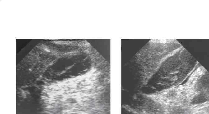
Findings
CASE 7.49. Sonogram. The gallbladder wall is irregularly thickened, and there are several linear filling defects in the gallbladder lumen.
CASE 7.50. Sonogram. The wall of the gallbladder is thickened, and there is a small amount of pericholecystic fluid. Echogenic debris fills the
gallbladder lumen. Echogenic linear filling defects also are seen within the lumen.
Di erential Diagnosis
Gangrenous cholecystitis
Diagnosis
Gangrenous cholecystitis
Discussion
Acute cholecystitis is caused by obstruction of the cystic duct by a calculus with secondary gallbladder distention, ischemia, wall edema, and transmural necrosis. Gangrenous cholecystitis is a designation used for severe gallbladder inflammation. At sonography, there is usually pronounced intramural edema or sloughing of the mucosa into the gallbladder lumen (as in these cases).
Gangrenous cholecystitis should be treated urgently to prevent gallbladder perforation and abscess from developing.
Gallbladder: Inflammatory Diseases
CASE 7.51
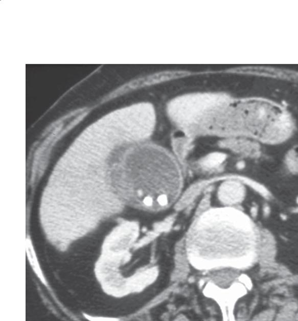
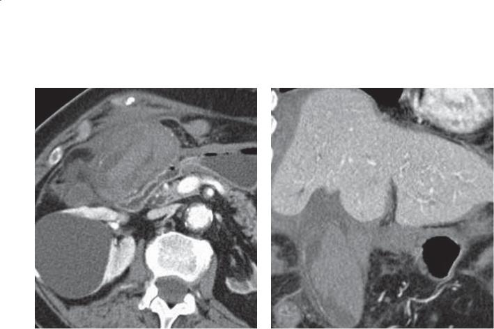
Findings
Contrast-enhanced CT. Cholelithiasis. The gallbladder wall is thickened, and there is a small pericholecystic fluid collection.
Di erential Diagnosis
1.Gangrenous cholecystitis
2.Gallbladder carcinoma
Diagnosis
Gangrenous cholecystitis with pericholecystic fluid collection
7. BILE DUCTS AND GALLBLADDER 613
Discussion
CT is usually not the primary imaging tool for the diagnosis of acute cholecystitis. It is optimally used when there are confusing findings either clinically or with sonography or cholescintigraphy or when complications of cholecystitis are suspected. The most common complication of cholecystitis is a pericholecystic fluid collection from perforation due to gangrenous cholecystitis. These fluid collections often are infected. Usually the wall of the gallbladder is of water attenuation (due to edema), and it may have a striated appearance. Attenuation changes can occur within the surrounding liver, and fluid may be identified in the posterior subhepatic space (Morrison pouch).
Gallbladder: Inflammatory Diseases
614 MAYO CLINIC GASTROINTESTINAL IMAGING REVIEW
CASE 7.52
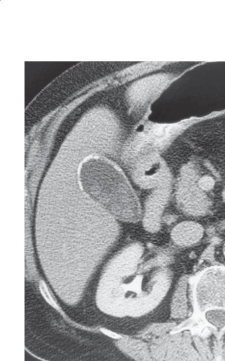
Findings
Contrast-enhanced CT. The gallbladder wall is thickened and of water attenuation. Several waterattenuation, rim-enhancing collections are present within the liver about the gallbladder.
Di erential Diagnosis
Acute cholecystitis with pericholecystic hepatic abscess
Diagnosis
Acute cholecystitis with pericholecystic hepatic abscess
Discussion
A complicating abscess from acute cholecystitis is often associated with a delayed diagnosis or inappropriate treatment. Inflammation and infection from the gallbladder can either directly extend or seed into the adjacent liver. The characteristic water-attenuation, multiloculated fluid collections with a hyperenhancing wall are characteristic of pyogenic abscess(es).
Gallbladder: Inflammatory Diseases
CASE 7.53
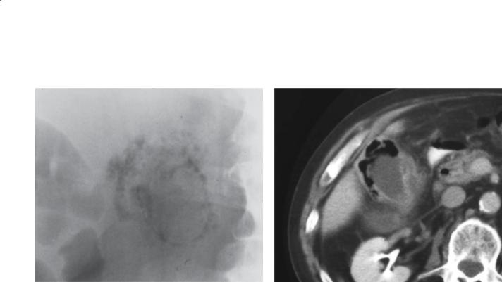
Findings
CASE 7.53. Abdominal plain radiograph. There is a collection of extraluminal gas within the right upper quadrant. Gas is seen within the wall of the gallbladder and surrounding the gallbladder.
CASE 7.54. Contrast-enhanced CT. The gallbladder wall is thickened, and gas can be seen within its wall.
Prominent inflammatory changes and fluid are present in the pericholecystic region.
Di erential Diagnosis
Emphysematous cholecystitis
Diagnosis
Emphysematous cholecystitis
7. BILE DUCTS AND GALLBLADDER 615
CASE 7.54
Discussion
Emphysematous cholecystitis is a severe form of cholecystitis caused by a gas-forming organism. Up to 30% of patients have diabetes, and a large number are elderly. Ischemia of the gallbladder may be an underlying cause or contribute to the condition in a large percentage of cases. Infection with Clostridium perfringens often is present, producing the gas that is pathognomonic of this disease. The gas can reside within the wall or the lumen of the gallbladder or
both. If gas is seen outside the lumen, it often indicates gallbladder perforation and a pericholecystic abscess. Cholelithiasis may or may not be present. The cystic duct is patent.
Gallbladder: Inflammatory Diseases
616 MAYO CLINIC GASTROINTESTINAL IMAGING REVIEW
CASE 7.55 |
CASE 7.56 |
Findings
CASE 7.55. Sonogram. The gallbladder wall is thickened. Dependently layering gallstones are present. A bright echogenic rim is seen along the nondependent wall
of the gallbladder (arrow) with “dirty” posterior shadowing.
CASE 7.56. Sonogram. Abnormal echogenicity is present along the anterior wall of the gallbladder with “dirty” posterior acoustic shadowing, making visualization
of the gallbladder difficult. Hypoechogenicity is seen within the liver parenchyma adjacent to the abnormal gallbladder.
Di erential Diagnosis
1.Emphysematous cholecystitis
2.Porcelain gallbladder
Diagnosis
Emphysematous cholecystitis
Discussion
Air within the wall of the gallbladder at sonography appears as echogenic foci that cause a “dirty” shadow. A porcelain gallbladder (cases 7.61 and 7.62) with wall calcification should cast a “clean” shadow,
or the gallbladder may be difficult to identify. If emphysematous cholecystitis is suspected, it is often helpful to confirm the finding with abdominal plain radiography or CT. The gas can extend from the wall into the gallbladder lumen and pericholecystic tissues.
Gallbladder: Inflammatory Diseases
7. BILE DUCTS AND GALLBLADDER 617
CASE 7.57
A
Findings
A.Contrast-enhanced CT. A soft tissue density mass is present within the gallbladder. The gallbladder wall enhances incompletely.
B.CT coronal reformatted image. The gallbladder is distended with the above-described intracholecystic tissue and pericholecystic-perihepatic fluid collection.
Di erential Diagnosis
1.Hemorrhagic cholecystitis
2.Gallbladder carcinoma
3.Gallbladder sludge
Diagnosis
Hemorrhagic cholecystitis
B
Discussion
Gallbladder inflammation or ischemia can result in sloughing of the mucosal lining of the gallbladder. Bleeding within the gallbladder can occur, leading to the findings in this case, referred to as hemorrhagic cholecystitis. The treatment is the same as that for any form of acute cholecystitis—operation. Other causes for hemorrhage into the gallbladder include trauma, anticoagulation, neoplasms, ruptured aneurysm, parasites, or ectopic gastric or pancreatic mucosa. In some patients, the clot will be high attenuation (most easily recognized on unenhanced scans). Gallbladder carcinoma usually focally thickens the gallbladder wall and enhances. Sludge is usually dependent within the gallbladder.
Gallbladder: Inflammatory Diseases
618 MAYO CLINIC GASTROINTESTINAL IMAGING REVIEW
CASE 7.58
A B
Findings
A.Sonogram. The wall of the gallbladder is thickened. No calculi are present.
B.Contrast-enhanced CT. The gallbladder wall is thickened, and the mucosa is hyperenhancing. No calculi are present.
Di erential Diagnosis
1.Chronic cholecystitis
2.Acalculous cholecystitis
3.Various metabolic disturbances
Diagnosis
Acalculous cholecystitis
Discussion
Acalculous cholecystitis refers to acute inflammatory changes of the gallbladder without stones. The actual cause of the disease is unclear; however, vascular insufficiency of the gallbladder is a likely cause.
The condition is often associated with functional obstruction of the cystic duct. Clinical settings associated with acalculous cholecystitis include hyperalimentation, trauma, prolonged care in an intensive care unit, infectious colitis, vascular disease of any cause, and an immunocompromised host. Sonography, CT, or cholescintigraphy can suggest the diagnosis by confirming either inflammatory changes in the wall of the gallbladder or occlusion of the cystic duct. At sonography, the gallbladder is thickened and has focal or striated sonolucency. Intramural wall edema and perihepatitis can be seen at CT. Nonfilling of the gallbladder is found at cholescintigraphy.
Gallbladder: Inflammatory Diseases
7. BILE DUCTS AND GALLBLADDER 619
CASE 7.59
A B
C D
Findings
A. Gallbladder sonogram. The gallbladder contains dependently layering echogenic debris (sludge), and the wall of the gallbladder is thickened. No stones are visible.
B through D. Cholescintigram. Nonfilling of the gallbladder on the 30-minute (B), 60-minute (C), and 4-hour (D) delayed images.
Di erential Diagnosis
1.Acalculous cholecystitis
2.Chronic cholecystitis
Diagnosis
Acalculous cholecystitis
Discussion
Th e diagnosis of acalculous cholecystitis can be difficult. Patients at greatest risk include patients in the intensive care unit requiring narcotics, those
receiving hyperalimentation, and those in whom the blood supply to the gallbladder could be jeopardized (hypotension, vasculitis, severe atherosclerosis).
Many of these patients have been fasting, and a distended gallbladder containing sludge will be present. It is unclear which imaging method is best for diagnosing this disorder. At sonography, findings of an inflamed gallbladder (thickened wall with striations, pericholecystic fluid, and a positive sonographic Murphy sign) without calculi are most helpful. Nonfilling of the gallbladder at cholescintigraphy is the primary finding. If the gallbladder has not filled at 60 minutes, morphine often is given to constrict the sphincter of Oddi and direct bile flow into the cystic duct. Alternatively, delayed imaging can be extended to 4 hours, as in this case. Nonfilling of the gallbladder at this point is consistent with cholecystitis.
Gallbladder: Inflammatory Diseases
620 MAYO CLINIC GASTROINTESTINAL IMAGING REVIEW
CASE 7.60
Findings
Contrast-enhanced CT. The wall of the gallbladder is markedly thickened and heterogeneous in attenuation. Multiple gallstones are present.
Di erential Diagnosis
1.Acute cholecystitis
2.Chronic cholecystitis
3.Adenomyomatosis
Diagnosis
Chronic cholecystitis
Discussion
It may be difficult to differentiate acute from chronic cholecystitis radiographically. In both cases the wall of the gallbladder is thickened and cholelithiasis usually is visible. Symptomatic evaluation usually differentiates acute disease from chronic disease. Chronic disease is associated with a history of multiple episodes of abdominal pain without the presence
of acute symptoms and signs that include fever and leukocytosis.
Gallbladder: Inflammatory Diseases
CASE 7.61
Findings
Contrast-enhanced CT. The wall of the gallbladder is calcified and contains calculi.
Di erential Diagnosis
Porcelain gallbladder with cholelithiasis
Diagnosis
Porcelain gallbladder with cholelithiasis
7. BILE DUCTS AND GALLBLADDER 621
Discussion
Porcelain gallbladder refers to calcification of the wall of the gallbladder. The calcification can be either linear or punctate. This gallbladder wall calcification develops as a result of a chronically obstructed cystic duct and changes of chronic cholecystitis. The importance of this condition is an increased risk of gallbladder carcinoma (case 7.75). In fact, in 25% of patients with porcelain gallbladder, gallbladder carcinoma develops without cholecystectomy.
Gallbladder: Inflammatory Diseases
622 MAYO CLINIC GASTROINTESTINAL IMAGING REVIEW
CASE 7.62
Findings
Sonogram. A thin echogenic arc (representing the anterior wall of the gallbladder) is present with complete “clean” shadowing of the remainder of the gallbladder lumen and back wall.
Di erential Diagnosis
1.Porcelain gallbladder
2.Gallstones (wall-echo-shadow complex)
3.Emphysematous cholecystitis
Diagnosis
Porcelain gallbladder
Discussion
Dystrophic calcification within the wall of an obstructed and chronically inflamed gallbladder produces a brittle wall and has led to the term porcelain gallbladder. The condition is associated with gallstones in more than 90% of cases. Stone disease often can be difficult to identify sonographically (as in this case) because of acoustic shadowing from the calcified gallbladder wall.
Porcelain gallbladder should be able to be differentiated clinically and sonographically from emphysematous cholecystitis (cases 7.55 and 7.56). The air within the gallbladder wall or lumen produces a “dirty” posterior shadow. The wall-echo-shadow triad of a gallbladder filled with stones (case 7.66) should give 2 echogenic lines separated by a thin line of sonolucency. “Clean” posterior shadowing should be seen posterior to the deep echogenic arc (gallstones). Unless there are medical contraindications, prophylactic cholecystectomy is recommended in patients with a porcelain gallbladder because of an increased risk of gallbladder carcinoma.
Gallbladder: Inflammatory Diseases
CASE 7.63
A
Findings
A.Abdominal plain radiograph. The gallbladder and cystic duct are opaque and contain multiple stones.
B.Unenhanced CT. High-attenuation material fills the gallbladder.
Di erential Diagnosis
1.Milk of calcium bile
2.Vicarious excretion of contrast material
3.Prior ERCP with cholelithiasis
Diagnosis
Milk of calcium bile
7. BILE DUCTS AND GALLBLADDER 623
B
Discussion
Milk of calcium bile is calcium carbonate, calcium phosphate, or calcium bilirubinate that is highly concentrated within the gallbladder because of cystic duct obstruction and chronic gallbladder
inflammation. Because it is dense enough to detect on plain radiography, it is referred to as milk of calcium bile or the limy bile syndrome.
Gallbladder: Inflammatory Diseases
624 MAYO CLINIC GASTROINTESTINAL IMAGING REVIEW
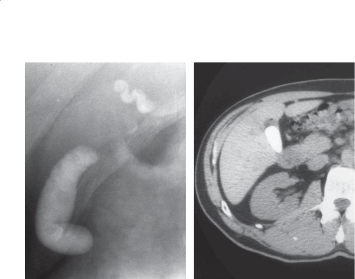
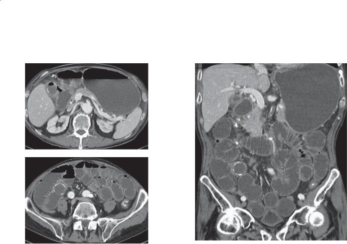
CASE 7.64
A C
B
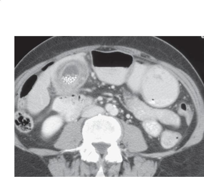
Findings
Contrast-enhanced CT. A. The gallbladder contains gas and has a calcified wall. There is extraluminal gas between the gallbladder and the duodenum.
B. Multiple dilated small bowel loops with a peripherally calcified object in the distal small bowel. C. Coronal view. The dilated small bowel loops and calcified object are well seen.
Di erential Diagnosis
1.Gallstone ileus
2.Foreign body obstruction and duodenal perforation
3.Obstructing enterolith with secondary small bowel perforation
Diagnosis
Gallstone ileus
Discussion
Gallstone ileus is due to chronic cholecystitis. Elderly women usually present to the emergency department with symptoms of bowel obstruction and no history of abdominal surgery. Patients usually do not have symptoms of acute cholecystitis. Chronic cholecystitis (note the porcelain gallbladder in this case) can lead to a cholecystoduodenal fistula. Gallstones can travel through the fistula into the small bowel. The stones will usually impact (if they are large) at the ileocecal valve and cause a mechanical small bowel obstruction. Identification of ectopic gallstones in the small bowel can be challenging, because some gallstones are near water attenuation. The stone in this case has peripheral calcification that aids in its diagnosis. The triad of biliary air, mechanical small bowel obstruction, and ectopic gallstone is considered pathognomonic for this condition; however, most commonly there are fewer than 3 findings. (See also cases 4.37 and 4.38.)
Gallbladder: Inflammatory Diseases

7. BILE DUCTS AND GALLBLADDER 625
TABLE 7.4
Inflammatory Diseases of the Gallbladder |
CASE |
|
|
|
|
Acute cholecystitis |
Due to cystic duct obstruction. Thickened gallbladder wall, |
7.46–7.48 |
|
cholelithiasis, and positive sonographic Murphy sign. Nonfilling |
|
|
of gallbladder at cholescintigraphy |
|
|
|
|
Gangrenous cholecystitis |
Severe inflammation of the gallbladder with sloughing |
7.49–7.52 |
|
of mucosa into the lumen. High risk of perforation or |
|
|
pericholecystic abscess |
|
|
|
|
Emphysematous |
Acute cholecystitis with gas in wall of gallbladder or lumen |
7.53–7.56 |
cholecystitis |
or both. Gas produced by Clostridium perfringens. More common |
|
|
in diabetics. High risk of perforation. Patent cystic duct |
|
|
|
|
Hemorrhagic cholecystitis |
Findings of acute cholecystitis plus solid (occasionally |
7.57 |
|
high-attenuation) intracystic component |
|
|
|
|
Acalculous cholecystitis |
Same findings as acute cholecystitis without cholelithiasis. |
7.58 and 7.59 |
|
Patent but functionally obstructed cystic duct |
|
|
|
|
Chronic cholecystitis |
Similar to acute cholecystitis. No fevers or leukocytosis. Porcelain |
7.60–7.63 |
|
gallbladder. Milk of calcium bile. Chronic cystic duct obstruction |
|
|
|
|
Gallstone ileus |
Biliary gas, mechanical small bowel obstruction, |
4.37, 4.38, 7.64 |
|
ectopic gallstone |
|
|
|
|
Gallbladder: Inflammatory Diseases
626 MAYO CLINIC GASTROINTESTINAL IMAGING REVIEW
CASE 7.65
Findings
Sonogram. Rounded echogenic filling defects are present dependently within the gallbladder with associated posterior acoustic shadowing. The gallbladder appears otherwise normal.
Di erential Diagnosis
Gallstones
Diagnosis
Gallstones
Discussion
Gallstones are common and found in approximately 8% of the general population. They are 4 times more common in women. The vast majority of patients
with gallstones are asymptomatic. The most common symptom is biliary colic. The pain usually ends when a stone dislodges from the gallbladder neck or passes through the cystic duct. Impacted stones in the cystic duct can lead to acute cholecystitis. Ultrasonography is
extremely useful for the detection of gallstones; reported sensitivity is 95% and specificity approaches 100%.
Gallstones can be differentiated from sludge balls (cases 7.68 and 7.69) because gallstones should exhibit posterior acoustic shadowing. Detection of gallstones sometimes is challenging in a collapsed gallbladder, and right upper quadrant ultrasonography should be performed with the patient in a fasting state. Because the majority of patients with gallstones remain asymptomatic, only symptomatic patients require surgical intervention.
Gallbladder: Masses and Filling Defects

CASE 7.66
Findings
Sonogram. Two curved echogenic lines (arrows) are separated by a thin hypoechoic space. Acoustic shadowing is seen behind the deeper echogenic line.
Di erential Diagnosis
1.Gallstones (wall-echo-shadow triad)
2.Porcelain gallbladder
3.Emphysematous cholecystitis
Diagnosis
Gallstones (wall-echo-shadow triad)
7. BILE DUCTS AND GALLBLADDER 627
Discussion
A gallbladder filled with stones often results in the sonographic appearance referred to as the wall-echo- shadow triad. The superficial echogenic arc represents the gallbladder wall, and the deeper echogenic arc with “clean” posterior acoustic shadowing represents the gallstones. The thin hypoechoic line between the two echogenic arcs represents fluid within the gallbladder lumen.
Th e wall-echo-shadow triad caused by gallstones usually can be differentiated from the single thin echogenic line with “clean” posterior acoustic shadowing seen with a porcelain gallbladder (case 7.62) or the “dirty” posterior acoustic shadowing seen in emphysematous cholecystitis (cases 7.55 and 7.56).
Gallbladder: Masses and Filling Defects
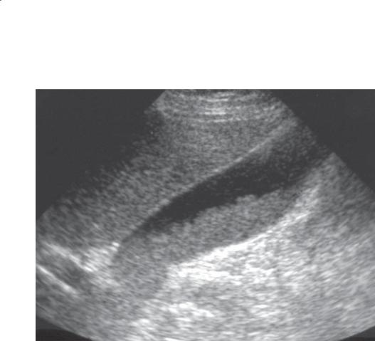
628 MAYO CLINIC GASTROINTESTINAL IMAGING REVIEW
CASE 7.67
Findings
Sonogram. Longitudinal view of the gallbladder shows dependent layering low-level echogenic material within the gallbladder with no posterior acoustic shadowing.
Di erential Diagnosis
Gallbladder sludge
Diagnosis
Gallbladder sludge
Discussion
Gallbladder sludge is concentrated, echogenic bile containing particulate material composed of cholesterol crystals and calcium bilirubinate
granules. Sludge usually layers dependently within the gallbladder. It should not result in posterior acoustic shadowing unless stones also are present. Sludge most commonly results from bile stasis and is frequent in patients who undergo prolonged fasting or hyperalimentation. It also occurs in patients with biliary obstruction. Gallbladder sludge may be a precursor to stone formation, but in most cases it disappears without forming stones.
Gallbladder: Masses and Filling Defects
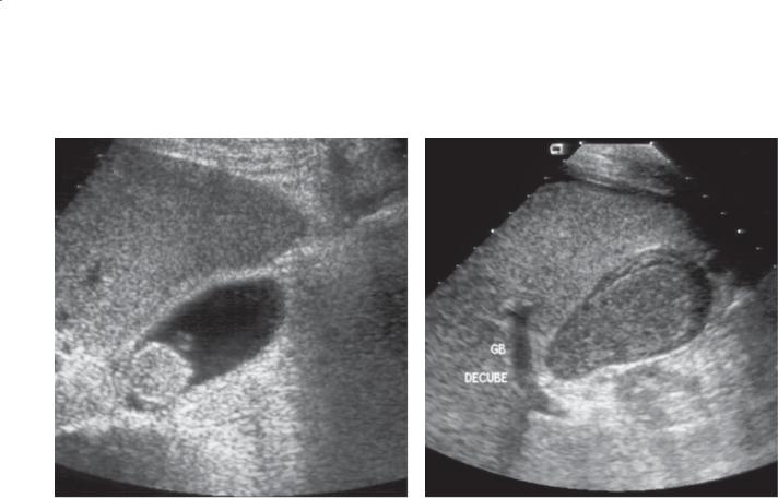
CASE 7.68
Findings
CASE 7.68. Sonogram. A rounded echogenic mass is present near the neck of the gallbladder. This mass was mobile within the gallbladder with repositioning of the patient.
CASE 7.69. Sonogram. A large mixed-echogenicity mass nearly fills the gallbladder lumen.
Di erential Diagnosis
1.Tumefactive sludge (sludge balls)
2.Gallbladder carcinoma
3.Gallbladder metastases
Diagnosis
Tumefactive sludge
7. BILE DUCTS AND GALLBLADDER 629
CASE 7.69
Discussion
Tumefactive sludge is a ball of sludge and can be mistaken for tumor without careful sonographic evaluation. Lack of shadowing distinguishes sludge balls from gallstones. Mobility within the gallbladder and lack of internal blood flow allow differentiation of sludge balls from polyps and tumors.
Gallbladder: Masses and Filling Defects
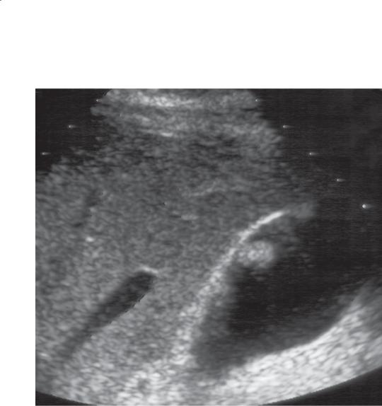
630 MAYO CLINIC GASTROINTESTINAL IMAGING REVIEW
CASE 7.70
Findings |
Discussion |
|
Sonogram. A solitary nonshadowing filling defect is |
Cholesterol polyps of the gallbladder are classified |
|
present within the gallbladder. |
as a form of cholesterolosis. Cholesterolosis is a |
|
|
|
condition of unknown origin in which cholesterol is |
Di erential Diagnosis |
deposited within the lamina propria of the gallbladder. |
|
1. |
Cholesterol polyp |
Cholesterol polyps are the commonest type of |
2. |
Adenoma |
gallbladder polyp. Because they usually are multiple |
3. |
Metastasis |
and small (<10 mm), it is common to detect only the |
|
|
largest at sonography. Gallstones shadow and move, |
Diagnosis |
whereas cholesterol polyps do not shadow and are |
|
Cholesterol polyp |
nonmobile. |
|
Gallbladder: Masses and Filling Defects
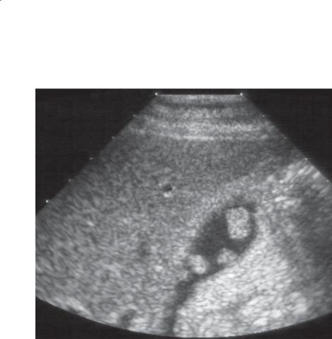
7. BILE DUCTS AND GALLBLADDER 631
CASE 7.71
Findings |
Discussion |
Sonogram. Several nonshadowing filling defects are |
Most polyps of the gallbladder are cholesterol polyps. |
present within the gallbladder lumen, arising from the |
These types of polyps are a form of cholesterolosis. |
gallbladder wall. |
Cholesterol polyps usually are pedunculated with |
|
a single layer of epithelium covering a core of |
Di erential Diagnosis |
cholesterol-filled cells. There are no known clinical |
1. Cholesterol polyps |
sequelae to this finding. At sonography, these polyps |
2. Gallbladder adenomas |
can be distinguished from calculi by the absence of |
|
an acoustic shadow and attachment to the gallbladder |
Diagnosis |
wall. Adenomatous polyps are usually solitary filling |
Cholesterol polyps |
defects and are much less common than cholesterol |
|
polyps. Polyps larger than 1 cm may be malignant. In |
|
most cases, when a gallbladder polyp larger than 1 cm |
|
is identified, the gallbladder is surgically removed to |
|
exclude malignancy. |
|
Multiple polypoid intraluminal masses resembling |
|
polyps also can be seen in patients with metastatic |
|
melanoma (case 7.77). Detection of gallbladder polyps |
|
should be viewed with suspicion in patients with |
|
a history of melanoma. Usually other evidence of |
|
metastases is present. |
Gallbladder: Masses and Filling Defects
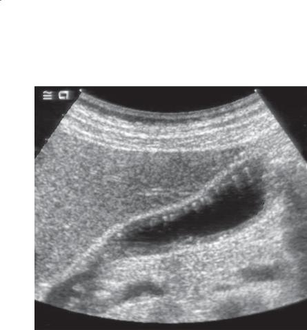
632 MAYO CLINIC GASTROINTESTINAL IMAGING REVIEW
CASE 7.72
Findings
Sonogram. The gallbladder wall is diffusely thickened. There are echogenic foci within the nondependent wall with associated comet-tail artifacts that project into the lumen.
Di erential Diagnosis
1.Adenomyomatosis (diffuse)
2.Acute cholecystitis
3.Chronic cholecystitis
Diagnosis
Adenomyomatosis (diffuse)
Discussion
Adenomyomatosis usually affects only a segment of the gallbladder (cases 7.73 and 7.74). Diffuse involvement can resemble either acute or chronic cholecystitis.
The finding of echogenic foci within the thickened wall with the comet-tail ring-down artifact is highly suggestive of adenomyomatosis. The cause of this condition is unknown.
In the past, adenomyomatosis was classified as one of the hyperplastic cholecystoses—a nonneoplastic, noninflammatory gallbladder abnormality. The term “hyperplastic cholecystoses” is not used widely today. Adenomyomatosis refers to convoluted infoldings of the normal gallbladder mucosa (Rokitansky-Aschoff sinuses) with associated smooth muscle proliferation. There is secondary wall thickening that can be diffuse or segmental. Diffuse involvement can resemble either acute or chronic cholecystitis. The finding of echogenic foci within the thickened gallbladder wall with associated comet-tail artifact is highly suggestive of adenomyomatosis. The comet-tail reverberation artifacts are due to precipitated cholesterol crystals trapped within the Rokitansky-Aschoff sinuses.
Gallbladder: Masses and Filling Defects

CASE 7.73
Findings
CASE 7.73. Sonogram. There is marked annular thickening of the gallbladder wall at the fundus with a thick septation that causes near complete segmentation of the gallbladder.
CASE 7.74. Sonogram. Focal masslike thickening of the wall of the gallbladder is present near the fundus.
Di erential Diagnosis
1.Focal adenomyomatosis
2.Gallbladder carcinoma
Diagnosis
Focal adenomyomatosis
7. BILE DUCTS AND GALLBLADDER 633
CASE 7.74
Discussion
Focal adenomyomatosis is characterized by hyperplasia of the mucosa and smooth muscle with secondary gallbladder wall thickening. Mucosal herniations
into the muscular layer, called Rokitansky-Aschoff sinuses, often contain precipitated cholesterol crystals. Adenomyomatosis is usually focal in the fundus but may be diffuse throughout the gallbladder
(case 7.72). These 2 cases show 2 common sonographic appearances of focal adenomyomatosis. Segmental involvement can narrow and distort the gallbladder lumen (case 7.73) or cause a filling defect in the fundus (case 7.74). Differentiation of focal adenomyomatosis from gallbladder carcinoma sometimes is difficult. Echogenic foci within the focal gallbladder wall thickening with comet-tail artifacts are due to precipitated cholesterol crystals within the RokitanskyAschoff sinuses and are nearly always pathognomonic for adenomyomatosis. This condition has no malignant potential. The cause of adenomyomatosis is unknown.
Gallbladder: Masses and Filling Defects
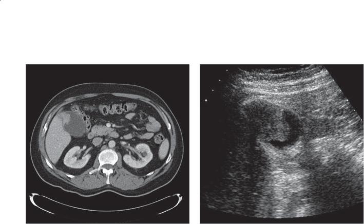
634 MAYO CLINIC GASTROINTESTINAL IMAGING REVIEW
CASE 7.75
A B
Findings
A.Contrast-enhanced CT. A soft tissue mass is visible in the gallbladder lumen, arising from the gallbladder wall.
B.Sonogram. The polypoid mass is present within the gallbladder lumen.
Di erential Diagnosis
Gallbladder carcinoma (polypoid)
Diagnosis
Gallbladder carcinoma (polypoid)
Discussion
Although the commonest manifestation of a gallbladder carcinoma is an infiltrating mass arising from the gallbladder fossa and extending into the liver (case 7.76), approximately one-quarter of gallbladder carcinomas appear as polypoid masses. These usually are well-differentiated tumors that are more likely to be resected for cure because of their well-circumscribed growth pattern.
Th e ability to demonstrate blood flow within polypoid gallbladder masses on sonography allows differentiation of gallbladder polyps (or carcinoma) from tumefactive sludge (cases 7.68 and 7.69).
Gallbladder: Masses and Filling Defects
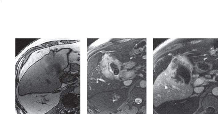
CASE 7.76
A B
Findings
A.T1-weighted MRI. Soft tissue mass within the liver surrounds the gallbladder fossa.
B.T2-weighted MRI. The abnormal soft tissue mass is of high signal intensity and surrounds the gallbladder, which contains stones. The mass abuts the liver capsule.
C.Contrast-enhanced MRI. Heterogeneous enhancement of the mass encasing the gallbladder.
Di erential Diagnosis
1.Gallbladder carcinoma
2.Hepatocellular carcinoma
3.Complicated cholecystitis
4.Metastases to the gallbladder
Diagnosis
Gallbladder carcinoma
7. BILE DUCTS AND GALLBLADDER 635
C
Discussion
Gallbladder carcinoma usually arises as a result of chronic cholecystitis. Most patients have gallstones, and porcelain gallbladder (caused by chronic cholecystitis) is a known risk factor. In the United States, the majority of patients are elderly women. There are 2 morphologic tumor types: scirrhous and polypoid. The scirrhous form infiltrates the liver from the gallbladder (as in this case), and the polypoid form (case 7.75) grows into and eventually fills the gallbladder lumen. The prognosis from this disease is poor, in that 85% of patients die within the first year.
Th ree main presentations are found at crosssectional imaging studies. These include a gallbladder fossa mass (most common), polypoid mass, or mural thickening (least common). When a nonspecificappearing liver mass is discovered, the finding of stones or a porcelain gallbladder is helpful for suggesting the correct diagnosis. These tumors can spread by various routes. Spread into the adjacent liver is most common, and often the tumor can spread locally into the
porta hepatis and cause biliary obstruction. Regional lymphadenopathy, hematogenous spread into the liver, and biliary invasion also can occur.
Gallbladder: Masses and Filling Defects
636 MAYO CLINIC GASTROINTESTINAL IMAGING REVIEW
CASE 7.77
A B
Findings
A.Sonogram. Two large polypoid masses are present within the gallbladder.
B.Color-Doppler sonogram. Blood flow is easily demonstrated within the largest polypoid mass.
Di erential Diagnosis
1.Gallbladder carcinoma
2.Gallbladder polyps (adenomas)
3.Gallbladder metastases
Diagnosis
Gallbladder metastases (melanoma)
Discussion
Metastatic disease to the gallbladder is uncommon. The most common tumor to metastasize to the gallbladder is melanoma (less commonly gastric and pancreatic carcinoma). Gallbladder polyps in patients with a history of melanoma should be viewed with suspicion. However, patients with melanoma metastatic to the gallbladder usually have other evidence of metastatic disease elsewhere in the abdomen. This same sonographic appearance can be seen with gallbladder adenomas or gallbladder carcinoma. However, gallbladder adenomas are almost always solitary.
Gallbladder: Masses and Filling Defects

7. BILE DUCTS AND GALLBLADDER 637
TABLE 7.5
Masses and Filling Defects of the Gallbladder |
CASE |
|
|
|
|
Benign |
|
|
Gallstones |
Stones are mobile with posterior acoustic shadowing. Gallbladder |
7.65 and 7.66 |
|
filled with stones results in wall-echo-shadow triad |
|
Sludge |
Dependently layering low-level echogenic material with no |
7.67 |
|
posterior acoustic shadowing |
|
Tumefactive sludge |
Sludge balls resemble polypoid tumors. Mobile without internal |
7.68 and 7.69 |
|
blood flow, no shadowing |
|
Gallbladder polyps |
Usually less than 1 cm. Most commonly cholesterol polyps. |
7.70 and 7.71 |
|
Nonmobile and no posterior acoustic shadowing. Internal blood |
|
|
flow sometimes is seen in larger polyps |
|
Adenomyomatosis |
Convoluted infoldings of gallbladder mucosa with smooth muscle |
7.72–7.74 |
|
proliferation. Gallbladder wall thickening and echogenic foci |
|
|
with ring-down artifact representing cholesterol crystals trapped |
|
|
within the Rokitansky-Aschoff sinuses. Focal adenomyomatosis can |
|
|
appear masslike |
|
|
|
|
Malignant |
|
|
Gallbladder carcinoma |
Polypoid form presents as a polypoid mass in gallbladder lumen. |
7.75 and 7.76 |
|
Scirrhous form infiltrates the liver from the gallbladder |
|
Metastasis |
Indistinguishable radiographically from a benign polyp or |
7.77 |
|
gallbladder carcinoma. Melanoma common |
|
|
|
|
Gallbladder: Masses and Filling Defects
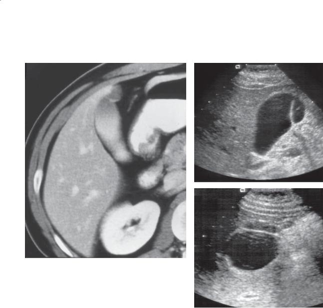
638 MAYO CLINIC GASTROINTESTINAL IMAGING REVIEW
CASE 7.78 |
CASE 7.79 |
|
A |
B
Findings
CASE 7.78. Contrast-enhanced CT. High-attenuation material is present within the gallbladder lumen. Near the tip of the gallbladder fundus is a region of focal narrowing.
Sonogram. A and B. Longitudinal (A) and transverse (B) views of the gallbladder show a thin infolding of the gallbladder wall near the fundus.
Di erential Diagnosis
1.Phrygian cap of the gallbladder
2.Circumferential adenomyomatosis
Diagnosis
Phrygian cap of the gallbladder
Discussion
A phrygian cap is a congenital infolding of the gallbladder wall that does not have any known clinical sequela. It is most likely to be confused with focal adenomyomatosis of the gallbladder (cases 7.73 and 7.74), in which there is localized, circumferential narrowing of the lumen of the gallbladder caused by a septum or annular thickening of the gallbladder wall. These 2 conditions can be distinguished because a phrygian cap is always located in the gallbladder fundus and is composed of a thin membrane of tissue, whereas adenomyomatosis is thicker and can be located anywhere in the gallbladder.
Gallbladder: Miscellaneous
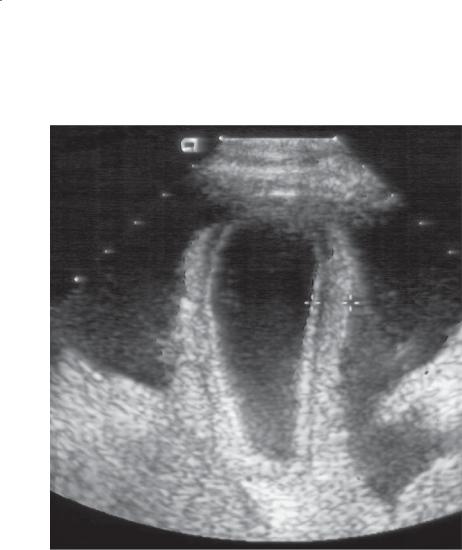
CASE 7.80
Findings
Sonogram. The gallbladder wall is markedly thickened. A large amount of ascites is present around the gallbladder.
Di erential Diagnosis
1.Cholecystitis
2.Adenomyomatosis
3.Hepatitis
4.Cirrhosis
5.Portal hypertension
6.Hypoproteinemia
7.Heart failure
8.Lymphatic obstruction
9.Renal failure
10.Pancreatitis
Diagnosis
Diffuse gallbladder wall thickening (due to ascites, cirrhosis, and portal hypertension)
7. BILE DUCTS AND GALLBLADDER 639
Discussion
Diffuse gallbladder wall thickening is present when the wall is 3 mm or more in diameter. The wall usually is uniformly echogenic without evidence of intramural edema (linear zones of sonolucency). As a solitary finding it has little specificity and must be viewed in context with other clinical findings. The most common cause of a thick gallbladder wall on sonography is a nondistended gallbladder (usually due to the patient having a recent meal). Abnormal gallbladder wall thickening can, therefore, not be confidently diagnosed unless the gallbladder is well distended.
Gallbladder: Miscellaneous
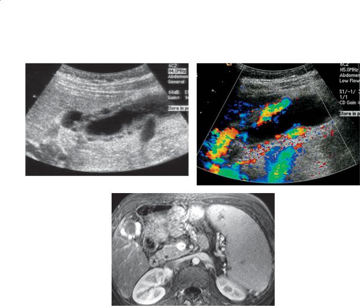
640 MAYO CLINIC GASTROINTESTINAL IMAGING REVIEW
CASE 7.81
A B
C
Findings |
Discussion |
A.Sonogram. Cystic thickening of the gallbladder wall is seen on this gray-scale image.
B.Color-Doppler sonogram. The cystic spaces within the gallbladder wall represent vessels.
C.MRI. This post-gadolinium image demonstrates large collateral vessels around the gallbladder in the right upper quadrant. Incidentally noted is splenomegaly, also consistent with changes of portal venous hypertension.
Di erential Diagnosis
Gallbladder varices
Diagnosis
Gallbladder varices
Gallbladder varices are a rare cause of gallbladder wall thickening, which can be diagnosed easily with the use of color Doppler sonography. They develop in patients with portal hypertension, particularly in those with chronic portal vein thrombosis. Gallbladder varices are almost always asymptomatic. In rare instances, they may result in life-threatening hemobilia or rupture of the gallbladder.
Gallbladder: Miscellaneous

7. BILE DUCTS AND GALLBLADDER 641
TABLE 7.6
Miscellaneous Gallbladder Conditions |
CASE |
|
|
|
|
Phrygian cap |
Thin membrane in gallbladder fundus |
7.78 and 7.79 |
|
|
|
Wall thickening |
Nonspecific: ascites, cirrhosis, portal venous hypertension |
7.80 |
|
|
|
Gallbladder varices |
Collateral vessels around gallbladder, usually with portal |
7.81 |
|
venous hypertension |
|
|
|
|
Gallbladder: Miscellaneous

642 MAYO CLINIC GASTROINTESTINAL IMAGING REVIEW
TABLE 7.7
Differential Diagnoses
Bile Duct Obstruction
BENIGN
Choledocholithiasis
Sludge balls, mucous plugs, blood clots
Mirizzi syndrome (edema from cystic duct stone) Benign stricture
Surgery or intervention Passed stone Pancreatitis
Trauma
Cholangitis (infectious, primary sclerosing cholangitis, Oriental cholangiohepatitis, AIDS)
Sphincter of Oddi spasm Biliary papillomatosis Biliary cystadenoma
MALIGNANT
Cholangiocarcinoma
Pancreatic carcinoma
Ampullary, duodenal carcinoma
Invasive gallbladder carcinoma
Adenopathy (benign and malignant)
Biliary cystadenocarcinoma
Gallbladder Masses and Filling Defects
Stones
Tumefactive sludge (sludge balls) Polyps (cholesterol, adenomatous) Adenomyomatosis
Gallbladder carcinoma Gallbladder metastases
Acoustic Shadowing From Gallbladder Fossa
Gallstones (wall-echo-shadow triad) Porcelain gallbladder (clean shadow) Emphysematous cholecystitis (dirty shadow
from wall)
Intraluminal gas (dirty shadow, usually due to interventional procedure)
Diffuse Gallbladder Wall Thickening (≥3 mm)
BILIARY
Acute cholecystitis
Chronic cholecystitis
Adenomyomatosis
Primary sclerosing cholangitis
NONBILIARY
Nonfasting gallbladder
Hepatitis, cirrhosis
Portal hypertension, ascites
Hypoproteinemia
Heart failure
Pancreatitis
Lymphatic obstruction
Gallbladder varices
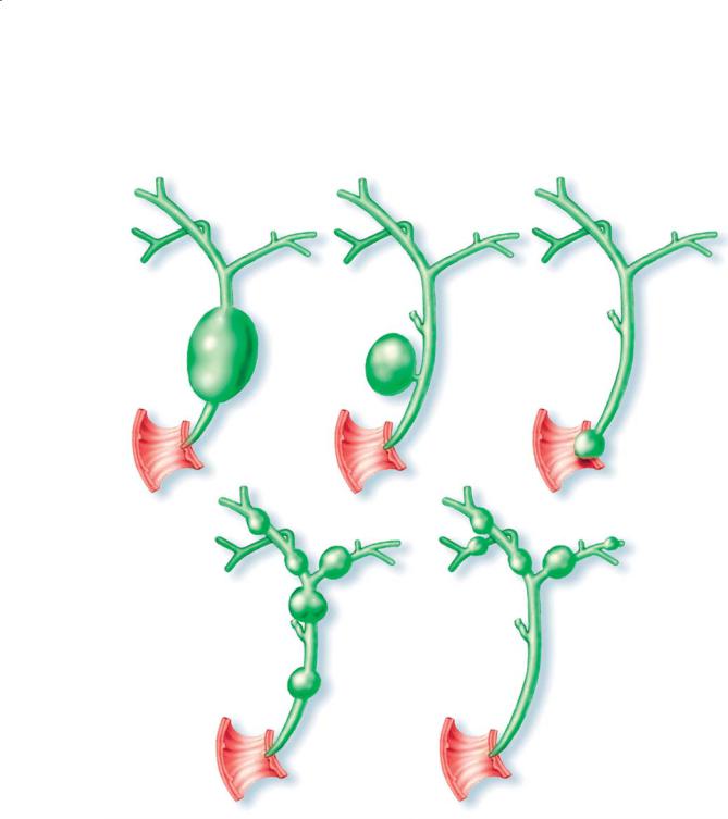
|
|
7. BILE DUCTS AND GALLBLADDER 643 |
|
|
|
|
CHOLEDOCHAL CYSTS |
|
Type I (fusiform) |
Type II (diverticulum) |
Type III (choledococele) |
Type IV (intraand |
Type V (Caroli) |
|
extrahepatic) |
||
|
D.F.
© MAYO 2013
Figure 7.82 (Used with permission of Mayo Foundation for Medical Education and Research.)
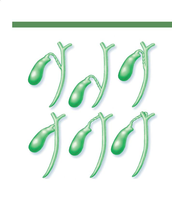
644 MAYO CLINIC GASTROINTESTINAL IMAGING REVIEW
CYSTIC DUCT ANOMALIES
|
Low insertion with |
Conjoined cystic and |
Normal |
common duct |
common duct |
D.F.
© MAYO 2013
Absent |
Insertion into right |
Insertion at |
cystic duct |
hepatic duct |
bifurcation |
Figure 7.83 (Used with permission of Mayo Foundation for Medical Education and Research.)
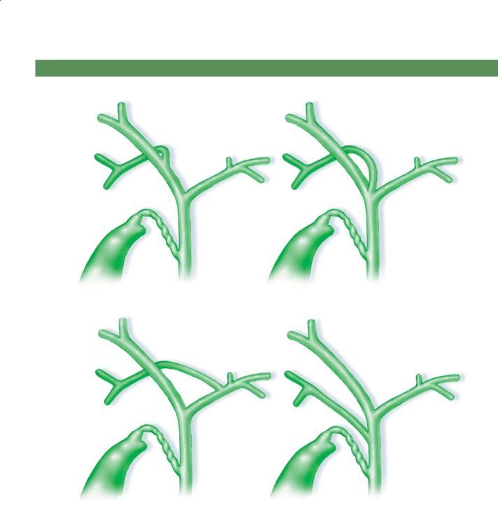
7. BILE DUCTS AND GALLBLADDER 645
INTRAHEPATIC DUCT VARIATIONS
Typical |
Triple confluence |
RA |
RA |
RP |
RP |
R |
|
L |
L |
Right posterior |
Right posterior |
into left |
into CHD |
RA |
|
RP |
RA |
|
L |
RP |
D.F. |
CHD |
© MAYO |
|
2013 |
|
CHD, common hepatic duct; L, left hepatic duct; R, right hepatic duct; RA, right anterior segmental duct; RP, right posterior segmental duct
Figure 7.84 (Used with permission of Mayo Foundation for Medical Education and Research.)
646 MAYO CLINIC GASTROINTESTINAL IMAGING REVIEW
BILE DUCT CONDITIONS
Bacterial cholangitis |
Primary biliary |
Oriental |
|
cirrhosis |
|
|
|
cholangiohepatitis |
Hilar cholangiocarcinoma
Primary sclerosing |
Extrinsic compression |
cholangitis |
Benign or malignant |
|
|
|
adenopathy |
Mirizzi
syndrome D.F.
© MAYO 2013
Papillomatosis
Choledocholithiasis
Pancreatic adenocarcinoma
Sphincter of Oddi spasm
Pseudocalculus
Figure 7.85 (Used with permission of Mayo Foundation for Medical Education and Research.)
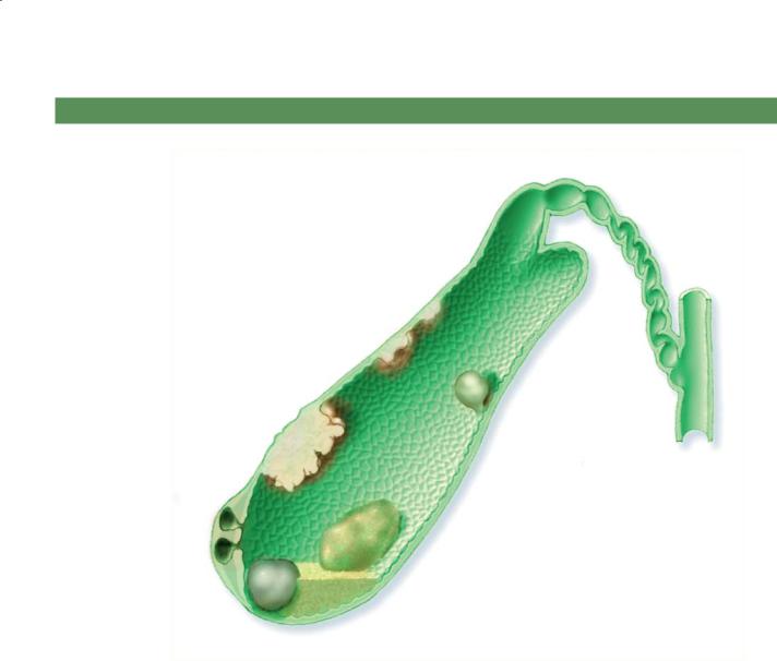
7. BILE DUCTS AND GALLBLADDER 647
GALLBLADDER MASSES
Metastases
|
D.F. |
Carcinoma |
© MAYO |
2013 |
|
|
Polyp |
Focal |
Tumefactive sludge |
|
adenomyomatosis |
||
(sludge ball) |
||
|
Stone Sludge
Figure 7.86 (Used with permission of Mayo Foundation for Medical Education and Research.)
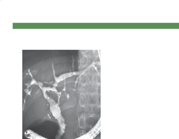
648 MAYO CLINIC GASTROINTESTINAL IMAGING REVIEW
QUESTIONS
Multiple Choice (choose the best answer)
7.1.Which of the following is the most likely diagnosis for the findings in the figure?
a.Primary sclerosing cholangitis
b.Caroli disease
c.Mirizzi syndrome
d.Oriental cholangiohepatitis
e.Cholangiocarcinoma
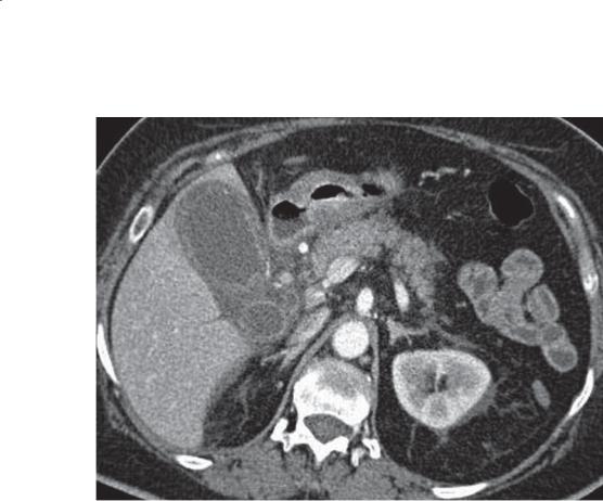
7. BILE DUCTS AND GALLBLADDER 649
7.2.Which of the following symptoms or signs is associated with the findings in the figure?
a.Skin eruption
b.Normal results of abdominal examination
c.Positive sonographic Murphy sign
d.Jaundice
e.Asterixis
7.3.For the case shown in Question 7.2, what is the usual next management step in patients with this condition?
a.Percutaneous drainage
b.Ultrasonography
c.Hepatobiliary iminodiacetic acid scanning
d.Surgery
e.Observation
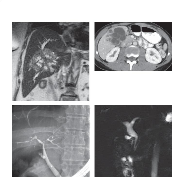
650 MAYO CLINIC GASTROINTESTINAL IMAGING REVIEW
A B
C D
Match images A through F with the responses listed below.
7.4. |
_____ Associated with chronic ulcerative colitis |
7.6. |
_____ Pancreatitis cause |
7.5. |
_____ Congenital |
7.7. |
_____ Delayed enhancement |
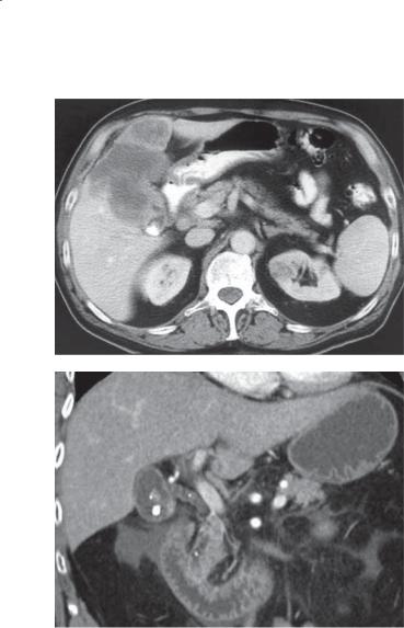
7. BILE DUCTS AND GALLBLADDER 651
E
F
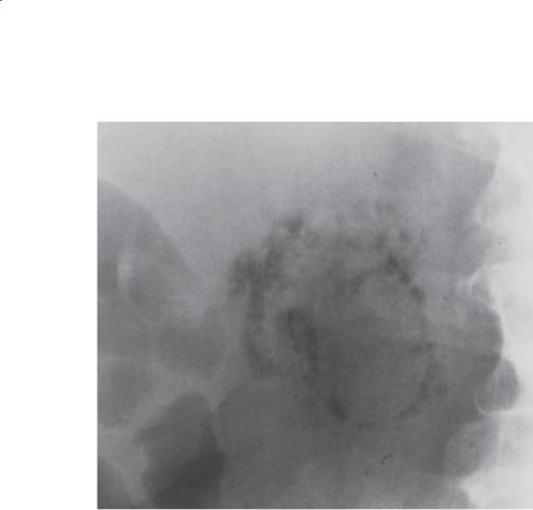
652 MAYO CLINIC GASTROINTESTINAL IMAGING REVIEW
7.8.What disease is most likely responsible for the condition shown in the figure?
a.Diverticulitis
b.Perforated peptic ulcer
c.Small bowel ischemia
d.Diabetes
e.Immunodefi ciency syndrome

7. BILE DUCTS AND GALLBLADDER 653
ANSWERS
7.1.Answer a.
Th e examination performed was ERCP. The common bile and common hepatic ducts are irregular in caliber and contour. The intrahepatic bile ducts are also irregular in caliber. These findings are typical for primary sclerosing cholangitis.
Caroli disease, a type of choledochol cyst, usually presents as saccular dilatation of the intrahepatic ducts.
Mirizzi syndrome occurs when a gallstone impacts in the neck of the gallbladder or cystic duct with secondary obstruction of the common hepatic duct. There is no mass effect in the region of the gallbladder, and the common hepatic duct is not obstructed.
Oriental cholangiohepatitis presents with multiple intrahepatic strictures due to prior infection with subsequent stasis, dilatation, and retained stones and debris within the ducts. There is no evidence of filling defects in the intrahepatic ducts, and the irregular extrahepatic ducts would be unusual in this condition.
Cholangiocarcinoma presents as an obstructing or infiltrating mass or filling defect within the bile ducts. These findings are not present in this case. Cholangiocarcinoma is a complication of primary sclerosing cholangitis and is often identified at ERCP by identification of a dominant high-grade stricture.
7.2.Answer c.
7.3.Answer d.
In patients with the diagnosis of acute cholecystitis, the next step is emergency cholecystectomy. Delay in diagnosis with other imaging findings runs the risk of gallbladder peforation, sepsis, and abscess formation. Percutaneous drainage would be considered only in patients who are not candidates for operation.
7.4.Answer c.
Primary sclerosing cholangitis, shown in figure c, can present with diffusely attenuated intrahepatic ducts, but they are always associated with segmental strictures and ductal dilatation (as in this case). Nearly all patients with primary sclerosing cholangitis have ulcerative colitis.
7.5.Answer d.
Figure d shows a choledochal cyst. Congenital dilatation of the biliary system (usually the common bile or hepatic ducts) is the key finding of a choledochal cyst. The various types of choledochal cysts are illustrated in the figure on page 643.
7.6.Answer f.
Figure f shows choledocholithiasis and cholelithiasis.
Multiple filling defects containing calcification can be visualized within the gallbladder and extrahepatic biliary tree. Gallstones and alcohol are the 2 main causes of pancreatitis.
7.7.Answer a.
Th is patient has thickening of the wall of the gallbladder with a pericholecystic fluid collection. The findings are most likely due to acute cholecystitis. Nearly always, patients with this condition have a positive Murphy sign with acute pain directly over the gallbladder when it is compressed at ultrasonography. This finding is helpful to differentiate gallbladder wall thickening due to other causes, such as hypoalbuminemia and cirrhosis.
Normal results of abdominal examination would not be expected in a patient with acute cholecystitis. Skin eruptions are not associated with this disease.
Jaundice usually is associated with either biliary obstruction or liver failure. No other evidence of biliary obstruction or cirrhosis is present on this image. Asterixis, a tremor causing wrist flapping when the wrists are extended, occurs in patients with various types of encephalopathy (including hepatic encephalopathy).
Figure a shows cholangiocarcinoma. There is a large central mass within the liver with dilated bile ducts coursing into the mass. These findings could also be due to a solitary metastasis, hepatocellular carcinoma, or gallbladder cancer. The central location and upstream biliary dilatation are characteristic of a Klatskin tumor (central cholangiocarcinoma). Because of the dense fibrotic nature of these tumors, they can enhance on delayed images at CT and MRI.
Figure b shows biliary cystadenoma. These lesions can resemble benign cysts, but often they contain internal septations. Because of the potential to become a cystadenocarcinoma, they are usually removed surgically. Nodularity of the internal contents of these tumors suggests malignant transformation.
Figure e shows gallbladder carcinoma. A heterogeneously enhancing mass fills the gallbladder fossa and extends into the adjacent left hepatic lobe.

654 MAYO CLINIC GASTROINTESTINAL IMAGING REVIEW
Findings are typical of a gallbladder carcinoma. Note the solid mass in the left kidney.
7.8.Answer d.
Th is is a case of emphysematous cholecystitis with air trapped within the wall of the gallbladder and within the pericholecystic tissues. This condition is usually
due to ischemia of the gallbladder due to small vessel disease in diabetes. Diverticulitis might be complicated by a hepatic abscess (usually without extraluminal gas). Perforated peptic ulcer would likely present with pneumoperitoneum. Small bowel ischemia would present with pneumatosis intestinalis and portal venous gas. An immunodeficiency is unlikely.