


234 Part VI – Differential Diagnosis
109 T2 Bright Liver Lesions
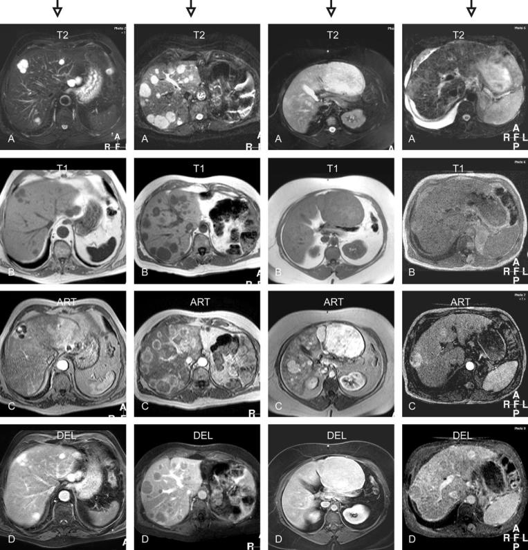
Evaluation of the T2-weighted sequences (Figs. 109.1A–109.4A):
–Lesions and the liver differ in appearance in all four examples. The first two types of lesions are however very bright, and multiple, in a non-cirrhotic liver. The third example shows a large moderately bright lesion with a non-cirrhotic liver, whereas in the final example a faintly bright nodule,
cirrhotic liver, and ascites constitute the image.
Evaluation of the T1-weighted sequences (Figs. 109.1B–109.4B):
–The first two types of lesions are similar in appearance. The third example shows isointense lesions, whereas in the final example a faintly T1 bright nodule is visible.
Evaluation of the arterial enhancement pattern (Figs. 109.1C – 109.4C):
–The lesions in the four examples show peripheral nodular, ring-shaped, almost homogeneous, and heterogeneous enhancement patterns, respectively.
Evaluation of the delayed enhancement pattern (Figs. 109.1D – 109.4D):
–The lesions in the four examples show persistent, washout without a capsule, homogeneous without a capsule, and washout with a capsular enhancement within a cirrhotic liver, respectively.
Based on the following pertinent combination of findings the lesions can be characterized as:
1.Multiple hemangiomas (T2 bright with peripheral nodular enhancement)
2.Multiple metastases (ring-shaped enhancement and washout without a capsule)
3.Multiple hepatocellular adenomas (T1 isointense, almost homogeneous enhancement without washout or capsule)
4.Multiple hepatocellular carcinomas (cirrhotic liver, slightly T2 bright lesion, heterogeneous enhancement, and washout with capsular enhancement)
|
|
|
109 T2 Bright Liver Lesions 235 |
|
|
|
|
T2 very bright lesions |
T2 very bright lesions |
T2 bright-isointense lesions |
T2 slightly bright-isointense |
T1 dark lesions |
T1 dark lesions |
T1 isointense lesions |
lesions |
Peripheral nodular |
Ring-shaped enhancement |
Homogeneous enhancement |
T1 slightly bright-isointense |
enhancement |
Washout |
Fade to isointensity |
Heterogeneous enhancement |
Persistent enhancement |
|
|
Washout with capsular |
|
|
|
enhancement |
Fig. 109.1. Multiple hemangiomas |
Fig. 109.2. Multiple metastases |
Fig. 109.3. Hepatocellular adenomas |
Fig. 109.4. Hepatocellular carcinomas |

236 Part VI – Differential Diagnosis
110 T1 Bright Liver Lesions
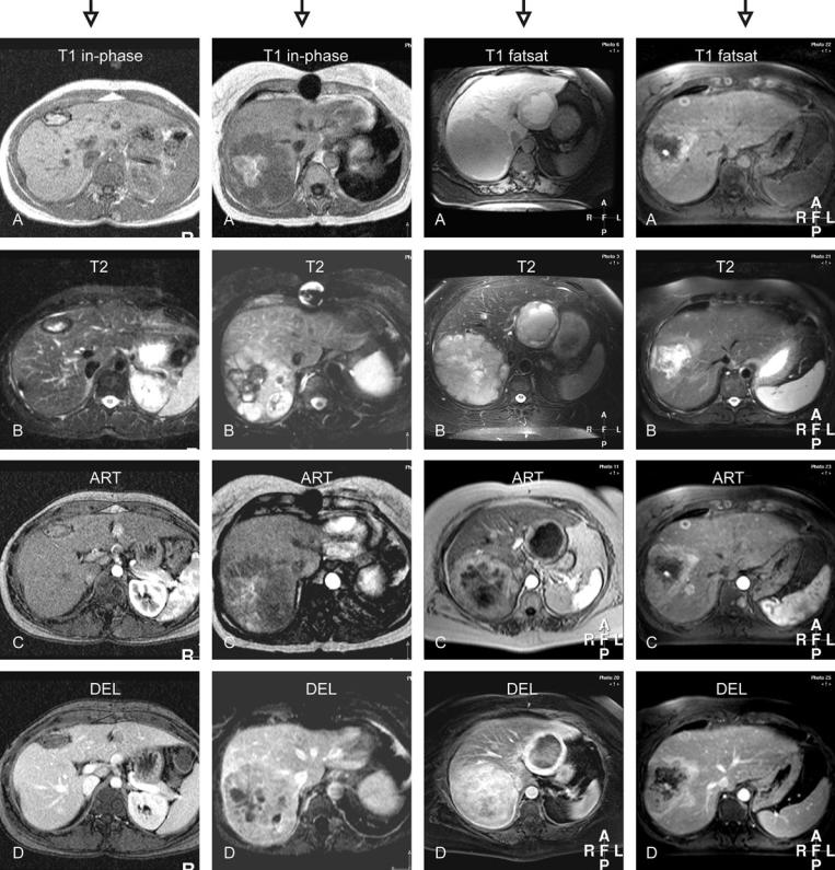
Evaluation of the T1-weighted sequence (Figs. 110.1A – 110.4A):
–All lesions have components with bright signal within a non-cirrhotic liver. In the first example, the lesion is surrounded by a dark rim. The second example shows a large
lesion with a central bright area. The third example shows a cystic lesion with bright contents in the left liver. The fourth example shows multiple, predominantly bright lesions, including one with a ring-shaped appearance.
Evaluation of the T2-weighted sequence (Figs. 110.1B – 110.4B):
–In the first example, the lesion is surrounded by a dark rim with increased thickness. The second example shows a large lesion with a central dark area. The third example shows two bright lesions with cystic and solid components. The fourth
example shows one large and small bright lesions. Evaluation of the arterial enhancement pattern (Figs. 110.1C – 110.4C):
–In the first example, the lesion does not show any enhancement (bright signal was present prior to contrast injection). The second lesion shows some heterogeneous enhancement. The third example shows lesions with a thick rim of en-
hancement. In the fourth example, the lesions show intense enhancement.
Evaluation of the delayed enhancement pattern (Figs. 110.1D – 110.4D):
–In the first example, the lesion shows lack of enhancement (mimics washout). The second lesion shows washout with some persistent heterogeneous enhancement. The third example shows lesions with a persistent rim of enhancement. In the fourth example, the lesions show washout and become less intense.
Based on the following pertinent combination of findings the lesions can be characterized as:
1.Hematoma (T1 bright due to methemoglobin with a rim of hemosiderin)
2.Hemorrhagic carcinoid metastasis (suggests metastases; non-specific findings; recommend clinical and somatostatinscintigraphy correlation)
3.Protein-producing carcinoid metastases (suggests metastases; non-specific findings; recommend clinical and somatostatinscintigraphy correlation)
4.Melanoma metastasis (T1 bright lesions in a patient with a history of uveal melanoma; intense enhancement and washout are typical for melanin-containing liver metastases)
|
|
|
110 T1 Bright Liver Lesions 237 |
|
|
|
|
T1 bright with a dark rim |
T1 very bright within a dark |
T1 bright and dark lesions |
T1 (predominantly) bright |
T2 bright with a dark rim |
lesion |
T2 mixed signal intensity |
lesions |
No enhancement |
T2 dark with a bright lesion |
Irregular ring-shaped |
T2 isointense to very bright |
No enhancement |
Heterogeneous enhancement |
enhancement |
Enhancement of the T1-bright |
|
within the lesion |
Heterogeneous and persistent |
parts |
|
Washout within the solid lesion |
enhancement |
Heterogeneous and persistent |
|
|
|
enhancement |
Fig. 110.1. Hematoma after surgery |
Fig. 110.2. Hemorrhagic carcinoid |
Fig. 110.3. Protein-producing |
Fig. 110.4. Melanoma metastases |
|
metastasis |
carcinoid metastasis |
|