

Книги по МРТ КТ на английском языке / Neurovascular anatomy in interventional neuroradiology Krings et al 2015
.pdfThe Radiculopial and Radiculomedullary Arteries
Fig. 40.4 A segmental artery injection in AP (a) and lateral (b) views demonstrates supply from the same segmental artery to both the dorsolateral spinal artery (arrows) and to the anterior spinal artery (arrowheads). Two separate vessels arise from the radicular artery that accompany the dorsal and the ventral nerve root to reach the cord at the anterior and posterior surface. The anterior artery reaches the midline, whereas the posterior vessel forms its hairpin more laterally and more acutely.
Fig. 40.6 Right T11 segmental artery angiogram in AP (a) and lateral
(b) views reveals the origin of the anterior spinal artery, with its classical hairpin curve, a small ascending branch (arrow), and a larger descending branch (thin double arrows). The white arrows denote the retrocorporeal anastomosis via which the left T12 segmental artery is supplied. On the lateral view, the anterior course of the ASA can be appreciated once it has reached the midline (arrowheads).
First is the anterior radiculomedullary artery, which runs with the anterior nerve root to join the longitudinal trunk on the anterior surface of the spinal cord (i.e., the anterior spinal artery). In addition to this main supply, a minor lateral contri-
Fig. 40.5 Vertebral artery angiograms in AP (a,c) and lateral (b,d) views in two different patients demonstrate the variable origin of the anterior spinal artery from the vertebral artery. It may arise from one or both vertebral arteries, and its junction in the midline is variable, ranging from early fusion in the anterior sulcus to a long unfused segment along the anterior surface of the cord. The upper row (a,b) demonstrates the bilateral unfused supply. The lower row (c,d) demonstrates, in addition to a unilateral supply to the anterior spinal artery (arrows), dorsal supply to the cord from the posterolateral spinal arteries (arrowheads) that may arise from the PICA (when the PICA origin is low or extradural) or the vertebral artery directly (when the PICA origin is high).
bution to a superficial pial network is also derived from the anterior radiculomedullary artery.
Second is the posterior radiculomedullary artery, which accompanies the posterior nerve root and joins the longitudinal systems of posterolateral and/or posterior spinal arteries. The first one is lying laterally, the second one medial to the posterior nerve root entry zone. These arteries mainly supply the pial superficial network of vessels but may give small branches to the gray matter of the posterior horns. One has to bear in mind that these latter arteries predominantly supply the surface of the spinal cord (i.e., the white matter), whereas the anterior radiculomedullary arteries predominantly supply the gray matter of the spinal cord ( Fig. 40.4).
Both the anterior and posterior radiculomedullary arteries supply a system of superficial longitudinal anastomoses that are called the anterior and posterior (or posterolateral) spinal arteries. The anterior spinal artery, with a diameter of 0.2 to 1 mm, travels along the anterior sulcus and typically originates from the two vertebral arteries ( Fig. 40.5), whereas the typi-
207

The Radiculopial and Radiculomedullary Arteries
Fig. 40.7 Three examples (a–c) of the anastomoses of the anterior and posterolateral spinal arteries. (a) The normal anatomy is visualized with a nonfused anterior spinal artery (arrowheads) descending to the level of the cone where the ASA connects through the arcade (curved arrows) to the two posterolateral arteries. (b) The arcade is dilated because of feeding of a hemangioblastoma. The anterior spinal artery (horizontal black arrow) is seen to anastomose not only via the arcade (vertical upward pointing arrows) but also via the vasocorona network (vertical downward pointing arrow) with the dorsolateral spinal arteries (white arrow). (c) Injection into one dorsolateral spinal artery opacifies the contralateral dorsolateral artery via a posterior rope ladder anastomosis (thin double arrows) in a patient with an arteriovenous malformation of the cone.
cally paired posterolateral spinal arteries originate from the preatlantal part of the vertebral artery or from the posteroinferior cerebellar artery (PICA) and typically have a diameter of 0.1 to 0.4 mm.
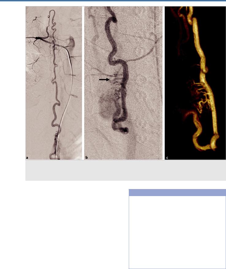
These three arteries run continuously from the cervical spine to the conus medullaris but are not capable of feeding the entire spinal cord. Instead, they are reinforced from the above-men- tioned radiculomedullary arteries that derive from various (and unpredictable) segmental levels by anterior and posterolateral radiculomedullary arteries. Therefore, the blood flow in these vessels may be both caudocranial and craniocaudal. The largest of the anterior radiculomedullary arteries is the arteria radiculomedullaris magna or Adamkiewicz artery, which arises close to the thoracolumbar enlargement (between T9 and L1 [exceptionally at L2 or L3], more often on the left side; Fig. 40.6).
Additional significant ventral feeders are unusual. In the cervical region, one of the ventral radicular feeders between C5 and C8 is often distinctly larger (0.4–0.6 mm) than the others and was termed the artery of the cervical enlargement. It is more often derived from the deep and ascending cervical arteries than from the vertebral artery. The average number of anterior radicular feeders to the cervical cord is two to three. Ventral feeders to the upper cervical cord, originating from the intracranial section of the vertebral artery, may be very small,
can be unior bilateral, and may or may not fuse with each other. Nonfusion of the anterior spinal artery over some distance is frequent in this region. The anterior radiculomedullary arteries branch in a very typical way to reach the spinal cord. The ascending branch continues along the direction of the radicular artery in the midline of the anterior surface. The descending branch, being the larger one at thoracolumbar levels, forms a hairpin curve as soon as it reaches the midline at the entrance of the anterior fissure. Anterior radiculomedullary arteries always reach the cord at the midline, whereas the posterior arteries reach the cord slightly o -midline. The posterior radiculomedullary arteries are connected to the anterior spinal artery through two anastomotic semicircles at the level of the cone, known as the “arcade” of the cone ( Fig. 40.7).
Apart from these conal anastomoses, both systems anastomose via an extensive pial network to which they both contribute. This superficial pial network encircles the spinal cord and has been called the vasocorona. As stated earlier, the major contribution to this superficial pial system is from the posterior radiculomedullary arteries, whereas the supply from the anterior radiculomedullary arteries is derived just before these arteries enter the subpial space in the central sulcus. There, branches to the pial system on the anterior and lateral surface supply the ventral parts of the vasocorona.
208
The Radiculopial and Radiculomedullary Arteries
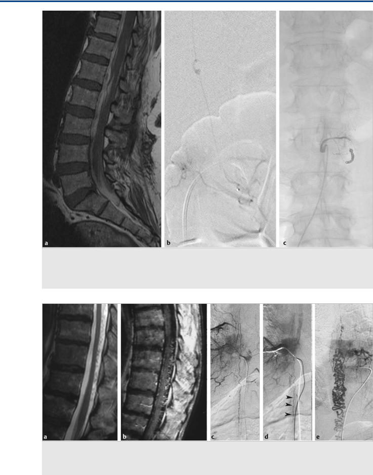
Fig. 40.8 This patient presented hyperacutely with severe back pain and paraplegia. T1-weighted MRI (a) demonstrated massive hemorrhage in the thecal sac (hyperintense subarachnoid signal on T1-weighted scans). Spinal angiography (b) revealed a dissecting aneurysm arising from the dorsolateral spinal artery in its intradural component. The feeding radicular artery was occluded with coils (c). As this was not a shunting lesion, there was no retrograde filling to the dissecting aneurysm from other segmental arteries, and the extensive collateral network, including the basket and the rope ladder anastomoses, prevented ischemic damage to the cord.
Fig. 40.9 T2-weighted (a) and contrast-enhanced T1-weighted (b) MRIs demonstrate abnormal flow voids in this patient, indicating the presence of a dural arteriovenous shunt. Spinal angiography at the level of the artery of Adamkiewicz (c,d) reveals persistent contrast stagnation in the ASA on the delayed images in the venous phase, indicating the presence of a shunt elsewhere (arrowheads). In this patient, a dural arteriovenous fistula was found
(e) at another level.
209
The Radiculopial and Radiculomedullary Arteries
Fig. 40.10 In this pediatric patient with diffuse cord swelling and abnormal vessels, angiography (a,b) did not reveal a shunt but, rather, cord hyperemia. The anterior spinal artery demonstrates increased tortuosity (arrows), a normal finding in the pediatric population that should not be mistaken for a vein.
tions in all segmental arteries that harbor cord supply are necessary. The predominant white matter supply of the dorsolateral arteries implies, in contrast to the predominant gray matter supply of the anterior spinal artery, that embolization via dorsolateral spinal arteries is considered a safer approach. In patients with isolated pathology of the dorsal spinal artery, parent vessel sacrifice may be considered, given the rich anastomotic network of the spinal cord supply ( Fig. 40.8). This can be important for presurgical embolization of spinal cord hemangioblastomas: As they are typically located on the dorsal surface of the cord, their usual supply is from the dorsolateral artery.
Pearls and Pitfalls
●Arteries always run along the anterior or posterior nerve roots to reach the cord. The location of the hairpin curve at the cord surface (lateral for the posterior spinal arteries and midline for the anterior spinal arteries) can be used to identify the type of feeding vessels if you do not use a lateral view.
●Contrast stagnation in the anterior spinal artery is a seen in cases of venous congestion and indicates the presence of a shunt elsewhere that interferes with the normal arteriovenous transit ( Fig. 40.9).
●In pediatric patients, in patients with cord hyperemia, or in patients with distal shunting lesions, the anterior spinal artery is often rather tortuous and should not be confused with a vein ( Fig. 40.10). In addition, pediatric patients and patients who have harbored a shunt from early childhood will have less pruning of the cord supply (i.e., more segmental arteries will still harbor cord supply).
40.3 Clinical Impact, Additional
Information and Cases
The rich anastomotic network implies that when embarking on embolization of spinal cord arteries, a liquid embolic material that is able to penetrate deep into this network may open collaterals via the vasocorona between the posterior and the anterior spinal arteries, which may lead to inadvertent embolization of healthy vessels. The rich network also implies that to “map” the entire supply of a spinal arteriovenous malformation, injec-
Further Reading
[1]Krings T, Geibprasert S, Thron AK. Spinal vascular anatomy. In: Naidich TP, Castillo M, Cha S, Raybaud C, Smirniotopoulos JG, Kollias S, Kleinman GM, eds. Imaging of the Spine: Expert Radiology Series, Expert Consult. Philadelphia: Elsevier Health Sciences; 2010:185–200
[2]Lasjaunias P, Berenstein A, ter Brugge KG. Surgical Neuroangiography. Vol. 1. 2nd ed. Berlin: Springer; 2006
[3]Thron AK. Vascular Anatomy of the Spinal Cord: Neuroradiological Investigations and Clinical Syndromes. Vienna: Springer; 1988
210
The Intrinsic Arteries of the Cord
41 The Intrinsic Arteries of the Cord
41.1 Case Description
41.1.1 Clinical Presentation
A 37-year-old patient presented with acute onset of stabbing back pain followed by paraplegia and complete loss of sensation below her midthoracic dermatomes.
41.1.2 Radiologic Studies
See Fig. 41.1, Fig. 41.2.
41.1.3 Diagnosis
Pial nidus-type arteriovenous malformation (AVM) with a false aneurysm arising from a sulcocommissural artery of the anterior spinal artery.
41.2 Anatomy
The main source of arterial supply to the cord is the anterior spinal artery (ASA) on its ventral axis. The arteries directly supplying the spinal cord that arise from the ASA are the central (sulcal and sulcocommissural) arteries that originate from the anterior spinal artery and the perforating branches arising from the superficial pial network, covering the spinal cord. Sulcal arteries are centrifugal, have a diameter between 0.1 and 0.25 mm, and supply the largest part of the gray matter. They penetrate the parenchyma to the depth of the anterior fissure, course to one side of the cord, and branch mainly within the gray matter ( Fig. 41.3).
These arteries can anastomose via transmedullary arteries with deep perforating arteries from the vasocorona or the posterolateral arteries. These perforating arteries arise from the superficial pial covering of the cord (vasocorona) and penetrate
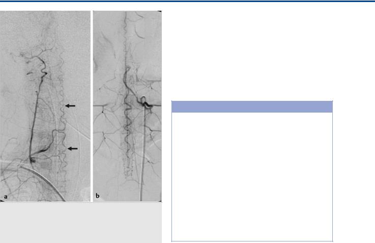
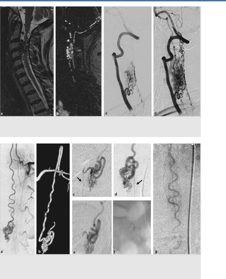
Fig. 41.1 Sagittal T2-weighted (a) and T1-weighted (b) MRIs demonstrate hematomyelia, cord edema, and posteriorly located perimedullary flow voids that enhance after contrast administration in the mid and lower thoracic region, raising suspicion of a spinal cord AVM. Left T10 segmental artery angiograms in AP (c) and lateral (d) views reveal a shunting lesion that was fed by both the anterior (arrow) and the posterior (white arrow) spinal arteries. As an anatomical variation, the supply to both the anterior and the posterior axis arose from the same segmental artery. An aneurysm that is located in the midline and centrally within the cord is noted as the rupture point. Case continued in Fig. 41.2.
211
The Intrinsic Arteries of the Cord
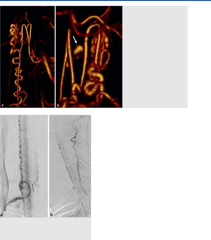
Fig. 41.3 Spinal angiograms in lateral view in two patients (a,b) with cord hyperemia demonstrate the sulcocommissural arteries that supply the anterior aspect of the cord. The arrow points to a transmedullary anastomosis that connects the anterior axis to the posterior axis through the cord.
Fig. 41.2 3D rotational reconstruction images (a, b) demonstrate that the aneurysm (arrow) arose from a sulcocommissural artery that interconnects the anterior and the posterior spinal arteries.
white matter tracts from the periphery, thus constituting a centripetal system. These vessels are numerous, with a diameter of up to 0.05 mm. The posterior and posterolateral spinal arteries distribute blood to the dorsal third of the vasocorona, and in this way, they share with central artery branches the supply of the posterior horn and marginal parts of the central grey matter. The posterior/posterolateral arteries do not have a distinct territory of supply similar to the anterior spinal artery, which means that they predominantly reinforce the rope ladder–like network of posterior pial arteries. The number of central arteries varies in the di erent levels: at the cervical enlargement, about five central arteries are present per (longitudinal) centimeter. They take a horizontal course. The number of central arteries is two to three per centimeter for the thoracic region, with a relatively high prevalence of steeply ascending and descending central artery branches. The densest concentration of central arteries is found in the thoracolumbar enlargement, where six to eight vessels per centimeter can be found ( Fig. 41.4).
41.3 Clinical Impact, Additional
Information and Cases
The dense connections between the anterior and posterior spinal arteries have to be kept in mind when embolizing with a liquid embolic material with high penetrance, as they will be opened and may lead to inadvertent occlusion of healthy vessels. Intramedullary spinal AVMs (i.e., nidusor glomus-type) will almost always be supplied from both the anterior and posterior spinal artery systems, given the rich transmedullary and perimedullary connections via the sulcocommissural and the vasocorona vessels, respectively ( Fig. 41.5).
212
The Intrinsic Arteries of the Cord
Fig. 41.4 Spinal angiogram of the ASA in AP (a) and oblique (b) views and 3D rotational reconstruction (c) reveal a high-flow fistulous AVM supplied by the dorsolateral arteries that is indirectly filled from the anterior spinal artery via the basket anastomosis. In addition, multiple enlarged sulcocommissural arteries (arrow) that course through the thoracolumbar enlargement to reach the dorsal surface are seen to be sumped into the fistula.
Similar to their counterpart, the pial brain AVMs, the spinal intramedullary AVMs may be monoor multicompartmental and can have multiple feeders or a single feeder ( Fig. 41.6).
Secondarily induced angiopathic changes with dilatation of normal vessels may be present and have to be di erentiated from true shunting lesions. In comparison with the intramedullary AVMs, the perimedullary fistulas are supplied (again similar to their cranial counterpart, the pial cerebral AV fistulaes) by only a single feeder. Depending on the location of the transition between the artery and the vein, this may be the anterior or posterolateral spinal artery, a rope-ladder anastomosis, or the vasocorona. In the latter two cases, supply to the fistula will be seen from multiple feeders; however, it has to be kept in mind that this is related to the described anastomoses between the intrinsic cord arteries. In these cases, one can choose the most direct and/or the safest access to the fistula to treat the shunt. The large variety of potential collateral pathways and anastomoses explains the wide variety of types of cord infarctions, as depicted in Fig. 41.7.
Pearls and Pitfalls
●The intrinsic arteries of the cord can be subdivided into those that arise from the anterior spinal artery, which supply the central cord and exhibit a centrifugal course, and those that arise from the posterior spinal artery, which supply the cord periphery (including the white matter) and have a centripetal course.
●They can anastomose via the vasocorona peripherally and via the sulcocommissural central arteries through the depth of the cord. The rich anastomoses explain the variability of cord infarctions on axial sections.
●Perimedullary fistulas have typically a single feeder, although indirect filling can be seen from various cord–supplying arteries, given their rich anastomotic network, whereas central intramedullary AVMs most commonly have multiple direct feeders.
213
The Intrinsic Arteries of the Cord
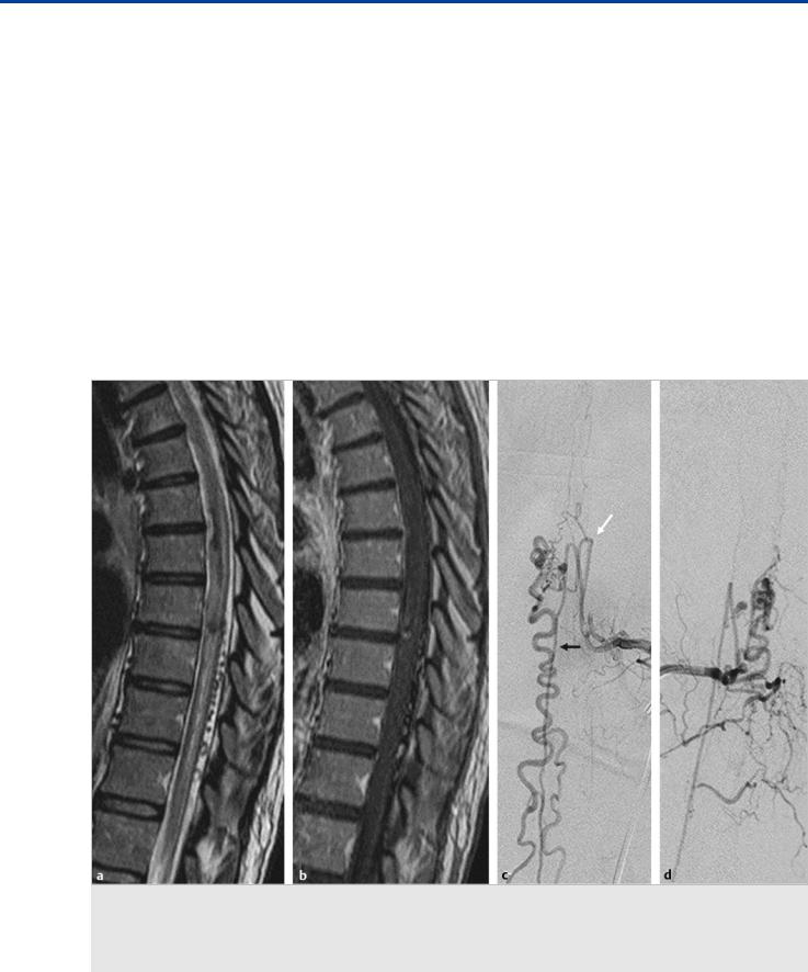
Fig. 41.5 Sagittal T2-weighted MRI (a) and contrast-enhanced MRA (b) demonstrate an intramedullary AVM. Left VA angiogram in lateral view in early
(c) and late (d) arterial phase confirms the presence of the AVM, which is seen to be primarily supplied by the ASA but transgresses the entire cord. Abnormally dilated vessels are also noted on the posterior cord surface, owing to a rich network of anastomoses between the anterior and posterior spinal arteries.
Fig. 41.6 Injection into the segmental artery harboring supply to the anterior spinal artery (a) reveals predominant supply to a pial intramedullary AVM by the ASA. The 3D rotational reconstruction image (b) demonstrates that a single sulcocommissural artery supplied the nidus. A microcatheter injection of the ASA just proximal to the feeding sulcocommissural artery in the lateral (c) and anterior (d) planes reveals the normal-caliber ASA running further caudally (arrows). Distal microcatheter injection (e) demonstrates the catheter location, which allowed for glue deposition just within the sulcocommissural artery (f) that enabled complete exclusion of the AVM and preservation of the normal ASA (g).
214
The Intrinsic Arteries of the Cord
Further Reading
[1]Krings T. Vascular malformations of the spine and spinal cord: Anatomy, classification, treatment. Clin Neuroradiol. 2010 Mar; 20(1):5-24
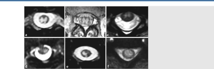
Fig. 41.7 Six different patients with anterior spinal cord infarctions on axial T2-weighted MRIs. The upper row (a,b,c) demonstrates the typical anterior spinal artery territory with the “owl-eye” appearance of T2 hypersignal in the central gray matter of the cord, predominantly supplied by the anterior spinal artery, whereas the white matter is still preserved, as it is primarily supplied by the superficial vasocorona network that is mainly supplied by the posterolateral arteries. The potential transmedullary anastomotic pathways and further distal ischemia of only the lateralized sulcocommissural arteries can explain the different patterns of ischemia, as seen in the lower row (d,e,f).
[2]Krings T, Thron AK, Geibprasert S et al. Endovascular management of spinal vascular malformations. Neurosurg Rev 2010; 33: 1–9
[3]Thron AK. Vascular Anatomy of the Spinal Cord: Neuroradiological Investigations and Clinical Syndromes. Vienna: Springer; 1988
215
The Artery of the Filum Terminale
42 The Artery of the Filum Terminale
42.1 Case Description
42.1.1 Clinical Presentation
A 62-year-old man presented with a 6-month history of progressive bilateral lower extremity paresthesias and weakness. Three months before admission, bladder incontinence developed, and 1 month later, the patient became impotent.
42.1.2 Radiological Studies
See Fig. 42.1, Fig. 42.2.
42.1.3 Diagnosis
Spinal arteriovenous fistula of the filum terminale.
42.2 Embryology and Anatomy
The primordia of the filum terminale forms during secondary neurulation. This process occurs after closure of the caudal neuropore. Degeneration, regression, and retrogressive di erentiations of the secondary neural tube then lead to the devel-
opment of the filum terminale, which remains as a fibrous structure connecting the conus medullaris to the coccyx. The filum terminale has two components. The filum interna is an extension of the primary pia mater that connects the conus medullaris to the dura mater of the terminal thecal sac. The filum externa extends from the termination of the thecal sac to the coccyx.
Classically, the arterial supply to the filum terminale was described as singular, fed by the artery of the filum terminale. This artery is the caudal extension of the anterior spinal artery, which arises at the conus medullaris. Most commonly, the anterior spinal artery bifurcates at the conus medullaris and forms the anastomotic arcade, and the artery of the filum arises from one of the branches of the bifurcation. Anatomic variants have been described, including a trifurcation of the anterior spinal artery, with one of the branches continuing caudally as the artery of the filum.
The argument for singular arterial supply to the filum terminale is supported by the fact that no nerve roots originate caudal to the conus medullaris, and thus all radiculomedullary arterial supply must be carried caudally from the conus to the filum via the artery of the filum terminale. However, this singular supply theory has come into question after a number of
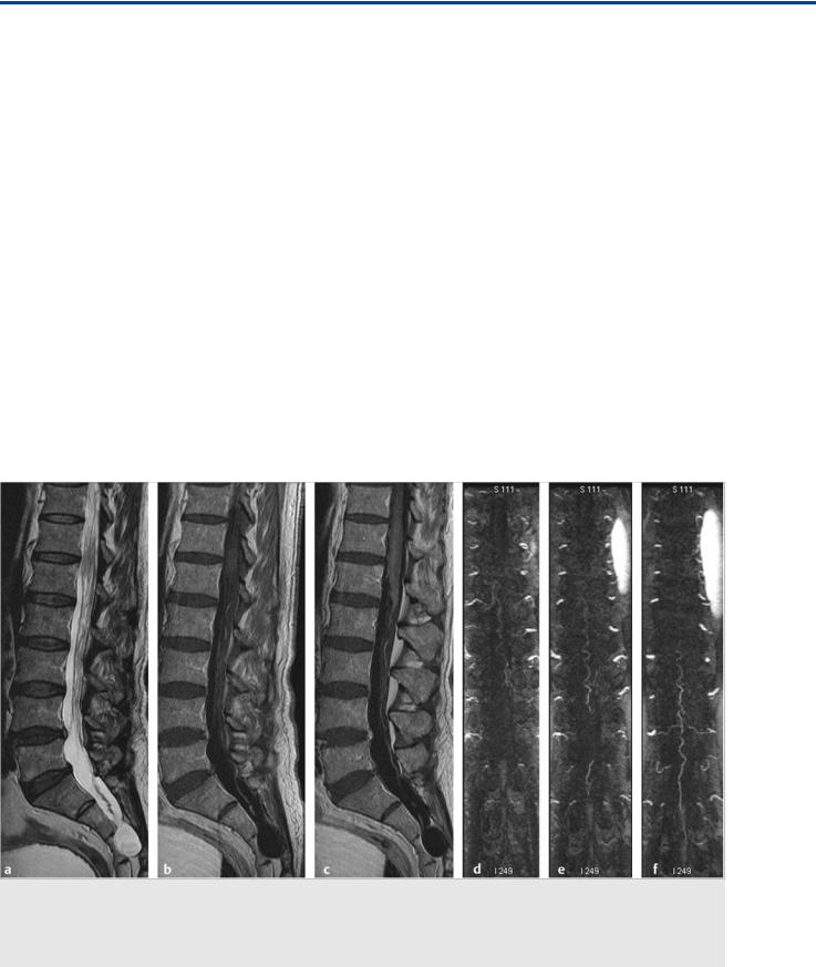
Fig. 42.1 Sagittal T2-weighted (a) MRI demonstrated venous congestion with vasogenic edema of the spinal cord and swelling from the lower thoracic levels to the conus medullaris, along with flow voids suggestive of enlarged perimedullary veins. Contrast-enhanced T1-weighted (b,c) sequences revealed a moderate degree of diffuse enhancement of the lower thoracic spinal cord, indicative of chronic venous ischemia. Contrast-enhanced MRA (d,e,f) of the spine showed a dilated midline vessel, likely the ASA, down to the filum terminale. The patient underwent conventional angiography for further evaluation. Case continued in Fig. 42.2.
216