

C H A P T E R 2
Development of the
Nervous System
Objectives
1.Describe the development of the neural tube, including the stages of development and the adult derivatives of each brain vesicle.
2.Trace the lineage of the cells of the neural tube wall, including the alar and basal plates.
3.Identify the derivatives of the neural crest.
4.Describe the development of the brainstem as well as the general arrangement of motor versus sensory components and somatic versus visceral components.
5.Describe the development of the pituitary (hypophysis).
6.List and characterize major congenital malformations of the central nervous system.
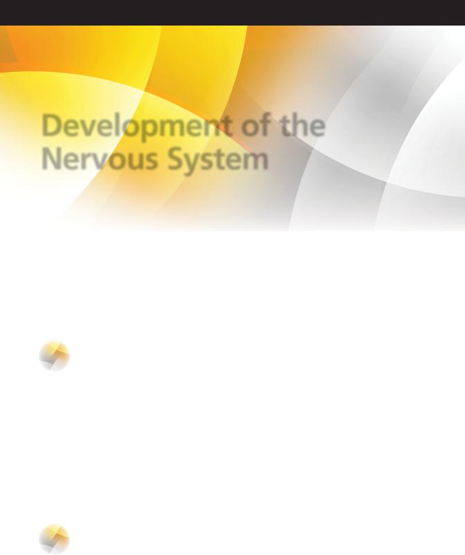
IThe Neural Tube (Figure 2-1) gives rise to the central nervous system (CNS)
(i.e., brain and spinal cord).
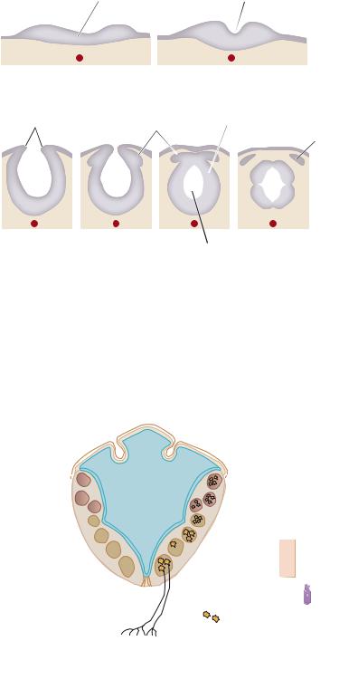
A.The brainstem and spinal cord are composed of plates separated by the sulcus limitans:
1.An alar plate—gives rise to sensory neurons.
2.A basal plate—gives rise to motor neurons (Figure 2-2).
3.Interneurons are derived from both plates.
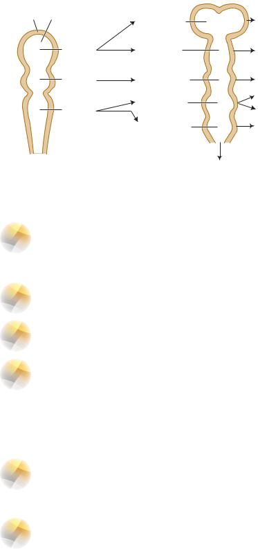
B.The neural tube gives rise to three primary vesicles (forebrain, midbrain, and hindbrain), which develop into five secondary vesicles (telencephalon, diencephalon, mesencephalon, metencephalon, and myelencephalon) (Figure 2-3).
C.Alpha-fetoprotein (AFP) is found in the amniotic fluid and maternal serum. It is an indicator of neural tube defects (e.g., spina bifida, anencephaly). AFP levels are reduced in mothers of fetuses with Down syndrome.
IIThe Neural Crest (see Figure 2-1) gives rise to:
A.The peripheral nervous system (PNS) (i.e., peripheral nerves and sensory and autonomic ganglia).
B.The following cells:
1.Pseudounipolar cells of the spinal and cranial nerve ganglia
2.Schwann cells (which elaborate the myelin sheath)
3.Multipolar cells of autonomic ganglia
10
Neural plate
 Notochord
Neural folds |
Neural crest |
Figure 2-1 Development of the neural tube and crest.
Development of the Nervous System |
11 |
Neural groove
 Surface ectoderm
Neural tube
Spinal (dorsal root) ganglion
 Alar plate (sensory)
 Sulcus limitans
 Basal plate (motor)
Central canal
4.Cells of the leptomeninges (the pia-arachnoid), which envelop the brain and spinal cord
5.Chromaffin cells of the suprarenal medulla (which elaborate epinephrine)
6.Pigment cells (melanocytes)
7.Odontoblasts (which elaborate predentin)
8.Cells of the aorticopulmonary septum of the heart
9.Parafollicular cells (calcitonin-producing C-cells)
10.Skeletal and connective tissue components of the pharyngeal arches
Roof plate (ependymal layer) |
Pia mater |
Tela choroidea |
|
Pial blood vessels |
Choroid |
Semicircular |
|
|
canals |
||
|
plexus |
|
|
SSA nuclei |
FOURTH VENTRICLE |
|
|
Alar plate |
|
||
GSA column |
|
||
Sulcus |
Ampullae |
||
SVA column |
|||
limitans |
Cochlea |
||
|
|||
GVA column |
Basal |
||
|
|||
GVE column |
plate |
|
|
|
|
||
SVE column |
|
Skin |
|
GSE column |
|
|
|
|
|
Taste bud cell |
|
Floor plate |
|
|
|
Somatic striated muscle |
|
|
|
(tongue) |
|
Visceral epithelium |
|
Branchial striated muscle |
|
Smooth muscle |
|
(larynx) |
|
||
|
|
Figure 2-2 The brainstem showing the cell columns derived from the alar and basal plates. The seven cranial nerve modalities are shown. GSA, general somatic afferent; GSE, general somatic efferent; GVA, general visceral afferent; GVE, general visceral efferent; SSA, special somatic afferent; SVA, special visceral afferent; SVE, special visceral efferent. (Adapted from Patten BM. Human Embryology. 3rd ed. New York: McGraw-Hill; 1969:298, with permission.)
12 |
Chapter 2 |
|
|
|
|
Three primary |
|
Five secondary |
Adult derivatives of: |
||
vesicles |
|
vesicles |
Walls |
Cavities |
|
Wall |
Cavity |
Telencephalon |
|
Cerebral |
Lateral |
|
|
|
|
hemispheres |
ventricles |
|
Forebrain |
Diencephalon |
|
Thalamus |
Third |
|
(prosencephalon) |
|
|
|
ventricle |
|
Midbrain |
Mesencephalon |
|
Midbrain |
Cerebral |
|
(mesencephalon) |
|
|
|
aqueduct |
|
|
Metencephalon |
|
Pons |
Upper part of |
|
Hindbrain |
|
Cerebellum |
fourth ventricle |
|
|
|
|
|||
|
|
|
|
||
|
(rhombencephalon) |
Myelencephalon |
|
|
|
|
|
|
Medulla |
Lower part of |
|
|
|
|
|
|
fourth ventricle |
Spinal cord
Figure 2-3 The brain vesicles indicating the adult derivatives of their walls and cavities. (Reprinted from Moore KL. The Developing Human: Clinically Orienting Embryology. 4th ed. Philadelphia, PA: WB Saunders; 1988:380, with permission.)
III |
The Cranial Neuropore—closure of the (cranial anterior) neuropore gives rise |
|
to the lamina terminalis. Failure to close results in anencephaly (i.e., failure of the brain to |
|
develop). |
IV |
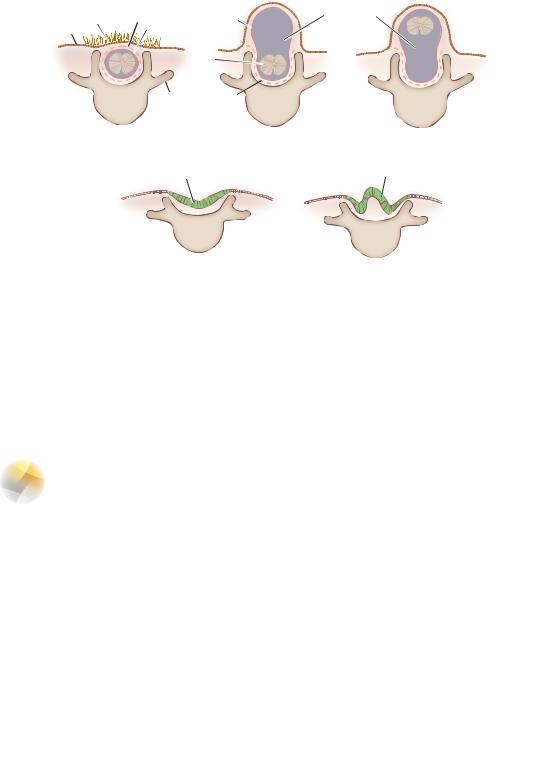
The Caudal Neuropore—failure to close results in spina bifida (Figure 2-4). |
VMicroglia arise from blood-born monocytes.
VI |
Myelination begins in the fourth month of gestation. Myelination of the corticospinal |
|
tracts is not completed until the end of the second postnatal year, when the tracts become |
|
functional. Myelination in the cerebral association cortex continues into the third decade of life. |
A.Myelination of the CNS—accomplished by oligodendrocytes.
B.Myelination of the PNS—accomplished by Schwann cells.
VII The Optic Nerve and Chiasma—derived from the diencephalon. The optic nerve fibers occupy the choroid fissure. Failure of this fissure to close results in coloboma iridis.
VIII The Hypophysis (pituitary gland)—derived from two embryologic
substrata (Figures 2-5 and 2-6).
|
|
|
|
|
Development of the Nervous System |
13 |
Hair |
Dura |
Arachnoid |
Subarachnoid space |
|
||
|
|
|
||||
Skin |
|
Arachnoid |
|
|
|
|
|
|
Spinal |
|
|
|
|
|
|
cord |
|
|
|
|
|
|
Transverse |
Dura |
|
|
|
A |
|
process |
B |
|
C |
|
|
|
|
|
|||
Spina bifida occulta |
|
Meningocele |
Meningomyelocele |
|
||
|
Neural tissue |
|
|
Folded neural tissue |
|
|
D |
E |
Rachischisis |
Rachischisis |
Figure 2-4 The various types of spina bifida. (Reprinted from Sadler TW. Langman’s Medical Embryology. 6th ed. Baltimore, MD: Williams & Wilkins; 1990:363, with permission.)
A.Adenohypophysis (anterior lobe)—derived from an ectodermal diverticulum of the primitive mouth cavity (stomodeum), which is also called Rathke pouch. Remnants of Rathke pouch may give rise to a congenital cystic tumor, a craniopharyngioma.
B.Neurohypophysis (posterior lobe) develops from an anterior (ventral) evagination of the hypothalamus (neuroectoderm of the neural tube).
IX Congenital Malformations of the CNS
A.Anencephaly (Meroanencephaly)—results from failure of the cranial neuropore to close. As a result, the brain does not develop. The frequency of this condition is 1:1,000.
B.Spina Bifida results from failure of the (caudal posterior) neuropore to close. The defect usually occurs in the lumbosacral region. The frequency of spina bifida occulta is 10%.
|
|
Lumen of |
|
Optic chiasma |
|
Infundibulum |
diencephalon |
|
|
|
|
|
||
|
|
|
|
Pars tuberalis |
|
Pharyngeal |
|
|
|
|
hypophysis |
Anterior |
|
|
|
|
Sphenoid |
Pars nervosa |
|
A Rathke's pouch Oral |
Notochord |
B |
lobe C |
|
|
|
Pars intermedia |
||
cavity |
|
|
|
|
Figure 2-5 Midsagittal section through the hypophysis and sella turcica.
14 |
Chapter 2 |
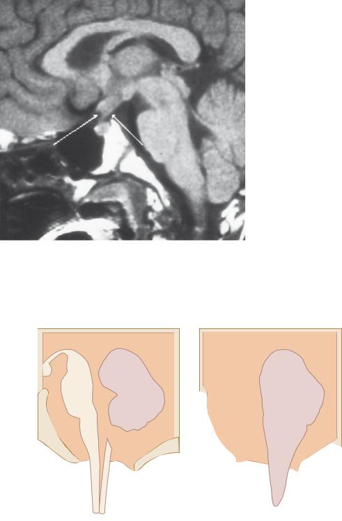
Figure 2-6 Midsagittal section through the brainstem and diencephalon. A craniopharyngioma (arrows) lies suprasellar in the midline. It compresses the optic chiasm and hypothalamus. This tumor is the most common supratentorial tumor that occurs in childhood and the most common cause of hypopituitarism in children. This is a T1-weighted magnetic resonance imaging scan.
C.Cranium Bifidum results from a defect in the occipital bone through which meninges, cerebellar tissue, and the fourth ventricle may herniate.
D.Chiari malformation has a frequency of 1:1,000 (Figure 2-7). It results from elongation and herniation of cerebellar tonsils through the foramen magnum, thereby blocking CSF flow.
III
1
2
IV
3
4
Foramen magnum
A B
Figure 2-7 Chiari malformation. Midsagittal section. A. Normal cerebellum, fourth ventricle, and brainstem. B. Abnormal cerebellum, fourth ventricle, and brainstem showing the common congenital anomalies: (1) beaking of the tectal plate, (2) aqueductal stenosis, (3) kinking and transforaminal herniation of the medulla into the vertebral canal, and (4) herniation and unrolling of the cerebellar vermis into the vertebral canal. An accompanying meningomyelocele is common. (Reprinted from Fix JD. BRS Neuroanatomy. Baltimore, MD: Williams & Wilkins; 1996:72, with permission.)

Development of the Nervous System |
15 |
E.Dandy–Walker malformation has a frequency of 1:25,000. It may result from riboflavin inhibitors, posterior fossa trauma, or viral infection (Figure 2-8).
F.Hydrocephalus—most commonly caused by stenosis of the cerebral aqueduct during development. Excessive CSF accumulates in the ventricles and subarachnoid space. This condition may result from maternal infection (cytomegalovirus and toxoplasmosis). The frequency is 1:1,000.
G.Fetal Alcohol Syndrome—the most common cause of mental retardation. It manifests with microcephaly and congenital heart disease; holoprosencephaly is the most severe manifestation.
Lateral ventricle
Cerebral aqueduct
Optic chiasm
A
Corpus callosum
Lateral ventricle
Massa intermedia
Thrid ventricle
Pons
Medulla
B
Third ventricle/ thalamus
Confluence of sinuses
Cerebellar vermis
Posterior fossa cyst
Mamillary body
Polymicrogyria
Superior sagittal sinus
Straight sinus
Confluence of sinuses
Cerebellar vermis
Posterior fossa cyst
Figure 2-8 Dandy–Walker malformation. Midsagittal section. An enormous dilation of the fourth ventricle results from failure of the lateral foramina (of Luschka) and median foramen (of Magendie) to open. This condition is associated with occipital meningocele, elevation of the confluence of the sinuses (torcular Herophili), agenesis of the cerebellar vermis, and splenium of the corpus callosum. (Reprinted from Dudek RW, Fix JD. BRS Embryology. Baltimore, MD: Williams & Wilkins; 1997:97, with permission.)

16 |
Chapter 2 |
H.Holoprosencephaly results from failure of midline cleavage of the embryonic forebrain. The telencephalon contains a singular ventricular cavity. Holoprosencephaly is seen in trisomy 13 (Patau syndrome); the corpus callosum may be absent. Holoprosencephaly is the most severe manifestation of fetal alcohol syndrome.
I.Hydranencephaly results from bilateral hemispheric infarction secondary to occlusion of the carotid arteries. The hemispheres are replaced by hugely dilated ventricles.
CASE 2-1
A mother brings her newborn infant to the clinic because the infant’s “legs don’t seem to work right.” The infant was delivered at home without antenatal care. What is the most likely diagnosis?
Relevant Physical Exam Findings
●Tufts of hair in the lumbosacral region
●Clubfoot (Talipes equinovarus)
●Chronic upper motor neuron signs, including spasticity, weakness, and fatigability
Diagnosis
●Spina bifida occulta results from incomplete closure of the neural tube during week 4 of embryonic development. This type of neural tube defect often affects tissues overlying the spinal cord, including the vertebral column and skin.