

C H A P T E R 1 0
Trigeminal System
Objectives
1.List the central and peripheral components of the trigeminal system, including the location and function of the appropriate nuclei.
2.Describe the divisions of the trigeminal nerve, including the fiber types in each division and their targets.
3.Diagram the ascending trigeminothalamic pathways, including the constituent primary, secondary, and tertiary neurons. List all cranial nerves that utilize these pathways.
IIntroduction. The trigeminal system provides sensory innervation to the face,
oral cavity, and supratentorial dura through general somatic afferent (GSA) fibers. It also innervates the muscles of mastication, tensors tympani and palati, anterior belly of digastric and mylohyoid through special visceral efferent (SVE) fibers.
II The Trigeminal Ganglion (semilunar or gasserian) contains pseudounipolar ganglion cells. It has three divisions:
A.The ophthalmic nerve (cranial nerve CN V1) lies in the lateral wall of the cavernous sinus. It enters the orbit through the superior orbital fissure and innervates the forehead, dorsum of the nose, upper eyelid, orbit (cornea and conjunctiva), and cranial dura. The ophthalmic nerve mediates the afferent limb of the corneal reflex.
B.The maxillary nerve (CN V2) lies in the lateral wall of the cavernous sinus and innervates the upper lip and cheek, lower eyelid, anterior portion of the temple, oral mucosa of the upper mouth, nose, pharynx, gums, teeth and palate of the upper jaw, and cranial dura. It exits the skull through the foramen rotundum.
C.The mandibular nerve (CN V3) exits the skull through the foramen ovale. Its sensory (GSA) component innervates the lower lip and chin, posterior portion of the temple, external auditory meatus,
and tympanic membrane, external ear, teeth of the lower jaw, oral mucosa of the cheeks and floor of the mouth, anterior two-thirds of the tongue, temporomandibular joint, and cranial dura. The motor (SVE) component of CN V accompanies the mandibular nerve (CN V3) through the foramen ovale. It innervates the muscles of mastication, mylohyoid, anterior belly of the digastric, and tensors tympani and palati (Figure 10-1).
87
88 Chapter 10
Motor cortex |
|
|
|
UMN |
||
|
|
|||||
|
|
|
|
|||
|
|
|
|
|||
Superior cerebellar peduncle |
|
4th ventricle |
||||
|
|
|
|
|||
Chief sensory nucleus |
|
|
CN V motor |
|||
|
|
|
|
|||
Motor nucleus CN V |
|
|
|
Pons |
||
|
|
|
||||
|
|
|
|
|||
Medial lemniscus
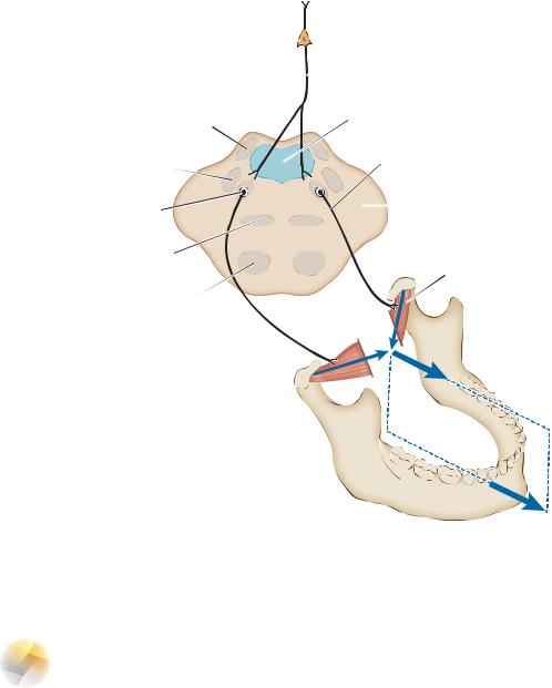
Lateral pterygoid
Corticospinal tract
LMN 
Condyloid process 
Figure 10-1 Function and innervation of the lateral pterygoid muscles (LPMs). The LPM receives its innervation from the trigeminal motor nucleus found in the rostral pons. Bilateral innervation of the LPMs results in protrusion of the mandible in the midline. The LPMs also depress the mandible. Denervation of one LPM results in deviation of the mandible to the ipsilateral or weak side. The trigeminal motor nucleus receives bilateral corticonuclear input. CN, cranial nerve; LMN, lower motor neuron; UMN, upper motor neuron.
IIITrigeminothalamic Pathways (Figure 10-2)
A.The anterior trigeminothalamic tract mediates pain and temperature sensation from the face and oral cavity.
1.First-order neurons are located in the trigeminal (gasserian) ganglion. They give rise to axons that descend in the spinal tract of trigeminal nerve and synapse with second-order neurons in the spinal nucleus of trigeminal nerve.
2.Second-order neurons are located in the spinal trigeminal nucleus. They give rise to decussating axons that terminate in the contralateral ventral posteromedial (VPM) nucleus of the thalamus.
3.Third-order neurons are located in the VPM nucleus of the thalamus. They project through the posterior limb of the internal capsule to the face area of the somatosensory cortex (Brodmann areas 3, 1, and 2).
B.The posterior trigeminothalamic tract mediates tactile discrimination and pressure sensation from the face and oral cavity. It receives input from Meissner and Pacinian corpuscles.
Trigeminal System |
89 |
Ventral posteromedial nucleus (of thalamus)
Facial area of |
Caudate nucleus |
postcentral gyrus |
|
Internal capsule (posterior limb)
Anterior trigeminothalamic tract
 Posterior trigeminothalamic tract
Midbrain |
Mesencephalic nucleus |
|
|
|
Chief sensory nucleus |
|
CN V1 |
|
CN V2 |
Pons |
Sensory branch of CN V3 |
|
|
|
Motor branch of CN V3 |
Motor nucleus of CN V |
Spinal trigeminal tract |
|
|
Spinal trigeminal nucleus |
Medulla |
|
Spinal cord
Figure 10-2 The anterior (pain and temperature) and posterior (discriminative touch) trigeminothalamic pathways. CN, cranial nerve.
1.First-order neurons are located in the trigeminal (gasserian) ganglion. They synapse in the principal sensory nucleus of CN V.
2.Second-order neurons are located in the principal sensory nucleus of CN V. They project to the ipsilateral VPM nucleus of the thalamus.
3.Third-order neurons are located in the VPM nucleus of the thalamus. They project through the posterior limb of the internal capsule to the face area of the somatosensory cortex (Brodmann areas 3, 1, and 2).
IV Trigeminal Reflexes
A.Introduction (Table 10-1)
1.The corneal reflex is a consensual and disynaptic reflex.
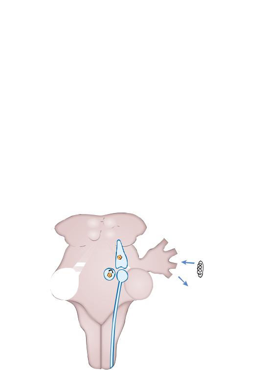
2.The jaw jerk reflex is a monosynaptic myotactic reflex (Figure 10-3).
90 Chapter 10
Table 10-1: The Trigeminal Reflexes
Reflex |
Afferent Limb |
Efferent Limb |
Corneal reflex |
Ophthalmic nerve (CN V1) |
Facial nerve (CN VII) |
Jaw jerk |
Mandibular nerve (CN V3)a |
Mandibular nerve (CN V3) |
Tearing (lacrimal) reflex |
Ophthalmic nerve (CN V1) |
Facial nerve (CN VII) |
Oculocardiac reflex |
Ophthalmic nerve (CN V1) |
Vagal nerve (CN X) |
aThe cell bodies are found in the mesencephalic nucleus of CN V. CN, cranial nerve.
3.The tearing (lacrimal) reflex occurs as a result of corneal or conjunctival irritation.
4.The oculocardiac reflex occurs when pressure on the globe results in bradycardia.
B.Clinical Correlation. Trigeminal neuralgia (tic douloureux) is characterized by recurrent paroxysms of sharp, stabbing pain in one or more branches of the trigeminal nerve on one side of the
face. It usually occurs in people older than 50 years, and it is more common in women than in men. Carbamazepine is the drug of choice for idiopathic trigeminal neuralgia.
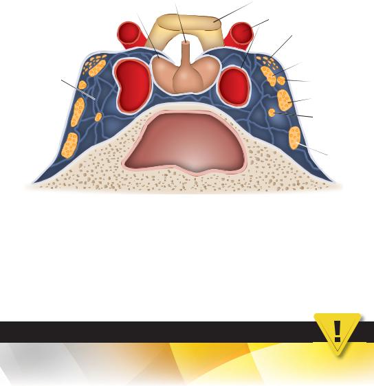
VThe Cavernous Sinus (Figure 10-4) contains the following structures:
A.Internal Carotid Artery (Siphon)
B.CNs III, IV, V1, V2, and VI
C.Postganglionic Sympathetic Fibers en route to the orbit
|
Mesencephalic nucleus |
|
|
V3 |
|
|
Muscle spindle |
|
|
from masseter |
|
|
Masseter |
|
Motor nucleus CN V |
Motor division CN V |
|
with secondary neuron |
||
|
||
|
Chief sensory nucleus |
|
|
Spinal trigeminal nucleus |
|
|
|
Figure 10-3 The jaw jerk (masseter) reflex. The afferent limb is V3, and the efferent limb is the motor root that accompanies V3. First-order sensory neurons are located in the mesencephalic nucleus. The jaw jerk reflex, like all muscle stretch reflexes, is a monosynaptic myotactic reflex. Hyperreflexia indicates an upper motor neuron lesion. CN, cranial nerve.
|
|
Trigeminal System |
91 |
Pituitary gland |
Infundibulum |
Optic chiasm |
|
(hypophysis)
Internal carotid artery
|
Anterior |
|
clinoid process |
|
CN III |
Cavernous |
|
sinous |
CN IV |
|
|
|
CN V1 |
|
CN VI |
|
Sphenoid sinus |
|
CN V2 |
Figure 10-4 The contents of the cavernous sinus. The lateral wall of the cavernous sinus contains the ophthalmic cranial nerve (CN V1) and maxillary (CN V2) divisions of the trigeminal nerve (CN V) and the trochlear (CN IV) and oculomotor (CN III) nerves. The siphon of the internal carotid artery and the abducent nerve (CN VI), along with postganglionic sympathetic fibers, lie within the cavernous sinus. (Modified from Fix JD. High-Yield Neuroanatomy. 3rd ed. Philadelphia, PA: Lippincott Williams & Wilkins; 2005:81, and Gould DJ, Fix JD. BRS Neuroanatomy 5th ed. Philadelphia, PA: 2014, Lippincott, Williams & Wilkins, a Wolters Kluwer business.)
CASE 10-1
A 50-year-old woman complains of sudden onset of pain over the left side of her lower face, with the attacks consisting of brief shocks of pain that last only a few seconds at a time. Between episodes, she has no pain. Usually, the attacks are triggered by brushing her teeth, and they extend from her ear to her chin. What is the most likely diagnosis?
Relevant Physical Exam Findings
●Neurologic exam was normal to motor, sensory, and reflex testing. Magnetic resonance imaging findings were normal as well.
Diagnosis
● Trigeminal neuralgia (tic douloureux)