

Книги по МРТ КТ на английском языке / Magnetic Resonance Imaging in Ischemic Stroke - K Sartor R 252 diger von Kummer Tobias Back
.pdfSpinal Infarcts |
251 |
17 Spinal Infarcts
Michael Mull and Armin Thron
CONTENTS
17.1Introduction 251
17.2 |
Vascular Anatomy of the Spinal Cord 251 |
17.2.1Arterial Supply 251
17.2.2Venous Drainage 254
17.3 |
Causes and Symptoms of Spinal Cord Ischemia 255 |
17.3.1 |
Pathogenetic Principles 255 |
17.3.2Clinical Aspects 256
17.4 |
MR Imaging in Spinal Infarcts 257 |
|
17.4.1 |
Imaging Principles and Differential Diagnoses |
257 |
17.4.2 |
MR Correlates of Acute Vascular Myelomalacia |
258 |
17.4.3MR Correlates of
|
Subacute/Chronic Vascular Myelopathy 261 |
17.4.3.1 |
Spinal Dural Arteriovenous Fistula 261 |
17.4.3.2 |
Arteriovenous Malformations 262 |
17.5Spinal Angiography 263
17.5.1 |
MR Angiography and Spinal Cord Vessels 263 |
17.5.2 |
Spinal Angiography (DSA) 264 |
17.6Treatment 264
17.7Summary 265 References 265
17.1 Introduction
The clinical diagnosis of spinal cord ischemia remains difficult and is mainly based on magnetic resonance imaging (MRI). In the last decade MRI has become the imaging modality of first choice in confirming the diagnosis of spinal vascular diseases. Spinal cord ischemia is a rare disease accounting only for 1%–2% of all vascular neurological pathologies. In contrast to the cerebrovascular system, the arteries directly supplying the spinal cord are not significantly affected by atherosclerotic vessel wall changes. Even in the first descriptions of the anterior spinal artery syndrome by Preobraschenski (1904) and Spiller (1909), the central arteries were affected in cases of luetic arteritis. With increasing
M. Mull, MD
A. Thron, MD
Department of Neuroradiology, Clinic for Diagnostic Radiology, University Hospital of the Technical University, Pauwelsstr. 30, 52074 Aachen, Germany
spatial and contrast resolution, MRI can show circumscribed regions of spinal cord ischemic affection. Moreover, the demonstration of blood vessels in and around the spinal cord based on MRI has substantially improved recently. Nevertheless, the spinal arterial system is seldom examined in detail by spinal digital subtraction angiography (DSA) during life resulting in a limited understanding and fragmentary knowledge of the vascular basis of spinal circulatory disorders. Our knowledge of the spinal cord vascularization remains based on anatomical and microradiographic studies.
The main purpose of this chapter is to define the impact of MRI on the diagnosis of vascular diseases of the spinal cord.
17.2
Vascular Anatomy of the Spinal Cord
To interpret MR findings in spinal cord ischemia, it is necessary to be aware of the normal arterial supply and venous drainage of the spine and spinal cord. Lesion patterns and concomitant signs such as vertebral body infarction highly depend on the underlying vascular pathology. Anatomical and microangiographic examinations remain the main basis of our understanding in this field (Kadyi 1889; Thron 1988).
17.2.1
Arterial Supply
Segmental arteries supply the spine with blood, including the vertebral bodies, paraspinal muscles, dura, nerve roots, and spinal cord. All these tissues, with the exception of the spinal cord, receive their blood supply from segmental arteries (one on each side) or their equivalents. In particular, the segmental supply of the thoracolumbar region is derived from intercostal and lumbar arteries arising from
252 |
M. Mull and A. Thron |
the aorta (Fig. 17.1). Sources of blood supply in the cervical and upper thoracic regions are longitudinal arteries following intrauterine vascular rearrangements. In this region the vertebral artery, the deep cervical artery, and the ascending cervical artery give rise to feeders supplying the spinal cord. In the sacral and lower lumbar region, sacral arteries and iliolumbar arteries arise from the internal iliac arteries and supply the caudal spine.
The radicular arteries are the first branches of the dorsal division of the segmental arteries and their regional equivalents. The blood supply of the bony spine consists of arteries, which come directly off the segmental and radicular arteries. Anterior central arteries derive from the segmental artery and supply the anterior and anterolateral portions of the vertebral body. The radicular arteries usually provide the blood supply to the posterior portion of the vertebral body via posterior central arteries and the arch via prelaminar arteries. Thus, the vertebral body is mainly supplied by posterior and anterior
central arteries, which derive from the segmental and radicular arteries.
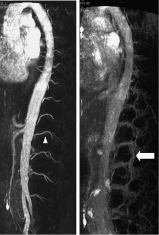
A spinal radicular branch is present at every segmental level. The number of spinal radicular arteries that supply as radiculomedullary arteries the spinal cord, is restricted. They follow the anterior and posterior nerve roots. When they reach the anterior or posterior surface of the spinal cord, they form the anterior or posterior spinal arteries. Thus, the arterial supply of the entire spinal cord depends on a single anterior spinal artery system and paired posterior spinal arteries. The number of anterior radiculomedullary arteries ranges from 2 to 17. The largest ventral radiculomedullary artery which supplies the thoracolumbar cord enlargement is the arteria radicularis magna of Adamkiewicz (Fig. 17.2). In this region large posterior radiculo-pial arteries are also frequently found and form an anastomotic circle around the conus medullaris (arcade of the conus; Kadyi 1889). The highest density of central arteries can be found here in microangiograms
Fig. 17.1a,b. Contrast-enhanced spinal MR angiography, sagittal views. a First phase: the segmental arteries are visible (arrowhead), note the bright enhancement of the aorta. b Second phase: the accompanying veins arise (arrow)
a |
b |
Spinal Infarcts |
253 |
b
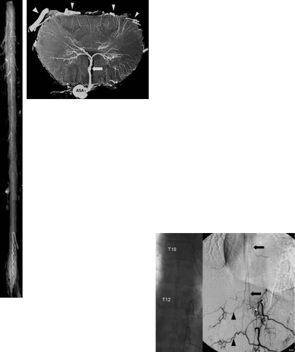
Fig. 17.2. a Superficial arteries of the spinal cord. X-ray film of an injected specimen in a.p. view. The anterior spinal artery system is visible on the ventral surface of the cord. b Intrinsic spinal cord arteries. X-ray microangiogram of a transverse section (lumbar enlargement). The sulcal or central artery within the anterior fissure (arrow) is dominant. Note the posterior and posterolateral spinal arteries at both sides of the posterior root entry zone (arrowheads). ASA, anterior spinal artery
a
median anastomotic trunc (anterior spinal artery) is 1 mm. The radicular arteries (< 0.4 mm) that run with the posterior nerve root, supply the posterior and posterolateral longitudinal arteries. Their size totals 0.1–0.2 mm, which is beyond the present spatial resolution of MR scanners. Together with transverse interconnections, these discontinuous trunks supply the network of pial arteries called the “vasocorona” (Lasjaunias et al. 2001; Thron 1988). The normal intrinsic arteries of the spinal cord are also too small (<0.2 mm) to be detected on CT or MR images.
The spinal cord is protected against ischemia by extraand intradural longitudinal and transverse anastomoses. Extradural longitudinal and transverse interconnections between segmental arteries can compensate for a focal vessel occlusion at the level of the radicular artery (Fig. 17.3). The anterior and posterior spinal arteries represent a system of longitudinal anastomoses that is reinforced from various segmental levels. The intrinsic anastomotic system is supplemented by secondary longitudinal connections between central arteries and by the pial network of the vasocorona. This specific vascular supply explains why spinal infarctions are highly variable, and distinct, predictable vascular territories as present in the brain do not exist (Fig. 17.4). Moreover, the normal arterial vascular system of the spinal cord is not detectable in MRI.
reflecting the metabolic demand of the thoracolumbar enlargement.
Those spinal radicular arteries that are radiculomedullary arteries, supplying nerve root, pial plexus and medulla, branch in a very typical way to form the anterior spinal artery. The ascending branch continues the direction of the radicular artery in the midline of the anterior surface. The descending branch, being the larger one at thoracolumbar levels, forms a hairpin curve as soon as it reaches the midline at the entrance of the anterior fissure (Fig. 17.3). The artery runs above the vein. The maximum diameter of a spinal radiculomedullary artery or the anterior
Fig. 17.3. Selective spinal DSA in a 59-year-old woman, p.a. projection. Injection of the 12th left thoracic segmental artery. Filling of the radiculomedullary arteries T12 and T10 on the left side (arrows) and the anterior spinal artery system. Collateral filling of the right-sided segmental arteries via retrocorporal anastomoses (arrowheads). These extradural anastomoses can compensate for focal vessel occlusions at the level of the radicular artery
254 |
M. Mull and A. Thron |
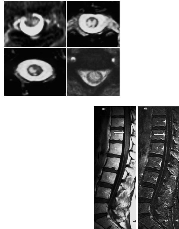
Fig. 17.4. Axial T2-weighted images in four patients with acute ischemic myelomalacia of the spinal cord. The hyperintense infarctions reflect different lesion patterns depending on occlusion site and collateral supply
17.2.2
Venous Drainage
Blood from radial veins of the spinal cord parenchyma and from the small superficial pial veins is collected in two large vessels, the anterior and posterior median spinal veins. The anterior median spinal vein accompanies the artery and can be distinguished by its larger diameter. Posterior, the superficial vein is the only midline vessel, as the arteries run in a more lateral position (Fig. 17.5). The transparenchymal anastomoses take a typical course around the central canal and show upand downward movements in the sagittal plane. They can be seen on MRI. This is also true for the veins of the thoracolumbar enlargement that are the largest vessels of the spinal cord. The transition of a median vein into a radicular vein shows the same hairpin shape like the artery. The superficial venous trunks drain into the epidural venous plexus via radicular veins. The number of radicular veins draining the spinal cord is only six to eleven for the anterior and five to ten for the posterior systems (Kadyi 1889, Suh and Alexander 1939). The radicular veins reach the inner extradural vertebral venous plexus. Drainage of blood from the spine occurs through the internal and external venous vertebral plexus and extends from the sacrum to the base of the skull. This system is valveless and connected with the azygos and hemiazygos venous systems.
Fig. 17.5. Normal anterior and posterior median spinal veins of the thoracolumbar cord. Sagittal contrast-enhanced T1weighted MR images (right side, subtraction image). The large anterior median vein (arrows) continues with the filum terminale

Spinal Infarcts |
255 |
17.3
Causes and Symptoms of Spinal Cord Ischemia
17.3.1
Pathogenetic Principles
Compared with brain ischemia spinal cord strokes are caused by more diverse etiologies. Up-to-now there is no satisfactory and accepted classification of spinal infarcts. Etiologies include circulatory arterial and venous disorders. From a clinical and pathoanatomical point of view it seems reasonable to differentiate between acute ischemic myelomalacia and subacute to chronic vascular myelopathy (Table 17.1). In most cases MRI enables the differentiation of these two main etiologies. A deficient spinal arterial blood flow generally has various causes, ranging from the occlusion of intercostal or lumbar arteries to affection of the intrinsic arteries of the spinal cord .
Many causes of acute spinal cord infarction (of arterial and venous origin) have been reported (Table 17.2). They include diseases of the aorta and aortic surgery, thromboembolic events and cartilaginous disc embolism, vasculitis, coagulopathy, radiation-induced vasculopathy, toxic effects of contrast medium, epidural anesthesia, periradicular nerve root therapy with crystalline corticoids, decompression illness, shock or cardiac arrest, lumbar artery compression and other etiologies
Table 17.1. Disorders resulting in primary vascular myelopathy
•Acute vascular myelomalacia
Acute ischemic myelomalacia of arterial and venous origin
•Subacute/chronic vascular myelopathy Dural AV fistula
AVM of the perimedullary fistula type (AVM of the glomerular type)
Table 17.2. Causes of acute spinal cord infarction
•Surgery for aortoiliac occlusive disease
•Dissection of the aorta
•Thrombosis/embolism
•Vasculitis
•Cardiac arrest and systemic hypotension
•Fibrocartilaginous embolism
•Caisson disease
•Lumbar artery compression
•Spinal tumor
•Spinal AVM/SDAVF
(Gravereaux 2001; Mawad et al. 1990; Mikulis et al. 1992; Naiman et al. 1961; Rosenkranz et al. 2004; Toro et al. 1994; Tosi et al. 1996; Weidauer et al. 2002).
Cartilaginous disc embolism, first described by Naiman and colleagues in 1961, is meanwhile a widely accepted cause of acute spinal cord ischemia mainly based on postmortem findings (Mikulis et al. 1992; Toro et al. 1994). Most reported postmortem cases are cervical, but conus medullaris infarctions are also reported. The exact mechanism of disc embolism remains unclear and speculative. An increased intraosseous pressure within the vertebral body, due to acute vertical disc herniations
– often after axial trauma – may explain the initial step in a process which ends up in spinal cord ischemia (Tosi et al. 1996). In the clinical course of the histologically established cases of fibrocartilaginous embolism three main characteristics could be identified: (1) the sudden severe pain at onset, (2) the free interval of neurological signs and symptoms (“spinal stroke in progress”), (3) no improvement of the neurological deficit. In our experience, there is a surprisingly high rate of disc herniations in close proximity to the vascular lesions of the cord (Mull et al. 1992). Intravital confirmation of this pathogenetic mechanism is difficult. Nevertheless it may be that the pathogenetic role of disc material has yet not been fully realized.
In contrast to disc embolism, most patients with thrombotic or embolic infarctions show at least partial recovery in the further course of the disease. Nevertheless, depending on the efforts in diagnostic work-up, the cause of spinal cord ischemia often remains undefined or speculative.
Lumbar artery compression by the diaphragmatic crus has been described as a new etiology for spinal cord ischemia (Rogopoulos et al. 2000). A prolonged hyperlordotic position may play a predisposing role in this new syndrome. The 1st right and left and the 2nd right lumbar arteries course through an osteotendinous passage between the vertebral body and the diaphragmatic crus. If spinal cord supplying arteries arise at the level of the first and second lumbar artery, compromise of the spinal cord arterial supply can result in spinal cord ischemia.
Suh and Alexander (1939) and later Zülch
(1954, 1976) postulated their traditional hypothesis of a watershed zone of increased ischemic vulnerability near T4. This model was based on the relative hypovascularity of this region.
Cheshire et al. (1996) described 44 patients with spinal cord infarctions. Surprisingly, the mean sen-
256 |
M. Mull and A. Thron |
sory level of the deficits was observed at T8 and T9 in cases of global ischemia. A review of the clinical literature by Cheshire and colleagues (1996) suggests that the most commonly sensory level found after spinal cord infarction is centered at T12.
Duggal and Lach (2002) analyzed clinical files and neuropathologic specimen in 145 patients with well-documented cardiac arrest or severe hypotension. In this group ischemic myelopathy was found in 46% of patients dying after either cardiac arrest or a severe hypotensive episode. Among the patients with myelopathy, predominant involvement of the lumbosacral level with relative sparing of thoracic levels was observed in 95% of cardiac arrest and hypotensive patients. None of the examined patients developed neuronal necrosis limited to the thoracic level only. These findings indicate a greater vulnerability of neurons in the lumbar or lumbosacral spinal cord to ischemia than other levels of the spinal cord (Duggal and Lach 2002).
Moreover, analysis of data coming from greater MR series and our own observations reveal no predominance of infarcts in the upper and mid-thoracic region (Mawad et al. 1990; Weidauer et al. 2002). Thus, the concept of a vulnerable watershed zone at T4 is no longer valid in acute spinal cord ischemia.
Subacute or chronic vascular myelopathy is the result of disturbances of microcirculation caused by compromised venous outflow or small-vessel disease.
Thus, acute, subacute, or chronic impairment of spinal blood supply can result from a deficient arterial supply and from venous circulatory problems (Mull and Thron 2004). Spinal vascular malformations like spinal dural arteriovenous (AV) fistulas and AV malformations (AVM) of the perimedullary fistula type are the typical disorders associated with venous congestion of the spinal cord. On the other hand, AVM of the glomerular type are seldom combined with a venous outflow disorder.
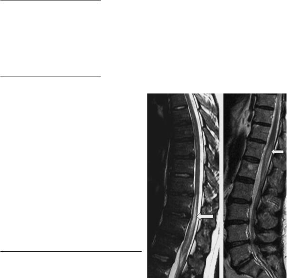
Spinal dural arteriovenous fistulas (SDAVF) are the most frequent (acquired) AVM of the spinal canal and its meninges. In 1977 Kendall and Logue were the first to recognize that this disease, which had formerly been described as so-called retromedullary angiomas, in fact is an AV fistula situated within the dura mater. This was confirmed by Merland et al. in 1980 who pointed out the angiographic characteristics of the disease in detail (Fig. 17.6).
The AV shunt is located inside the dura mater close to the spinal nerve roots. The arterial blood enters a radicular vein where it passes the dura (Thron et al. 1987; Hassler et al. 1989). Flow in the radicular
vein is reversed and directed to the perimedullary veins. The dural shunt is supplied by radiculomeningeal arteries which normally supply the segmental nerve roots and dura. They arise from the segmental vessels that persist as intercostal or lumbar arteries in the thoracolumbar region. Depending on the location of the fistula, the supply may also come from the sacral or hypogastric arteries (Burguet et al. 1985; Heindel et al. 1975; Larsen et al. 1995;
Partington et al. 1992; Reinges et al. 2001; Stein et al. 1972). In addition, intracranial dural fistulas have been described to drain to spinal cord veins and evoke the same symptoms as spinal dural fistulas. The cause of the fistulas is unknown so far.
Functional disturbances within the spinal cord are caused by the venous congestion due to arterialization and elevated pressure of the medullary veins (Aminoff et al. 1974). In addition, venous outlets are insufficient and thus reinforcing impairment of the venous circulation and chronic spinal hypoxia. In particular, Merland and coworkers (1980) introduced the concept of blocked venous drainage into the epidural space.
17.3.2
Clinical Aspects
Vascular diseases of the spinal cord are characterized by transverse cord symptoms and can be differentiated by their clinical features and course. Neurologic deficits and imaging findings depend on the level and extent of spinal cord damage. Acute ischemic myelomalacia has an acute onset and pain is a common initial complaint. Symptoms may include nerve root deficits or sphincter weakness (Mull and Thron 1999). But typical clinical syndromes (i.e., anterior spinal artery syndrome) are exceptional presentations in clinical practice. A spinal hemorrhage presents with peracute signs and symptoms and always requires prompt diagnostic investigation, if necessary on an emergency basis.
In most cases congestive myelopathy takes a subacute to chronic course, although acute worsening may be observed. SDAVF is the prototype of a congestive myelopathy. The symptoms in SDAVF are slowly progressive and consist of sensory loss and paraparesis both of which reflect the extent of the cord involvement. They are independent of the location of the fistula (Koenig et al. 1989). An acute onset of the disease (11%) or a progressive development interrupted by intermediate remissions (11%) are the rare forms (Symon et al. 1984). Almost all
Spinal Infarcts |
|
257 |
a |
b |
c |
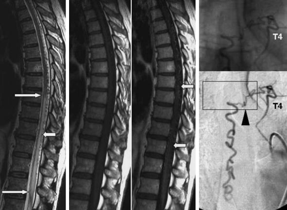
Fig. 17.6a–c. Thoracic SDAVF in a 50-year-old man with chronic progressive paraparesis due to congestive myelopathy. a Sagittal T2-weighted MRI. The hyperintense lesion (long arrows) involves the thoracic cord indicating vascular damage. b Dilated perimedullary veins are detectable in the thoracic region (right image, arrows) after paramagnetic contrast application (T1weighted MRI). c DSA, p.a. projection. Selective contrast injection in the 4th left-sided intercostal artery shows the dural AV fistula (arrowhead) draining slowly in the caudal direction
patients have additional sphincter disturbances at the time of diagnosis. The disease affects men more often than women, most of them being in the middle or older age group. In some cases a relapsing and remitting course of the disease can be observed. The symptomatology in SDAVF can cause confusion with other disorders like polyneuropathy, spinal stenosis, transverse myelitis and multiple sclerosis.
17.4
MR Imaging in Spinal Infarcts
17.4.1
Imaging Principles and Differential Diagnoses
The imaging work-up should be guided by a clinical assessment of the lesion level.
MRI is the diagnostic modality of choice, T1and T2-weighted images are obligatory. Strongly T2-weighted images (e.g., a CISS sequence) may help to detect pathologic vessels within the bright cerebrospinal fluid. Sagittal and transverse images are more important than coronal views. Fat can be suppressed by spectral fat saturation or by using the inversion recovery technique. These sequences are essential in acute spinal cord ischemia to detect associated abnormal bone signals indicating vertebral body infarction. Fluid-attenuated inversion recovery (FLAIR) sequences suppress signals from cerebrospinal fluid and, therefore, help to detect intraspinal hemorrhage. Contrast-enhanced T1weighted images can reveal abnormal intraspinal vessels. Acute spinal cord ischemia is characterized by a typical time-dependent pattern of contrast enhancement. MRI can directly depict the cause of spinal cord ischemia in dissecting aneurysms of the
258 |
M. Mull and A. Thron |
aorta. The use of diffusion-weighted MRI (DWI) has been limited in the spine thus far due to very strong susceptibility artefacts. Nevertheless, diffusion imaging has been successfully applied also to spinal cord ischemia (Gass et al. 2000). MRI can directly detect ischemic spinal cord lesions as well as intramedullary, subdural, or epidural hemorrhage. Myelitis and tumor-associated lesions are the most important clinical and imaging differential diagnoses in acute spinal cord ischemia (Table 17.3). CSF examinations are necessary if inflammatory lesions of the spinal cord (transverse myelitis) and multiple sclerosis have to be excluded in the acute phase (de Seze et al. 2001).
Table 17.3. Differential diagnosis in acute non-traumatic transverse lesion
•Hematomyelia
•Multiple sclerosis
•Spinal cord compression
–Neoplasm
–Disc herniation
–Sub-/epidural hematoma
•Abscess/empyema
•Delayed radiation myelopathy
•Acute transverse myelitis
•Spinal AVM
•Ischemic infarction
In the presence of an intraspinal hemorrhage spinal vascular malformation, cavernoma, coagulopathy and tumor have to be differentiated.
In spinal vascular malformations abnormally thick, often tortuous vessels on the surface of the spinal cord can be identified as well as the angiomalike nidus of the AVM. MRI facilitates the differentiation between SDAVF and intramedullary AVM.
Myelography and post-myelographic computed tomography should only follow MRI if there is a high clinical index of suspicion for a SDAVF. Myelography is more sensitive than MRI to detect pathologic perimedullary vessels especially in cases of low-flow fistulas (Gilbertson et al. 1995).
Spinal angiography remains the gold standard in spinal vascular malformations and can help to elucidate the underlying pathology of acute spinal cord ischemia in selected cases (Di Chiro and
Wener 1973; Djindjian et al. 1970; Lasjaunias et al. 2001).
Fig. 17.7. Acute ischemic myelomalacia of the thoracolumbar region in two patients, T2-weighted MR images. The pencilshaped zone of increased signal represents the acute infarcted cord
17.4.2
MR Correlates of Acute Vascular Myelomalacia
Ischemic infarction of the spinal cord is difficult to establish in the early phase, only 50% of the patients show early demarcation within 24 h. The role of MRI in the acute phase is to exclude hematomyelia, spinal vascular malformation (which requires spinal angiography in special cases) or a compressive lesion.
Common sites of spinal cord infarction are the thoracolumbar enlargement and the conus medullaris in 65%, the cervical region is less commonly affected (only 11% in our own series). The damage of the spinal cord is most accurately demonstrated by MRI in T2-weighted sequences (Brown et al. 1989). Hyperintensity in T2-weighted images may be demonstrated as early as 8 h after onset (Yuh et al. 1992). In most cases pencil-shaped hyperintensive lesions are visible predominantly in the anterior spinal artery system at least in follow-up examinations (Fig. 17.7). Circumscribed infarctions in the posterior spinal artery system are rare (Mascalchi et al. 1998). Moderate swelling is generally present in the acute stage followed by contrast enhancement of the cord and the cauda equina in the subacute stage – usually after 5 days (Amano et al. 1998; Casselman et al. 1991; Friedman and Flanders 1992). Con-
Spinal Infarcts |
259 |
trast-enhanced images demonstrate enhancement of the infarct up to 3 weeks after the onset (Berlit et al. 1992; Hirono et al. 1992; Yuh et al. 1992).
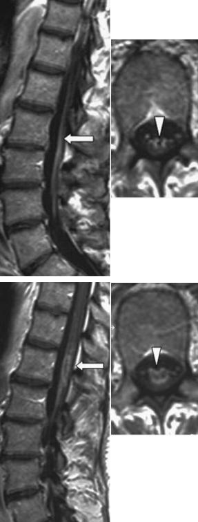
In most cases of the thoracolumbar infarction, the swollen cord shows peripheral enhancement of the central gray matter. The concomitant enhancement of the cauda equina was reported first by Friedman and Flanders in 1992 (Fig. 17.8). This phenomenon is a characteristic finding in the course of spinal cord ischemia which might involve the cord itself and the ventral cauda equina as well, which is composed of motor fibre bundles (Amano et al. 1998). It indicates disruption of the blood–cord barrier as well as reactive hyperemia (Friedman and Flanders 1992; Amano et al. 1998). The differential diagnosis of contrast enhancement of the cauda equina includes transverse myelitis, bacterial or viral meningitis, and spinal metastasis.
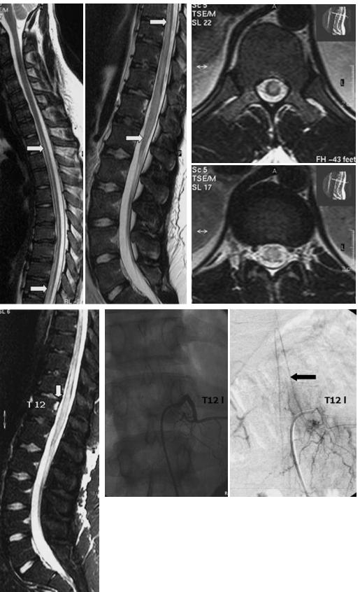
Abnormal signal or evolution of signal abnormality in a vertebral body associated with a cord lesion is highly suspicious of an acute spinal infarction (Haddad et al. 1996; Yuh et al. 1992). Due to the vascular supply to the vertebral body, the pattern of signal changes is distinct from degenerative disease. The deep medullary portion and the region of the endplates are most vulnerable to ischemia. Yuh et al. (1992) reported early hyperintensity in T2-weighted images due to vertebral body infarction after 8 h. As described before, the radicular arteries supply the vertebral bodies and – inconsistently – the spinal cord, if there is a radiculomedullary artery at the same level. Abnormal bone signals indicate this special vascular condition and can be regarded as a confirmatory sign in spinal infarction (Faig et al. 1998; Yuh et al. 1992). In our patient group, the spinal angiography demonstrated that infarction of the vertebral body indicated the origin of the anterior spinal artery (Fig. 17.9). Bone marrow abnormalities are also detectable in posterior spinal cord syndrome (Suzuki et al. 2003). In the chronic stage the affected cord segment may shrink.
Early detection of the ischemia is desirable with regard to therapeutic implications.
In few studies, DWI is reported to detect spinal cord ischemia in the acute phase (Gass et al. 2000;
Bammer et al. 2000; Stepper and Lövblad 2001).
Küker and coworkers (2004) recently reported the time course of diffusion abnormality in three patients with acute spinal cord ischemia. They found diffusion abnormality earliest after 8 h. At this time there was only a faint hyperintensity visible in the central part of the conus medullaris. Diffusion abnormality did not last for longer than 1 week. Further stud-
a
b
Fig. 17.8a,b. Acute ischemic myelomalacia of the lumbar enlargement 3 weeks after onset of symptoms. Contrastenhanced T1-weighted images in sagittal and axial orientation at different levels. a Enhancement of the ventral cauda equina (arrow, arrowhead). b Peripheral ring-like enhancement of the affected lumbar enlargement (arrow, arrowhead)
260 |
M. Mull and A. Thron |
a |
b |
d
|
Fig. 17.9a–d. Acute ischemic myelomalacia in an 18-year-old man with acute |
|
paraplegia and sphincter disturbances. a Sagittal T2-weighted images show |
|
centromedullary multisegmental hyperintensities from T2 to the conus |
|
(arrows). b Axial T2-weighted images. c On day 2, a fat-suppressed T2-weighted |
|
sequence reveals abnormal signal in the dorsal part of the vertebra T12 cor- |
|
responding to vertebral body infarction (arrow). d DSA, p.a. projection, 10 |
|
days after onset of symptoms. The radiculomedullary artery originates at T12 |
|
level and supplies the anterior spinal axis. At this time the anterior spinal |
c |
artery is patent. The origin of the cord supplying radiculomedullary artery |
corresponds well to the level of the vertebral body infarction |