

NUCLEAR POWER PLANTS
.pdfRadiobiological Characterization Environment Around Object "Shelter" |
255 |
deeper degenerative processes in the parenchyma with formation of lymphoid infiltrates. In a more favorable course of the process next sections of lesions appear regeneration areas. At high intensity the process changes in the tissues may be irreversible.
Fig. 29. Dystrophia of hepatocytes in violation of the architectonics of organ and the destruction of the nuclei. 1 - a violation of the architectonics of the lobules, blurred boundaries (irradiated mouse).
Fig. 30. Around blood vessels (or periportal) small clusters of mostly round the nuclear cell (lymphoid) elements. Stasis of blood within the vessel. More intensively painted the nucleus of cells (irradiated mouse).
Thus it was shown that exposure to IR can modify the immune status of the organism with the breakdown of immune tolerance and the subsequent emergence of autoimmune hepatitis. Its characteristic feature is the launch of autoimmune reactions against their own membrane antigens not only in the liver but also in other organs. The presence of long-term in excess of the background radiation pressure associated with the abolition of immune tolerance and the creation of conditions for the development of autoimmune reactions, in particular against antigens of liver tissue. As well known in turn the activity of autoimmunity is a favorable condition for the transfer of persistent infections in the active state and stimulation of vegetation as a saprophyte and pathogenic microorganisms was increased.

256 |
Nuclear Power Plants |
Fig. 31. Granulomas in the periportal areas of the parenchyma. 1 - Formation of granuloma on site infiltrate, 2 - Regeneration and repair of hepatocytes. (Irradiated mouse).
The impact of single external irradiation at a dose of 5.0 Sv for the transfer of intercellular signals in the bone marrow of mice Balb/c.
Nowadays it is known that hematopoietic stem cells (HSC) have higher radiation sensitivity than other cellular self-renewal systems. This conclusion was made by scientists on the basis of experimental works that showed the presence of the damage to hematopoietic progenitor cells not only due to the effects of the high doses but also upon action of the low doses of IR [Muksinova K.N., Mushkacheva G.S. 1995, Serkiz Ya.I. 1992].
It was shown that regeneration of the maturing cell pool and renewal of their quantity in the peripheral blood is determined by the completeness of the progenitor cell clone recovery. During the bone marrow regeneration period it was determined that both the proliferation of HSC is increased and the transition time of the maturing cell elements shortens [Bilko N.M. 1998, Bilko N.M., Klimenko S.V., Velichko E.A. 1999, Tavassoli M.V. 2008]. After acute period of the damage to the hematopoietic system regeneration phase follows, which is dependent on the viability of the stem cells, their migration ability in to the most affected areas of the hematopoietic tissues, time of proliferation and maturation of the committed progenitors and quantity of the functional mature cells [Shouse S.S., Warren A.S.L., Whipple G.H. 2004]. Critical for recovery of the hematopoiesis are quantity and quality of the HSC that recovered after irradiation. Possibility of hematopoiesis recovery is observed if more than 5% of the stem and progenitor cells remain intact and carry on proliferation and differentiation [Bond V.P., Fliedner T.M., Archambeau J.O. 2007]. If their level falls below this critical value, hematopoietic system can be exhausted due to lack of the stem cells capable of regeneration [Down J., Van Os. R., Ploemacher R. 1991]. Main proliferation stimulating factor of the HSC, which remain in the dormant state, is reduction of their quantity [Serkiz Ya.I., Pinchuk L.B. 1992]. Decisive role in the regulation of the recovery of the polipotent hematopoietic progenitors belongs to the microenvironment that upon interaction with HSC supports stability of their quantitative parameters in the physiological conditions and supports its recovery in case of injures [Hall E.J. 1991, Hall Mauch P., Constine L., Greenberger J. 2005]. Increase in the proliferation activity of the HSC is observed starting after irradiation exposure at the doses of 0.2 – 0.3 Gy [Grande T., Varas F., Bueren J.A. 2000].
Radiobiological Characterization Environment Around Object "Shelter" |
257 |
The mechanism of the microenvironment influence on the hematopoietic system is still not fully determined. However, today it is known that its elements control the processes of hematopoiesis via production of the cytokines as well as by direct cell-to-cell contacts between HSC and microenvironment. Membrane-associated contacts serve for the transfer of the required molecules, homing and migration of the progenitor cells to the specific sites of the hematopoietic tissue and transport of the hematopoietic growth factors [Cronkite E.P., Inoue T., Hirabayashi Y. 2003]. Cultural investigations of the bone marrow (BM) indicated that despite the normalization of the quantitative parameters changes of the ability of hematopoietic elements to colony-forming had reduced character during prolonged periods of time with prevailing eosinophilic and neutrophilic colonies [Bilko N.M. 1998].
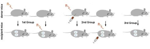
For the determination of the distant intercellular transfer of the post-radiation signals between the cells of irradiated animals a novel method of in vivo culture using diffusion capsules (DC) was described [Bilko N.M., Votyakova I.A., Vasylovska S.V., 2005]. Investigations were done of the Balb/c mice (Figure 32). The 16-hour model of exposure was used (see Protocol modes of mice irradiation). Animals were divided in to three groups: 1st group was irradiated without the use of radioprotector, 2nd group of animals received melanin – glucan complex prior to irradiation and the 3rd group was non-irradiated control.
Fig. 32. Groups of experimental animals
Further each group was separated into the subgroups of the donors (3 animals) and recipients (3 animals for each donor and 2 capsules per recipient). Donor animals were sacrificed on the day 1, day 7, and day 30 after exposure and bone marrow cells were extracted from the femur. In each case, colony-forming activity (CFU) in the culture was determined by injection of the 1x10⁵ cells into the inner cavity of the diffusion capsule in the semisolid (0.33%) agar Difco. Diffusion chambers (DC) permit free diffusion of the peptide factors; however, they allow avoiding any contact of the cultured material with the immune system of the recipient. Each animal was implanted with two DC into the peritoneal cavity under Sagatal narcotization. Animals were retained in the conventional vivarium conditions with 12-hour light/dark cycle illumination and free access to food and water. After 12 days implantation DC were extracted from the recipients and investigated under the inverted microscope for the CFU activity as indicated by formation of the colonies or clusters of the proliferated HSC.
The groups of cells less than 20 cells were considered as clusters, while groups of cells from 20-40 were considered as large clusters, and all cellular aggregates above 40 cells were counted as colonies (see Figure 33 and 34).

258 |
Nuclear Power Plants |
Fig. 33. Granulocyte-macrophage colony of mouse bone marrow culture of Balb/c, irradiated with a dose of 5.0 Sv. Inverted microscope, increase 200.
Fig. 34. Granulocyte cluster of mouse bone marrow culture of Balb/c, irradiated with a dose of 5.0 Sv. Inverted microscope, increase 400.
Cultured material was extracted from the inner cavity of the DC and individual colonies were picked up for the preparation of the cytospin slides and Pappenheim staining for identification of the cell types. Obtained data indicated that BM of the animals of the 1st group was affected by the IR. Cell aggregate numbers were on average 24 colonies and 48 clusters in the cultures of the 1st day after exposure. Almost no colony-forming activity was observed in the culture of 7th and 30th day after exposure, that was indicative of significant suppression of the bone marrow function by the IR. At the same time in the culture of bone marrow cells that were obtained from the animals, which received MGC prior to exposure, the average colony count was 32 with 86 clusters at the 1st day after exposure, 45 colonies and 112 clusters on the 7th day after exposure, and 80 colonies and 136 clusters on the 30th day after exposure.
These results may indicate that MGC is able to protect the population of the bone marrow stem cell population for the influence on the IR and stimulate recovery after irradiation at

Radiobiological Characterization Environment Around Object "Shelter" |
259 |
dose comparable to the LD50/30. This conclusion can also be supported by increase of the quantity of the CFU in comparison to the 1st group of animals that have not received MGC.
Implantation of the normal BM into the organism of the irradiated recipients at the 1st day after exposure after culturing resulted in formation of 114 colonies and 386 clusters on average. In the cultures of the 7th and 30th days such proliferation activity was observed, that it was not possible to determine individual colonies or clusters. There is indicative of a significant stimulation of the release of the compensatory signal substances by the radioresistant stromal cells of the BM, which highly stimulated the recovery of the radiationdamaged BM of the recipient. Normal bone marrow cells implanted into the irradiated mice treated with MGC yielded in 84 colonies and 192 clusters in the cultures of the 1st day after exposure, 52 colonies and 548 clusters on the 7th day, and 106 colonies and 302 clusters in the 30th day post exposure cultures (Figure 35).
Fig. 35. Determination of the CFU activity of the bone marrow derived stem cells: Group 1A - irradiated bone marrow culture in normal recipients; Group 2A - irradiated BM protected with MGC in normal recipients; Group 1C - normal bone marrow culture in irradiated recipients; Group 2C - normal bone marrow culture in irradiated recipients treated with MGC.
Such decrease in the CFU activity is indicative of less apparent stimulation of the compensatory factors release as a result of the radiation exposure due to protective effects of the MGC on the bone marrow cells so that less factors are required for reparation and therefore less factors are available for stimulation of the CFU activity in the DC.
Results of the in vivo culture of the bone marrow cells indicated that colony-forming activity of the hematopoietic progenitor cells of the animals non-treated with MGC prior to radiation exposure was significantly lower if compared to the control while treatment of the animals with MGC had increased the functional activity of the bone marrow cells.
Therefore upon irradiation exposure quantity of the hematopoietic stem cells is decreased and as a result, stromal component of the BM starts to secrete large quantities of cytokines
260 |
Nuclear Power Plants |
and growth factors, which are able to stimulate HSC to active proliferation [Goldberg E.D., Dygai A.M. 2001]. Clearly, in animals irradiated at doses of 5.0 Sv, production of growth factors was blocked by excessive amounts of free radicals which are formed when passing through biological tissue of ionizing particles [Timofeev-Resovskii N.V. 1963]. At the same time, free radicals can quickly neutralized by the MGC in mice receiving radioprotector. Improved products of growth factors caused the hyperproduction of bone marrow stem cells. On the 7th day after exposure in the bone marrow culture of the irradiated animals, a deep depression in the CFU activity was observed. Finally pretreatment of animals with MGC resulted in the significant increase of the CFU activity. These data indicates that in the BM of irradiated animals the pool of later mononuclear cells was rapidly exhausted that resulted in the decrease of the CFU activity [Bilko N.M., Klimenko S.V., Velichko E.A. 1999]. In the group of animals pretreated with MGC prior to exposure, CFU activity in the BM increased during the experiment. On the 30th day post exposure, CFU activity of the bone marrow stem cells remained low and in the animals that have not received MGC, and in the animals treated with this radioprotector these values were significantly higher.
Obtained data may be indicative of the gradual recovery of the BM by implementation of the so called “golden reserve” of the stem cells that during radiation exposure were positioned in the crypts of the stroma and therefore remained undamaged [Tsyb A.F., Budagov R.S., Zamulaeva A.I. 2005]. Such significant effects of the MGC on the numbers of colonies and clusters in the DC cultures indicates decrease quantity of the stimulating bystander signals and strong radioprotective properties of the MGC, however the exact mechanism of action remains to be fully elucidated.
Concluding on the obtained experimental data it is clear that MGC is a strong radiation
protector that helps to avoid consequences of the LD50/30 dose of irradiation at the level of HSC by affecting quantity of available growth stimulating factors that are commonly
associated with radiation-induced damage to the BM.
2.4 Influence radionuclide fallout to plant grown around object “Shelter” in Chernobyl alienation zone
Contaminated of the wide territories in Ukraine not only with radionucludes 137Cs and 90Sr, and with fission products of uranium and transuranium elements is an essential consequence of the accident at the IV block of Chernobyl Nuclear Power Plant that is classified as a global ecological catastrophe. The biota behaviors and adapt in this areas captured dose from radionuclide with long half-value period decay isotopes. As dose related amount of the isotopes 137Cs and 90Sr during long time after accident were decreased. But only the amount of the radioactive isotope 241Am depend of time is increasing exactly in environmental alienation zone of Chernobyl. Radionuclide 241Am as -emitter is a daughter product of 241Pu isotope appeared after β-decay. The activity in environment of the isotope 241Am is increasing with during time owing to β-decay of the 241Pu isotope. The biota behavior in this areas captured dose from radionuclide with long half-value period decay isotopes. The peculiarity of radionuclides contamination associated with the Chernobyl accident is verified of physical and chemical forms of radioactivity elements through out into the environment [Rashydov N.M. 1999, Rashydov N.M., Konoplyova A.A., Grodzinsky D.M. 2004, Rashydov N.M., Kutsokon N.K. 2005, Rashydov N.M., Grodzinsky D., Berezhna V. 2006]. A part of the radioactivity isotopes is registered in water soluble droplets-liquid
Radiobiological Characterization Environment Around Object "Shelter" |
261 |
state, an other part – as “hot” particles, the interrelation between there forms being unstable and change under the influence of biotic and abiotic environmental factors. As rule in this conditions accumulation of radionuclides in plants which occurs mainly at the expense of their water-soluble and exchangeable forms, reflects rather complicated transitional processes in the soil, the rate and direction of these ones is determined by biological activity of all component of the plant rhizosphere inhabited layer of the soil. After Chernobyl accident already during 25 year a lot of “hot” particles transferred into fine dispersive conditions, which easy movements in outdoors where captured by biota which could characterize by help of transfer coefficient (TC) radionuclide ongoing. The transfer coefficient is ratio specific activities (kBq/kg) of plant to specific activity of soil (kBq/kg) where its grow that characterize go over a radionuclide from soil to vegetative plant on experimental plot. Necessary mentioned that the TC not constant and it differed on depend of parts of plant were determinate. For radionuclide 241Аm observed the value TC a lot of plants and mushrooms several order less than for isotopes 137Cs, 90Sr. Especially for matured seed the value of the TC observed less than for other vegetative parts of the plant [Rashydov N., Berezhna V., Kutsokon N. 2007, Rashydov N.M., Kutsokon N.K. 2008, Rashydov N.M., Berezhna V.V., Grodzinsky D.M. 2009, Rashydov N., Berezhna V. 2010, Rashydov N.M. 2010, Rashydov N.M. 2011]. To study of the TC peculiarity modification is reason elucidation of our field research in alienation Chernobyl zone around object “Shelter”.
Contamination of plant in natural experimental fields at the alienation zone of Chernobyl significant added by flying dust with very small size radioactivity particles less than “hot particles” in environment. The results received for plants soybean (content: 137Cs - 3.6 kBq/kg and 90Sr – 11.84 kBq/kg) and flax (content: 137Cs - 0.78 kBq/kg and 90Sr – 3.55 kBq/kg) which grown in Chistogalovka (specific activity of soil is 20.65 kBq/kg and 5.18 kBq/kg for radionuclide 137Cs and 90Sr, accordingly) and Chernobyl confirmed this hypothesis. The value TC for above mentioned seeds specimens collected from plant which grow on Chistogalovka was approximately 22.3 (soybean) and 6.63 (flax) times (for isotope 137Cs) and 13.97 and 4.71 (for isotope 90Sr) times higher by comparison with control variants which grown in Chernobyl where specific activity was 1.41 kBq/kg for radionuclide 137Cs and 0.55 kBq/kg for isotope 90Sr, correspondingly.
The peculiarity distribution in controlled laboratory conditions the radionuclide 241Аm in Arabidopsis thaliana plant on high level first layer leaves, in petiole and in carry out fascicles of the leaves significantly that go into this isotope from root system to top of plant very slow and membrane of cells played as discrimination barrier in this processes as mentioned in our previously investigations [Rashydov N.M., Berezhna V.V., Grodzinsky D.M. 2009].
In laboratory conditions for autoradiography investigation purpose the seedlings Arabidopsis thaliana were aseptically grown in hard agar cultured medium containing 241AmCl3 in concentration with specific activity 50 kBq/kg. After 25 days some leaves and top of stems of plants witch had not direct contact with medium were carefully cut off so that to avoid contact with medium. Selected parts of plants Arabidopsis thaliana settled down on the microscopic glass slides and dried a few days. During this process they were gluing to the slides themselves. The slides with parts of plants were coated with photo emulsion LM-1 in gel (Amersham – Biosciences UK) and exposure during time 20 days at temperature +40 C. After development the samples of slides were observed of the track of α-particles from radionuclide 241Аm with light microscope. A lot of datum confirmed that the coefficient

262 |
Nuclear Power Plants |
uptake very small for radionuclide 241Am and this element maldistribution by organs and tissues. We observed that accumulation the radionuclide of 241Am depended of carry out fascicles system of the leaves and localization of the layer leaves not far from length root collar of plant which grow in laboratory conditions. The first layer leaves were taken up high-level amount radionuclide 241Аm. As result the capture dose also may tissues of plant distribute no uniform. It is known that mineral nutrients are transported apoplastically, i.e. in the wall system outside the plasma membrane, or symplastically, i.e. in the cytoplasm from cell to cell deal with through plasmodesmata. The nutrient elements that penetrate into the cytoplast can also be shuttled into the vacuole via various mechanisms depending of biological function in cell life behaviors for mentioned isotope.
For field experiments we use plant white blow (Erophila verna (L.) Bess.) for autoradiography investigation from Chistogalovka and Yaniv contaminated soil sites the distribution radionuclide essential differs in spite of above-mentioned experiment. On the top shoot apex leaves and flower observed a lot of tracks of the particles α- and β- decays [Rashydov N.M., Berezhna V.V. 2010] (Figure 36).
Our experimental data confirms that radioactivity fallout in environment essentially differed important amendment of the TC. Thus extra-root nutrition that included microor nanosize “hot” particles had essential role of plant behavior in environment. But for plants that harvested from contaminated sites distribution of the radionuclide 241Am by tissue and organs essentially differed from plants which grown in laboratory conditions.
(a) |
(b) |
Fig. 36. Appear inside radioactivity of petal of bud (a) and also tracks of α-particles on surface of sepal (b) flower of the plants white blow (Erophila verna (L.) Bess.).
Contamination with radionuclide in natural experimental fields significant added tracks elementary particles from flying in air very small dust such as nanoand micro-size with radioactivity similarly “hot” particles in environment by help foliar pathway uptake into top leaves and aboveground apical apex of plants, especially around the object “Shelter”.
2.5 Flowering flax plant under chronic irradiation
Flowering plant and seed development have long held the interest of general biologists because they represent a critical sensitivity changes phases in the pattern of shoot development and have significant consequences in creating yield seed [Hopkins W.G.,

Radiobiological Characterization Environment Around Object "Shelter" |
263 |
Huner N.P.A., 2009]. The generative phase growth of plant include appear the floral organ with flower and filling seed event is involve important question: Why do plant this sensitivity processes controlled in shot during flowering spring and/or summer under stress factors? The flax plant flowering under chronic irradiation is a complex event that involves genome destabilizations as well as posttranslational regulation, signal transduction and epigenetic regulation metabolism inside living cell [Kutsokon N.K., Rashydov N.M., Grodzinsky D.M. 2007, Kutsokon N., Rashydov N.M. & Grodzinsky D. 2003, Kutsokon N., Rashydov N.M. & Berezhna V. 2004, Kutsokon N., Lazarenko L.M. & Bezrukov V.F. 2004]. For shed light this problem we carry out especially experiment by help of pretreatment by melanin-glucan complex flax seeds which growth under chronic irradiation in Chernobyl zone. As well known the melanin-glucan complex has high gene protective response against of chronic IR in a wide range of doses due to capturing free radical regulation and influencing on epigenetic changes in cells. The curve bloom rate of flax plant depend of during of term observe and treatment by the melanin-glucan complex under influenced chronic irradiation shown in figure 37.
Fig. 37. Flowers per day depend of observed time and approach of treatment flax plants: with melanin-glucan complex plus chronic irradiation ( +mel), irradiated by only chronic radiation ( ), with melanin-glucan complex (mel) and control variant (con) without any treatments.
On base above given curves we were calculated for all variants for the experiment curve parameters that characterized the altitude rate bloom and the time of flower appear necessary for realization of the half of peak flowering. The height of the altitude curve of bloom H (%) and half height of the peak flowering term L (days) of first and second bloom peak of flax plants which grown under different specific chronic irradiation with rate 2.6 0.3 mcSv/h and 25.4 0.4 mcSv/h to versus of control variants depend of melanin-glucan complex treatments shown at tables 1 and 2.
264 |
|
|
|
|
|
|
|
|
|
|
|
|
|
|
Nuclear Power Plants |
|||||||
|
|
|
|
|
|
|
|
|
|
|
|
|
|
|
|
|
|
|
|
|
|
|
|
|
Variants |
|
First flowering peak |
|
Second flowering peak |
|
|||||||||||||||
|
|
|
|
|
|
|
|
|
|
|
|
|
|
|
|
|
|
|
|
|
||
|
|
H, % |
|
|
L, days |
|
|
R2 |
|
|
H, % |
|
|
L, days |
|
|
R2 |
|
|
|||
|
|
|
|
|
|
|
|
|
|
|
|
|
|
|||||||||
|
|
|
|
|
|
|
|
|
|
|
|
|
|
|
|
|
||||||
|
|
|
|
|
|
|
|
|
|
|
|
|
|
|
|
|
|
|
|
|
|
|
|
|
+ melanin- |
|
7.2 |
|
14 |
|
0.91 |
|
3.3 |
|
15 |
|
0.96 |
|
|
||||||
|
|
|
|
|
|
|
|
|
|
|||||||||||||
|
|
glucan complex |
|
|
|
|
|
|
|
|
||||||||||||
|
|
|
|
|
|
|
|
|
|
|
|
|
|
|
|
|
|
|
|
|
|
|
|
|
|
|
|
|
|
|
|
|
|
|
|
|
|
|
|
|
|
|
|
|
|
|
|
- irradiated |
|
9.4 |
|
12 |
|
0.95 |
|
2.1 |
|
11 |
|
0.96 |
|
|
||||||
|
|
|
|
|
|
|
|
|
|
|||||||||||||
|
|
|
|
|
|
|
|
|
|
|
|
|
|
|
|
|
|
|
|
|
|
|
|
|
melanin-glucan |
|
9.2 |
|
13 |
|
0.89 |
|
1.5 |
|
12 |
|
0.94 |
|
|
||||||
|
|
complex |
|
|
|
|
|
|
|
|
||||||||||||
|
|
|
|
|
|
|
|
|
|
|
|
|
|
|
|
|
|
|
|
|
|
|
|
|
|
|
|
|
|
|
|
|
|
|
|
|
|
|
|
|
|
|
|
|
|
|
|
Control |
|
9.2 |
|
12 |
|
0.92 |
|
1.5 |
|
12 |
|
0.96 |
|
|
||||||
|
|
|
|
|
|
|
|
|
|
|
|
|
|
|
|
|
|
|
|
|
|
|
Table 1. The altitude curve of bloom H (%) and half height time L (days) of first and second flowering peak flax plants which grown under chronic irradiation with specific rate of radiation 2.6 0.3 mcSv/h and control variants depend of melanin-glucan complex treatments
|
Variants |
|
First flowering peak |
|
Second flowering peak |
|||||||||||||||
|
|
|
|
|
|
|
|
|
|
|
|
|
|
|
|
|
|
|
||
|
H, % |
|
|
L, days |
|
|
R2 |
|
|
H, % |
|
|
L, days |
|
|
R2 |
|
|||
|
|
|
|
|
|
|
|
|
|
|
|
|||||||||
|
|
|
|
|
|
|
|
|
|
|
|
|
|
|
||||||
|
|
|
|
|
|
|
|
|
|
|
|
|
|
|
|
|
|
|
|
|
|
+ melanin- |
|
6.6 |
|
9 |
|
0.91 |
|
5.5 |
|
8 |
|
0.96 |
|
||||||
|
|
|
|
|
|
|
|
|||||||||||||
|
glucan complex |
|
|
|
|
|
|
|
||||||||||||
|
|
|
|
|
|
|
|
|
|
|
|
|
|
|
|
|
|
|
|
|
|
|
|
|
|
|
|
|
|
|
|
|
|
|
|
|
|
|
|
|
|
|
- irradiated |
|
8.4 |
|
10 |
|
0.89 |
|
3.0 |
|
8 |
|
0.96 |
|
||||||
|
|
|
|
|
|
|
|
|||||||||||||
|
|
|
|
|
|
|
|
|
|
|
|
|
|
|
|
|
|
|
|
|
|
melanin-glucan |
|
7.2 |
|
12 |
|
0.87 |
|
2.4 |
|
7 |
|
0.74 |
|
||||||
|
|
|
|
|
|
|
|
|||||||||||||
|
complex |
|
|
|
|
|
|
|
||||||||||||
|
|
|
|
|
|
|
|
|
|
|
|
|
|
|
|
|
|
|
|
|
|
|
|
|
|
|
|
|
|
|
|
|
|
|
|
|
|
|
|
|
|
|
Control |
|
7.6 |
|
10 |
|
0.92 |
|
3.7 |
|
8 |
|
0.94 |
|
||||||
|
|
|
|
|
|
|
|
|||||||||||||
|
|
|
|
|
|
|
|
|
|
|
|
|
|
|
|
|
|
|
|
|
Table 2. The altitude curve of bloom H (%) and half height time L (days) of first and second flowering peak flax plant which grown under chronic irradiation with specific rate of radiation 25.4 0.4 mcSv/h and control variants depend of melanin-glucan complex treatments
As shown from figure 37 after 10-15 days since started flowering period the percent of flax flower per day increased until maximal magnitude and the curve passed first peak. During 10-12 day bloom probable decrease until zero and keeping in spontaneous level a few days. But after that during 10-12 days probable of bloom increased and on the curve appear second late flowering peak. For treatment melanin-glucan complex reveal more quantity flower release during second bloom period depend of chronic irradiation: yield proximally 3.3% under chronic irradiation with specific dose 2.6 mcSv/h, in case specific dose 25.4 mcSv/h it increased until 5.5%, accordingly. Increasing the second peak inflorescence appear for both curves variant treatment with melanin-glucan complex at dose 2.6 mcSv/h as well as under chronic irradiation specific dose 25.4 mcSv/h deal with involve this subtract in epigenetic changes in genome regulations flowering process during ontogenesis of flax plants. Necessary mentioned that the bloom of second peak for several plant usually