

Sehu - Ophthalmic Pathology-2005
.pdf170 C H A P T E R 8
Microscopic
At the earliest stages, focal granulomatous cellular infiltration occurs on the surface of the exposed lens matter (Figure 8.36). There may be evidence of rupture of the lens capsule. A zonal distribution of inflammatory cells is often evident. Neutrophils line the lens matter and are surrounded by macrophages (epithelioid and multinucleate giant cells), lymphocytes, and plasma cells (Figure 8.37). Eosinophils may also be identified – these cells are thought to be a critical component of anaphylaxis, hence the usage of the term “phacoanaphylaxis”. The capsule is also
eroded by inflammatory cells once the reaction is established (Figure 8.38).
The most vigorous reaction is encountered when the lens matter comes to rest against the ciliary body or is adherent to the iris. The iris often contains a dense lymphoplasmacytoid infiltrate.
Any reaction in the choroid should raise the suspicion of concomitant sympathetic ophthalmitis. Care should be taken to exclude the presence of a bacterial endophthalmitis by using a Gram stain.
inflammatory |
remnant lens matter |
partially |
|
cell infiltration |
intact lens |
||
|
|||
|
|
capsule |
retina
*
iris
ciliary body
granulomatous inflammatory reaction
Inflammatory disease - Lens induced uveitis
Phacoanaphylactic endophthalmitis
Figure 8.36
lens capsule
macrophages
giant cell
eosinophils lymphocytes
Inflammatory disease - Lens induced uveitis
Phacoanaphylactic endophthalmitis
Inflammatory disease - Lens induced uveitis Phacoanaphylactic endophthalmitis
lens fibres
neutrophils
epithelioid
macrophages
giant cells
chronic inflammatory cells
Figure 8.37
Figure 8.36 A granulomatous mass fuses the exposed lens matter to the iris and ciliary body epithelium. The inflammatory cells have cleaved a space between the capsule and lens matter. The iris stroma contains an inflammatory cell infiltrate. The angle is open and is filled by a hyphaema. The area marked with an asterix is magnified and shown in Figure 8.37.
Figure 8.37 The magnified area (marked with an asterix in Figure 8.36) shows erosion of lens matter by a giant cell granulomatous reaction consisting of epithelioid macrophages, neutrophils, and lymphocytes.
Figure 8.38 As a rarity, the lens capsule is consumed by the giant cell granulomatous process. In this example, the infiltrate contains eosinophils, hence the adoption of the term “phacoanaphylactic endophthalmitis”.
Figure 8.38

I N F L A M M AT I O N 171
Scleritis
Destruction of the corneoscleral envelope by a granulomatous or non-granulomatous inflammatory process is the primary abnormality. The corneal manifestations are described in Chapter 4. A milder form of the disease, episcleritis, should be differentiated from anterior scleritis as the aetiology and management differ.
Scleritis is an idiopathic autoimmune disease either confined to the eye or associated with systemic disease. The most common association is rheumatoid disease, but the following conditions should be considered in the differential diagnosis:
•Wegener’s granulomatosis
•relapsing polychondritis
•polyarteritis nodosa
•inflammatory bowel disease (Crohn’s disease)
•sarcoidosis
•systemic lupus erythematosus
•ankylosing spondylitis
•Reiter’s syndrome
•psoriatic arthritis.
Scleritis is subgrouped to the following categories which
are very different both clinically and pathologically:
1Anterior scleritis: which may be non-necrotising (diffuse or nodular) or necrotising (with or without inflammation). Scleromalacia perforans is a rare non-inflammatory variant of the latter and is a specific painless necrotising condition in untreated rheumatoid disease (see below).
2Posterior scleritis: located behind the equator of the eye (brawny scleritis).
Clinical presentation
Scleritis is more common in females and is often bilateral. The onset of this condition is gradual and the main symptom is often a severe ocular ache along with visual loss.
1Anterior scleritis: the affected areas may be translucent revealing the underlying purplish choroid. Staphylomas may develop over previously necrotic areas. The cornea may be affected with sclerokeratitis and marginal keratolysis (see Chapter 4). It is common to find a concomitant anterior uveitis. It is important to emphasise that surgical interventions in the anterior segment (for example trabeculectomy, an intraocular procedure, and penetrating keratoplasty) may all be complicated by scleritis.
2Posterior scleritis: the diagnosis presents many more difficulties than anterior scleritis. The patient may be asymptomatic, although pain and visual disturbance are still common. The sclera is usually thickened by inflammation resulting in exudative retinal and choroidal detachments, chorioretinal inflammation, and disc swelling. The severe thickening of the sclera and retro-ocular tissue can produce a proptosis. Ultrasound or CT imaging is required to confirm the thickened sclera.
With the high visual morbidity and mortality in many of the associated systemic conditions, it is common to have a protocol of investigations that would help to differentiate the many possible causes of scleritis.
Pathogenesis
The pathogenesis is commonly thought to be secondary to the formation of antigen–antibody complexes resulting in a vasculitis. In some cases of posterior scleritis, T cells have also been implicated in the inflammatory reaction.
Genetics
Genetics may have relevance depending on any underlying systemic disease.
Possible modes of treatment
Systemic non-steroidal anti-inflammatory medications, corticosteroids, and immunosuppressive therapy may be required. Topical steroids may be used to treat the anterior uveitis. Reinforcement of a weakened sclera may occasionally require patch grafting with sclera or fascia lata.
Macroscopic and microscopic
It is very rare to receive a scleral or corneal biopsy to confirm a scleritis due to the risk of perforation or stimulation of the disease process. Enucleations are performed for intractable symptoms, irretrievable damage after perforation, in misdiagnosis of malignancy, and for cosmesis.
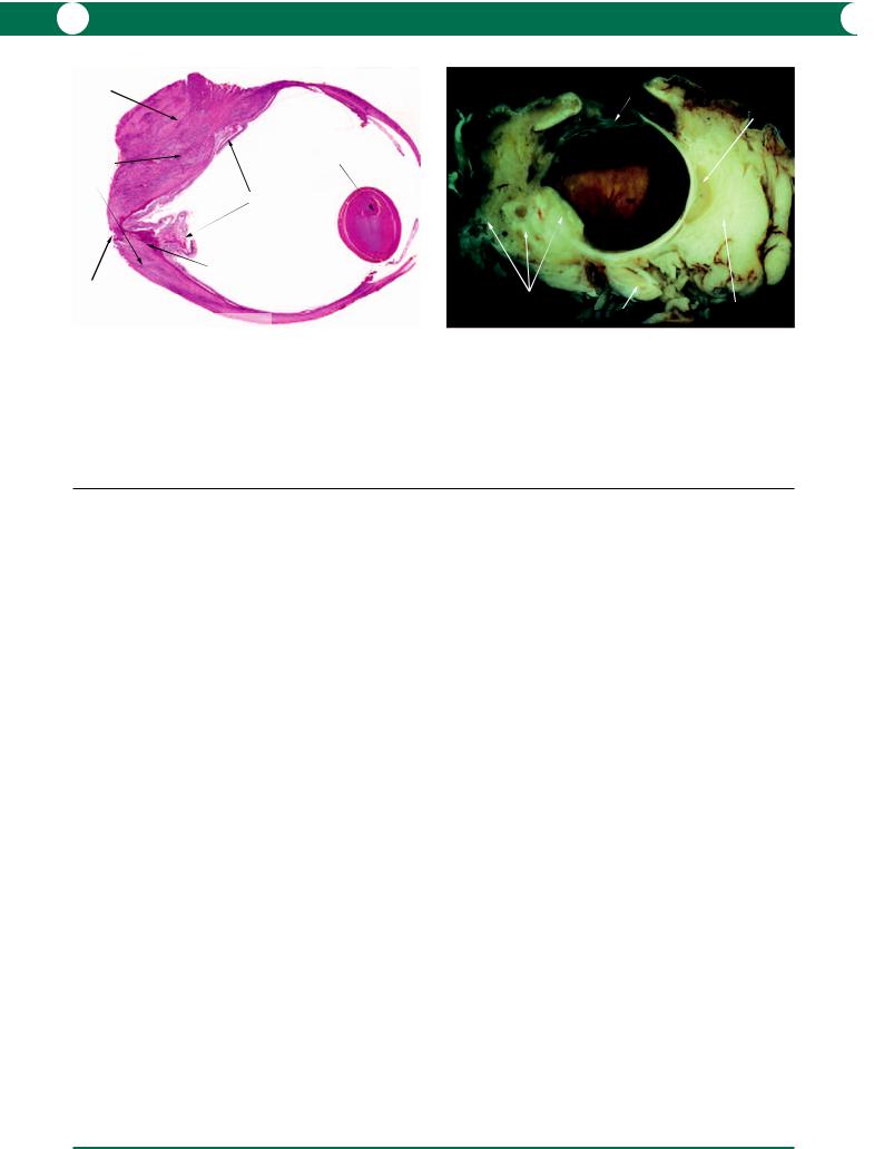
1 Anterior scleritis: at a macroscopic level, the necrotic sclera has a creamy yellow appearance (Figure 8.39). On section, the normal white condensed scleral tissue is replaced by partially pigmented granular tissue (Figure 8.40). Corneoscleral collagenolysis with a predominant lymphocytic infiltrate is characteristic; giant cell granulomatous reactions are inconspicuous (Figures 8.41, 8.42). A dense lymphocytic infiltration may replace the normal sclera (Figure 8.43).
2Posterior scleritis: in contrast with the anterior variant, the progress of posterior scleritis is much more chronic
and the inflammatory cell infiltration is accompanied by massive reactionary fibrosis (Figure 8.44). When the process is diffuse, the term brawny scleritis is appropriate (Figures 8.45, 8.46).
The fibrous reaction can be so extreme that an episcleral mass causes a proptosis (Figures 8.47, 8.48). The mass may also extend into the globe and may simulate an intraocular melanoma (Figure 8.49).
Complications/secondary effects
Severe inflammation can result in scleral thinning with staphyloma formation as well as perforation. Bacterial superinfection may result.

172 C H A P T E R 8
Inflammatory disease - Necrotising scleritis Postoperative cataract surgery
remnant of tripod lens
conjunctival congestion
Figure 8.39
frayed end of sclera
necrotising scleritis with perforation
opaque cornea
hypopyon
dense lymphocytic infiltrate
Inflammatory disease - Necrotising anterior scleritis
Figure 8.41
Figure 8.39 A massive anterior necrotising scleritis developed after a lens extraction and insertion of a tripod lens into the posterior chamber. The necrotising process extends into the cornea which is opaque due to iridocorneal contact. One haptic of the lens is identified externally.
Figure 8.40 A cut across the anterior segment of the globe shown in
Figure 8.39 reveals the intraocular lens and the scleral thinning due to scleritis. The vitreous is filled with a gelatinous exudate.
SUPERIOR |
|
INFERIOR |
intraocular lens |
iris |
cornea |
necrotising |
|
|
scleritis with |
|
normal ciliary body |
perforation |
|
normal sclera
|
proteinaceous exudate |
|
within vitreous |
|
Inflammatory disease - Necrotising scleritis |
|
Postoperative cataract surgery |
Figure 8.40 |
|
Inflammatory disease - |
CONJUNCTIVAL SIDE |
Necrotising anterior scleritis |
episcleral fibrosis |
|
|
fibrinous eosinophilic exudate |
|
|
thinned sclera |
macrophages
giant cell
CHOROIDAL SIDE
Figure 8.42
Figure 8.41 Anterior scleritis is characterised by a dense lymphocytic reaction in the region of the scleral disruption and destruction. It is frequently difficult to demonstrate giant cells and macrophages in such cases.
Figure 8.42 In some cases of scleritis, a granulomatous reaction with epithelioid macrophages and multinucleate giant cells can be demonstrated. Leakage of fibrin into the necrotic sclera gives the tissue a brick-red appearance.

I N F L A M M AT I O N 173
Inflammatory disease - Necrotising scleritis
open angle
pupillary membrane
mild disc oedema
necrotising scleritis
(scleromalacia perforans)
closed angle
|
lens removed |
|
before sectioning |
Figure 8.43 |
|
Inflammatory disease - Posterior scleritis |
swollen opaque lens |
|
|
|
shallow anterior |
|
chamber |
optic nerve |
|
thickened choroid |
|
oedematous folds in retina
thickened sclera
thickened |
detached |
||
episclera |
|||
organised vitreous |
|||
|
|||
Figure 8.45 |
|
|
|
|
episcleral mass |
|
|
|
retinal haemorrhages |
opaque |
|
diffuse |
|
lens |
|
thickened sclera
exudative
retinal detachment
shallow anterior
chamber
Inflammatory disease - Posterior scleritis
Figure 8.47
Inflammatory disease - Early posterior scleritis
inflammatory infiltration of the choroid |
artefactual detachment |
|
|
|
of proteinaceous |
|
exudate under retina |
early inflammatory |
inflammatory |
|
cell infiltrate |
||
infiltrate in sclera and |
||
in meninges |
||
fibrosis |
||
|
Figure 8.44
Inflammatory disease - Posterior scleritis
optic nerve
|
thickened sclera |
|
|
|
thickened lens - |
lens nucleus |
closed |
thickened |
angle |
||
showing |
|
|
|
episclera |
calcification |
|
|
Alizarin stain
Figure 8.46
Figure 8.43 A patient suffering from rheumatoid arthritis had intractable pain due to extensive anterior necrotising scleritis with imminent perforation. Enucleation was the only option available. Note the dense cellular infiltrate in the pars plana region with severe scleral thinning. Ocular hypotonia has resulted in retinal and optic disc oedema.
Figure 8.44 In the early stage of posterior scleritis, there is a lymphocytic infiltration within the sclera and this is accompanied by a fibrovascular proliferation in the episclera. Lymphocytic infiltration in the choroid interferes with the integrity of the choriocapillaris and retinal pigment epithelium, and the release of proteinaceous fluid detaches the retina. Note the involvement of the meninges.
Figure 8.45 The macroscopic appearance of posterior scleritis. The posterior sclera and episclera are diffusedly thickened. Involvement of the choroid has led to exudation in the retina which is thrown into folds. The vitreous is semiopaque and is detached posteriorly. The cataractous lens is unusually white – see Figure 8.46.
Figure 8.46 A low power photomicrograph of the specimen shown in Figure 8.45 reveals the extent of inflammatory cell infiltration in the choroid,
sclera, and episclera. The white macroscopic appearance of the lens is due to calcification. The inset is stained to demonstrate the presence of calcium in the lens (alizarin is a red stain which chelates calcium).
Figure 8.47 A patient presented with a proptosis. In this more advanced case, the fibrous proliferation extends internally into the globe and the secondary complications are exudative detachment and haemorrhage in the retina. Enucleation of such a deformed globe can be difficult, and in this case the surgeon did not excise the anterior part of the optic nerve with the globe.
174 C H A P T E R 8
episcleral mass
diffuse |
cataract |
thickened |
|
sclera |
|
|
retinal folds |
subretinal haemorrhage due to close excision
of optic nerve
optic canal
Inflammatory disease - Posterior scleritis
Figure 8.48
Figure 8.48 A section of the same globe illustrated in Figure 8.47 shows that there is massive inflammation and fibrosis throughout the posterior sclera. The haemorrhage around the optic nerve is a surgical artefact.
Inflammatory disease - Posterior scleritis simulating a melanoma Exenteration
extraocular muscle
fibrovascular |
|
inflammatory mass |
orbital fat |
|
optic nerve |
Figure 8.49 |
|
Figure 8.49 The patient presented with a proptosis which appeared to be due to an intraocular mass with extension into the orbit. A diagnosis of an amelanotic melanoma with extraocular extension was made. The possibility of posterior scleritis was excluded as the changes were so localised and the remainder of the ocular tissues appeared normal. An exenteration was performed in view of the possibility of extraocular spread into the orbit.
Vogt–Koyanagi–Harada (VKH) syndrome
A systemic granulomatous condition characterised by bilateral panophthalmitis of unknown aetiology. It is more common in pigmented races and was formerly divided into two different groups according to systemic findings:
1Vogt–Koyanagi:
•skin: alopecia, poliosis, and vitiligo
•anterior uveitis.
2Harada:
•neurological: headache, encephalopathy, inner and middle ear symptoms (deafness, tinnitus, and vertigo), cerebrospinal fluid pleocytosis
•posterior uveitis: in conjunction with disc swelling and exudative retinal detachments.
VKH is now commonly thought of as being one condition with a wide spectrum of presentations, although bilateral ocular involvement occurs in every case.
Clinical presentation
Ophthalmic findings will depend on the stage of the disease:
1Prodromal stage: flu-like illness. Eye signs are non-specific with visual disturbance, photophobia, ocular pain, and conjunctivitis.
2Uveitic phase: bilateral uveitis, disc swelling, and exudative retinal detachment.
3Chronic phase: fundus depigmentation, subretinal and optic disc neovascularisation.
Investigations are extensive to exclude other causes. Fluorescein angiography demonstrates multiple retinal pigment epithelium (RPE) window defects.
An important differential diagnosis is sympathetic ophthalmitis and a history of penetrating trauma should be sought.
Pathogenesis
An autoimmune granulomatous reaction against melanocytes, melanin, and retinal pigment epithelium. A viral agent has been suggested although not proven.
Genetics
Females are more commonly affected. There is an association with HLA DR4.
Possible modes of treatment
Principles involve high dose systemic corticosteroids with a long tapering off period. Immunosuppressants may also be used.
Macroscopic and microscopic
The appearances within a globe are indistinguishable from sympathetic ophthalmitis (see above).
Immunohistochemistry
The infiltrating lymphocytes are of T-cell type (suppressorcytotoxic subsets).
Behçet’s syndrome
A systemic vasculitic condition that manifests itself in the eye as a panuveitis. It is most common in Japan and Turkey.
Clinical presentation
Diagnostic criteria is recurrent oral ulceration with any two of the following:
1Eye lesions: anterior and posterior uveitis.
2Recurrent genital ulcerations.
3Skin lesions: for example erythema nodosum.
4Positive pathergy test.

I N F L A M M AT I O N 175
Pathogenesis and genetics
The aetiology is unknown. The geographical distribution of the disease follows that of the “silk road”. Association with HLA-B51 for ocular involvement is recognised.
Possible modes of treatment
Despite treatment, visual prognosis is poor with progression to blindness within 4 years of disease onset.
Macroscopic and microscopic
By the time enucleation is required, secondary pathology obscures the nature of the primary disorder.
Retinal haemorrhage, exudation, and detachment are the result of retinal vasculitis. The uveal tract is thickened by a non-granulomatous lymphocytic infiltrate with a prominent CD4 T-cell population.
Chorioretinitis
Many infective organisms can lead to chorioretinitis. As a rule of thumb, a viral infection results in a diffuse necrotising retinitis, while parasitic and fungal infections are focal (see section on microbiology under “Stains for microscopy” in Chapter 1).
Viral: herpes simplex virus (HSV), herpes zoster virus (HZV), and cytomegalovirus (CMV)
The clinical patterns of disease in viral infections vary and have become of great importance due to the increase in
incidence of AIDS and immunosuppression therapy. Thus it is convenient to consider viral retinitis in terms of the patient’s immune status.
Immunocompetent
Acute retinal necrosis (ARN)
In HSV and HZV infections, there is a unilateral necrotising retinitis, initially patchy and subsequently becoming confluent, accompanied by a non-granulomatous choroiditis – this is classified as acute retinal necrosis (ARN). Severe vitritis, vasculitis, and papillitis are important features of this condition. The emphasis for prompt antiviral treatment is to avoid bilateral involvement (BARN).
Macroscopic and microscopic
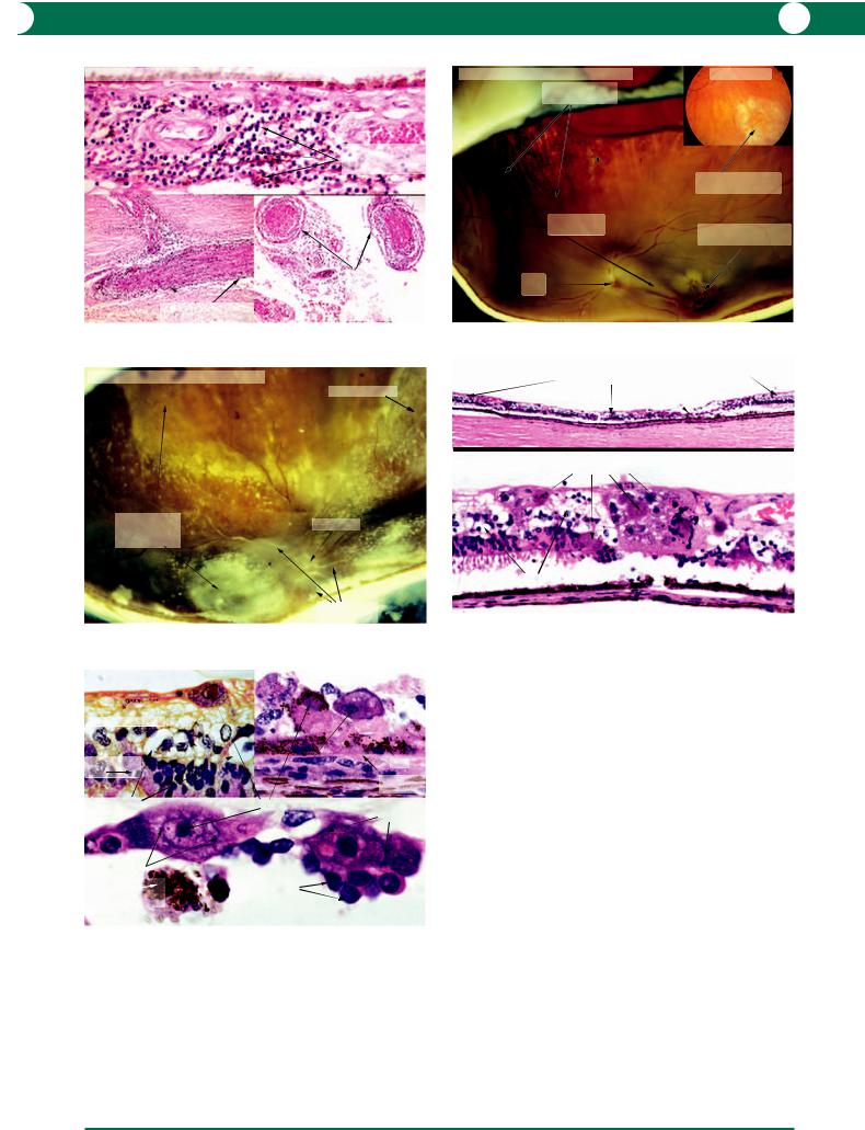
The likely specimen submitted to the pathologist would be an enucleation for end-stage ARN (Figure 8.50). Microscopic examination reveals total retinal infarction, vitreous exudation, and infiltration by inflammatory cells (Figure 8.51). A retinal biopsy may be taken at the earlier stages of the disease and it is possible to demonstrate intranuclear inclusions formed by proliferating herpes simplex viral particles (Figure 8.52). The ultrastructural characteristics of HSV are identical to those of CMV and consist of a central core surrounded by a capsid (Figure 8.53).
Inflammatory disease - Acute retinal necrosis (ARN)
mildly thickened choroid
*
vitreous haemorrhage and exudation
necrotic detached retina
clear anterior chamber
minor lens opacities
Figure 8.50
Figure 8.50 An immunocompetent patient known to be suffering from acute retinal necrosis (ARN) was treated with systemic acyclovir which protected against infection in the opposite eye. The disease in the affected eye however progressed to a haemorrhagic retinitis with detachment and an enucleation was performed for discomfort. A section through the centre of the eye reveals a normal anterior segment, early lens opacities, and a haemorrhagic exudate in the vitreous. The necrotic retina is grey in colour. The minor thickening of the choroid is due to lymphocytic infiltration. The asterix shows the region shown histologically in Figure 8.51.
Inflammatory disease - ARN
sub-hyaloid space
inflammatory cells in vitreous
necrotic |
|
cells in |
|
retina |
surviving outer nuclear layer |
|
surviving RPE and
photoreceptors
Figure 8.51
Figure 8.51 In the higher power view of the area marked with an asterix in Figure 8.50, there is total destruction of the retinal cells which appear as smudgy pink structures. Some of the cells in the outer nuclear layer have survived (in contrast with progressive outer retinal necrosis). The vitreous is detached by a proteinaceous exudate and the vitreous gel contains an inflammatory cell infiltrate which would be minimal in an immunocompromised patient.

176 C H A P T E R 8
Inflammatory disease - ARN |
Inflammatory disease - ARN |
|
HSV infection |
|
Transmission electron microscopy |
photoreceptors |
|
intranuclear |
|
inclusions |
|
Bruch’s membrane |
envelope |
capsid
lymphoplasmacytoid |
core |
|
infiltration in choroid |
||
|
Figure 8.52 |
Figure 8.53 |
Figure 8.52 In the early stages of infection, herpes virus can be identified by intranuclear inclusions within the retinal pigment epithelium cells and macrophages. Compared with cytomegalovirus (CMV) inclusions, the parent cell in a herpetic infection is much smaller and similar in size to a macrophage. Degeneration and fragmentation is present in the photoreceptor layer. The choroid contains a dense lymphoplasmacytoid infiltrate and the choriocapillaris is obliterated.
Figure 8.53 This electron micrograph illustrates the architecture of viral particles within a retinal cell infected with herpes simplex virus (HSV). Note that the viral morphology is identical in HSV and CMV infections. The viral particles vary in appearance because replication requires migration into the cell nucleus for the formation of viral DNA. The envelope is derived from the nuclear membrane.
Herpes zoster ophthalmicus (HZO)
HZO involving the fifth cranial nerve (shingles) can cause a mild neuritis in the ciliary nerves and a non-granulomatous nodular choroiditis (Figure 8.54) which is evident on fundoscopy.
Immunocompromised
The hallmark feature of viral retinitis in the immunocompromised patient (for example when CD4 T cells are reduced below 50 cells/mm3 in peripheral blood) is an absence of the inflammatory response, particularly in the vitreous. It is important to note that it is not always possible to reliably differentiate clinically between the infections caused by herpes simplex virus (HSV), herpes zoster virus (HZV), cytomegalovirus (CMV), and toxoplasmosis. Simultaneous infections may also occur. Thus, it is important in biopsies to perform PCR for the entire herpes family to achieve a precise diagnosis, because the treatments can vary.
Cytomegaloviral (CMV) retinitis
Small areas of retinal necrosis increasing in size, with or without haemorrhage, in any region of the retina are characteristic (Figure 8.55). As the infection progresses, the retina becomes thickened and linear perivascular infiltrates extend laterally from the vessel (frosted branch angiitis – Figure 8.56). Untreated, the condition is progressively destructive to the whole retina resulting in blindness with the further complication of retinal detachment.
Destruction of the neurones in the retina by the virus leaves a relatively thin strand of tissue containing inflammatory cells and scattered surviving neurones (Figure 8.57). Extremely large infected cells, diagnostic for CMV infection, may contain obvious viral inclusion bodies within the nuclei (Figure 8.58).
Progressive outer retinal necrosis (PORN)
This is the result of HSV/HZV retinitis in the absence of an active immune response. The lack of vitritis, vasculitis, and papillitis are important differentiating features from ARN.
The condition is characterised by extensive destruction of the outer retina (compared with ARN), which can be located in any region of the fundus. The clinical appearance is characteristic: it begins with a blanching of the fundus which later progresses to extensive destruction of the outer retina. The histological features are similar to ARN, but the destruction of the outer retina predominates. There is also relative sparing of the retina adjacent to retinal vessels.
Human immunodeficiency virus (HIV)
Infection by HIV alters the immune system by selective destruction of the CD4 helper and inducer cells with an increased incidence of secondary infections apart from those described above. An awareness of the complexities which occur in the management of AIDS patients will be apparent from the following list of ocular opportunistic infections and malignancies (refer to relevant chapters):
•Toxoplasma gondii (see below)
•fungal chorioretinitis (Histoplasma capsulatum, Coccidioides immitis, Pneumocystis carinii, Candida sp.)
•endogenous bacterial endophthalmitis (Mycobacterium intracellulare and M. avium)
•molluscum contagiosum
•Kaposi’s sarcoma
•lymphomas.
NB: HIV causes a specific microvasculopathy which is manifest in early cases as retinal microinfarcts due to viral parasitisation of vascular endothelial cells.
I N F L A M M AT I O N 177
Inflammatory disease - Herpes zoster ophthalmicus
perineural lymphocytic infiltrate in sclera
Figure 8.54
Inflammatory disease - CMV retinitis
active retinal inflammation & necrosis
Figure 8.56
Inflammatory disease - CMV retinitis
inner nuclear layer
non-granulomatous lymphocytic infiltrate in choroid
perineural lymphocytic infiltrate
in posterior ciliary artery
low-grade vitritis
optic disc
frosted branch angiitis
outer nuclear |
|
RPE |
layer |
|
Bruch’s |
|
|
|
|
phloxine-tartrazine stain |
membrane |
degeneration |
intranuclear inclusions |
limits |
of retina |
|
|
|
of nucleus |
|
|
|
limits of nucleus
macrophage
containing lymphocytes RPE melanosomes
Figure 8.58
Figure 8.54 The histological features of herpes zoster ophthalmicus include a non-granulomatous lymphocytic infiltrate in the choroid (upper), and in the perineurium of intrascleral nerves (lower left) and posterior ciliary nerves (lower right). Intranuclear viral inclusions are not demonstrated in lymphocytes but are present in the epithelium in shingles.
Inflammatory disease - CMV retinitis
inferonasal sectorial necrosis
active inflammation
optic disc
Figure 8.55
Inflammatory disease - CMV retinitis widespread retinal necrosis
enlarged infected cells
widespread retinal necrosis
Figure 8.57
Fundus photo
area of active retinal necrosis
coexisting Toxoplasma infection at the macula
unaffected retina
Figure 8.55 A patient suffering from Hodgkin’s disease was immunosuppressed and died as a result of disseminated toxoplasmosis in addition to cytomegaloviraemia. Five weeks prior to his death, an area of active inflammation and necrosis was noted in the inferior nasal quadrant of his right eye consistent with CMV retinitis (inset). In the right eye enucleated at autopsy, a triangular defect in the retina exposed the underlying choroid (marked sectorial necrosis). Thickened white areas in the retina indicate more recent infection. The necrotic focus at the macula was found on histology to be due to toxoplasma chorioretinitis. Note that the vitreous is clear.
Figure 8.56 A middle aged man with a long history of HIV infection lost vision due to massive CMV retinitis. His T-cell CD4 counts were less than
20 cells/mm3. After death, the globes were removed at autopsy. The affected areas in the retina appear to contain yellow granular material which is becoming confluent. Vascular sheathing away from necrotic areas is evident and the appearance is consistent with frosted branch angiitis as seen clinically.
Figure 8.57 Histology from the necrotic area shown in Figure 8.55 illustrates widespread retinal necrosis with sharp demarcation at the edge of the unaffected retina (upper). At higher magnification, the necrotic retina contains extremely large cells indicative of cytomegaloviral infection (lower) – see Figure 8.58.
Figure 8.58 With a phloxine-tartrazine stain, intranuclear inclusions in CMV are easily demonstrated (upper left). In this example the infected cell is present in the inner retina and only thin strips of cells represent the surviving inner and outer nuclear layers. Extremely large infected cells are present in the debris on the inner surface of the retinal pigment epithelium (RPE) (upper right). The size of the large infected cells (with intranuclear inclusions) should be compared with that of the adjacent lymphocytes (lower).

178 C H A P T E R 8
Parasites
Toxoplasma
Toxoplasma gondii is the commonest protozoal parasite to infect the eye. This organism is neurotrophic and infection is limited to the neural retina and the brain. Whereas it was previously thought that toxoplasma retinochoroiditis was due to reactivation of congenital infections, there is growing evidence for acquired disease.
Clinical presentation
Congenital In this category, acute infection of the mother during pregnancy proceeds to transplacental infection of the fetus. If infection occurs during the first trimester, stillbirth usually results. The fetus can survive a later infection but often the triad of convulsions, cerebral calcifications, and chorioretinitis are clinical manifestations after birth. With this potential outcome, mothers who have toxoplasma chorioretinitis during pregnancy may choose to have an elective termination of pregnancy.
Reactivation or acquired infection in adulthood The patient presents with reduced vision and floaters. Examination reveals a moderate to severe vitritis with underlying single or multiple patches of necrotising retinitis. A pre-existing chorioretinal scar is evident in cases of reactivation of infection – “headlight in the fog” appearance. Satellite lesions are common. The parasitisation is usually situated in the inner retina leading to retinal oedema.
The surrounding inflammation may be severe enough to affect other regions producing choroiditis, scleritis, and anterior uveitis of differing severity.
Pathogenesis: life cycle of Toxoplasma gondii
The cat is the definitive host in which the organism undergoes sexual reproduction (Figure 8.59). Oocysts are passed in cat faeces which may be directly ingested by domestic animals. Human infection occurs either from direct ingestion of oocysts in contaminated soil or from uncooked meat of infected animals. The organism can exist in a free form (trophozoites) or an encysted form (bradyzoites). Parasitisation within neural cells provides protection from the immune system. Multiplication within intermediate hosts is by asexual reproduction.
Possible modes of treatment
Infections are usually self limiting and treatment is conservative. The indications for treatment are:
1Sight threatening: involving macula, papillomacular bundle, and optic disc.
2Vasculitis: overlying a major retinal vessel.
3Persistence: more than a month.
4Severity: severe vitritis leading to significant visual loss.
5Immunosuppression.
Treatment involves multiple drug combinations:
1Folic acid antagonists – sulfadiazine and pyrimethamine: the Toxoplasma organism has an inability to transport folate which is an essential component for DNA synthesis, and antagonists inhibit synthesis in the active trophozoite. Folinic acid supplement is added to prevent patient folate deficiency.
2Clindamycin, azithromycin, and atavaquone: although many of these agents are found to have cysticidal properties in vitro, recurrence of infection has been known to occur following treatment.
3Corticosteroids: introduced systemically at a later stage to modulate the inflammation and secondary damage.
4Other treatment modalities: these include argon laser ablation, cryotherapy, and vitrectomy.
The difficulty in management lies in the treatment resis-
tance of the parasite when it is in an encysted form.
Macroscopic
Acute Most cases of acute disease are observed in autopsy material obtained from aborted fetuses or stillborn infants.
Chronic or healed Sites of previous infection may be observed as an unrelated finding in an enucleated eye (Figure 8.60). This has the appearance of an irregular white chorioretinal scar with clumps of pigmentation usually in the periphery.
The associated uveitis may lead to secondary glaucoma.
Microscopic
Acute In the centre of the infected sector in the retina, there is a thickened strip of hypocellular pink-staining tissue. Adjacent to this, there is a more cellular zone containing inflammatory cells. The adjacent recognisable retina is thickened by oedema and contains a scattered infiltrate of inflammatory cells – lymphoplasmacytoid cells and macrophages (Figure 8.61). Bradyzoite cysts are most commonly identified at the edge of the surviving retina: freeform trophozoites are identified less easily (Figure 8.62). The complex morphology of Toxoplasma gondii is best appreciated at the ultrastructural level (Figure 8.63).
Chronic A healed scar consists of a thin strip of gliotic retina separated by clumps of retinal pigment epithelium from a fibrotic choroid.
Immunohistochemistry
Labelled antitoxoplasma antibodies identify the organisms (Figure 8.62). PCR may also be used.

I N F L A M M AT I O N 179
Inflammation - Toxoplasmosis
Life cycle
oocysts in litter trays, |
|
|
|
garden, sandpits, |
sporulated |
tissue cysts in infected |
|
unwashed fruit |
|||
oocysts |
undercooked meat |
||
or vegetables |
|||
|
|
||
|
|
oocysts in feed, |
|
|
|
water or soil |
vertical transmission of tachyzoites
through placenta
to foetus |
unsporulated |
|
|
|
Intermediate |
oocysts |
|
Intermediate |
|
passed in faeces |
|
|||
hosts |
|
|||
host |
|
|
|
|
|
|
|
|
|
|
Definitive host |
|
|
|
|
|
tissue cysts in flesh |
|
|
|
|
of infected rodents |
|
|
Figure 8.59 |
|
|
|
|
Inflammatory disease - Acute toxoplasmosis |
|
|
||
|
|
|
area |
|
|
vitritis |
|
containing |
oedematous |
|
|
|
cysts |
retina |
total retinal |
subretinal |
necrosis coagulative retinal |
exudate |
necrosis |
choroiditis |
|
Figure 8.61
Inflammatory disease - Toxoplasma gondii
Transmission electron microscopy
organisms cut in
longitudinal section organisms cut in transverse section
Figure 8.63
Inflammatory disease - Adult toxoplasmosis
Healed chorioretinal scar
proliferation of retinal pigment epithelium
chorioretinal scar
optic disc
Figure 8.60
Inflammatory disease -
Toxoplasma gondii
free forms
(trophozoites)_ encysted organisms (bradyzoites)
encysted organisms (bradyzoites)
encysted organisms |
Anti-toxoplasma antibody |
(bradyzoites) |
immunolabelling - PAP |
Figure 8.62
Figure 8.59 Diagram to show the lifecycle of Toxoplasma gondii. (Courtesy of Dr Fiona Roberts.)
Figure 8.60 This globe was enucleated for angle closure glaucoma secondary to recurrent anterior uveitis. A typical chorioretinal scar is identified in the midperiphery. It was not possible to histologically identify Toxoplasma cysts in the retina adjacent to the scar, and this is often the case in healed disease.
Figure 8.61 An eye from a stillborn infant was found to contain an area of acute retinal necrosis associated with dense choroiditis. Toxoplasma cysts are found at the edge of the necrotic area outlined by a box. An exudative retinal detachment is evident.
Figure 8.62 Identification of Toxoplasma organisms requires the high magnification of an oil immersion lens. Free forms or trophozoites (top left) are identified by a basophilic spot within an elongated eosinophilic tail. Most commonly the organisms are identified within cystic structures containing blue or purple dots (bradyzoites – top right and bottom left). The organisms can also be identified with antitoxoplasma antibody immunolabel (bottom right).
Figure 8.63 Electron microscopy demonstrates the complex structure of
Toxoplasma organisms within a retinal cyst. The organisms are cut in different planes.