

Sehu - Ophthalmic Pathology-2005
.pdf150 C H A P T E R 7
Secondary closed
Neovascular
Neovascular glaucoma is commonly seen in enucleation specimens.
Clinical presentation
Early
1 The development of neovascularisation first occurs in the angle and this can be seen by gonioscopy; later new vessels are seen on the anterior iris surface.
2The initial pressure rise is due to a neovascular membrane on the inner surface of the trabecular meshwork.
Late
1Contraction of the fibrovascular tissue in the angle leads to iridotrabecular and iridocorneal contact. Gonioscopy reveals closed angles.
2Fibrovascular proliferation on the iris surface rotates the pupil margin forward so that the pigmented epithelium is visible (ectropion uveae).
3There may be evidence of glaucomatous decompensation in the cornea (see “Tissue effects” below).
4A primary cause for ischaemia must always be sought (for example for central retinal vein occlusion or diabetes).
Pathogenesis
Any process which leads to the release of vascular endothelial growth factor (VEGF) into the aqueous could potentially cause neovascularisation of the iris. The subcategories of possible sources of VEGF are:
1 Ischaemic tissue: for example central retinal vein occlusion, diabetic retinopathy, and prolonged retinal detachment.
2 Neoplasia: for example intraocular melanomas or retinoblastomas.
3 Inflammation: prolonged uveitis of any cause.
Possible modes of treatment
This depends on the primary disease, but the following options are available:
1Removal of cause: for example panretinal photocoagulation of an ischaemic retina.
2Medical treatment for inflammation and glaucoma.
3Ciliary body ablation.
4Surgical treatment by drainage procedures with or without setons.
5Enucleation of the painful blind eye for comfort.
Macroscopic
In an enucleation specimen the following changes may be identified (Figure 7.38):
•bullous keratopathy
•peripheral corneal pannus
•ectropion uveae
•cataract
•retinal changes relevant to vascular disease or tumour.
Microscopic
It is important to appreciate that the neovascular membrane consists of small capillaries and fibroblasts. At the earliest stage, the capillaries and fibroblasts are identified on the iris surface and on the inner surface of the trabecular meshwork (Figure 7.39). Fibroblasts proliferate in the chamber angle and subsequently contract to pull the peripheral iris towards the meshwork (Figure 7.40).
The angle eventually closes and the peripheral iris comes into contact with the cornea (Figure 7.41). Contraction of the fibrovascular membrane on the iris surface results in flattening. NB: Any loss of normal iris folds or the presence of ectropion uveae should alert the examiner to the possibility of neovascularisation.
In some cases, the corneal endothelium migrates across the surface of the neovascular membrane (Figure 7.42).
Pathological evidence of treatment
Evidence of surgery or laser as described above, especially pan-retinal photocoagulation (see Figure 10.48).
Tumour
Mechanical displacement of the iris and lens by tumours of the ciliary body, choroid and retina can lead to a pupil block (see Figures 11.9, 11.10, 11.33, 11.43).
Uveitis/inflammation
In inflammation of the anterior uvea, release of a sticky fibrinous exudate promotes the formation of peripheral anterior and central posterior synechiae with resultant secondary angle closure glaucoma. With prolonged inflammation, neovascularisation may also contribute to a rise in intraocular pressure (see above).
Trauma
With civil or surgical trauma involving decompression and perforation of the cornea or limbus, hypotonia and reactionary fibrosis result in anterior synechia formation.

G L A U C O M A 151
Glaucoma - Neovascular |
opaque cornea |
Glaucoma - Neovascular |
|
Ectropion uveae |
closed angles |
artefactual |
Schlemm’s canal |
|
|||
pannus |
|
||
|
separation of |
|
|
|
|
trabecular |
|
|
iris pigment |
meshwork |
|
|
epithelium |
|
|
|
|
|
narrowing |
|
|
|
of angle |
|
|
neovascular membrane |
|
|
artefactual adhesion |
|
|
|
of pigment epithelium |
|
|
|
to lens |
|
|
Figure 7.38 |
|
Figure 7.39 |
|
Glaucoma - Neovascular |
|
Glaucoma - Neovascular |
|
pannus
iris
neovascular Schlemm’s canal ectropion membrane
uveae
|
contractile |
compressed |
|
|
trabecular |
||
|
spindle cells |
||
hyalinised acellular |
meshwork |
||
|
|||
meshwork |
|
|
|
neovascular membrane |
|
stromal |
|
|
|
||
|
|
fibrosis |
|
|
|
|
Figure 7.40
Glaucoma - Neovascular |
PAS stain |
|
edge of Descemet’s membrane |
endothelial downgrowth |
|
|
chronic |
neovascular |
inflammatory |
membrane |
cells |
PAS positive vacuoles - diabetic
Figure 7.42
Figure 7.41
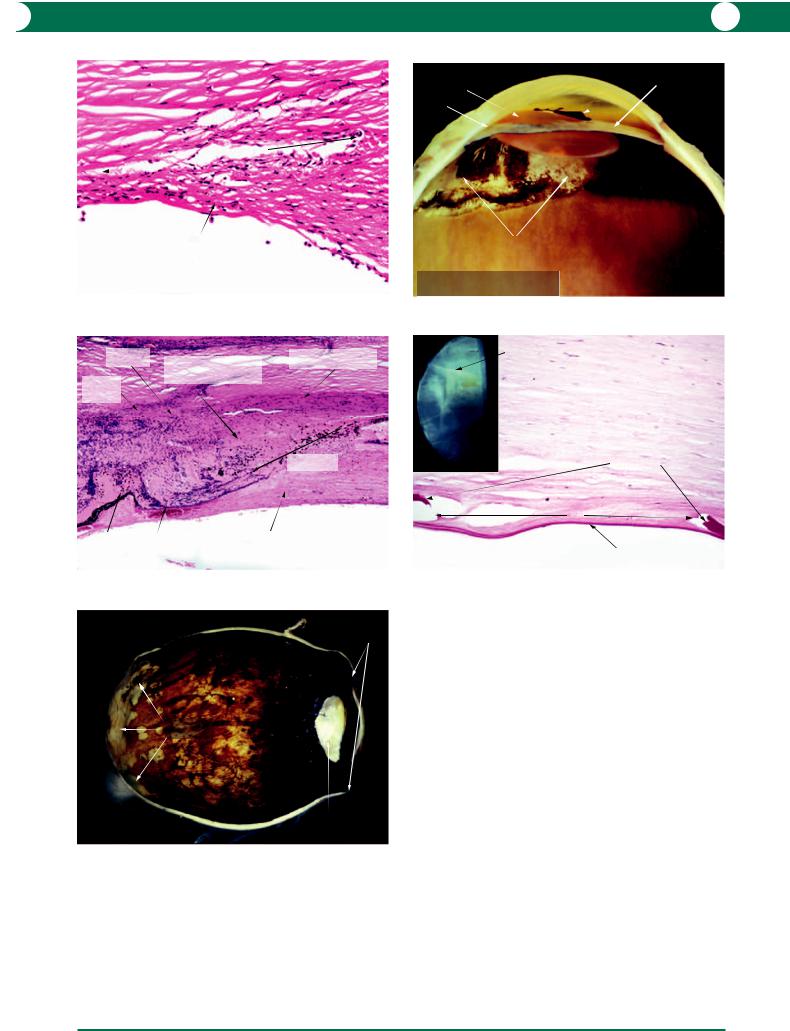
Figure 7.38 The macroscopic appearance of the anterior segment in neovascular glaucoma. The iris surface is smooth and the angles are closed. The pigment epithelium of the iris is retracted around the pupil (ectropion uveae). Pigmentation on the lens surface is artefactual but, in vivo, occurs in inflammation in the anterior segment.
Figure 7.39 In early neovascular glaucoma, the aqueous outflow is obstructed by a layer of fibrovascular tissue on the inner surface of the meshwork. Similar capillaries are present on the iris surface. This example shows the early stages of secondary angle closure.
Figure 7.40 As the disease progresses, the fibroblastic proliferation and contraction in the chamber angle pulls the peripheral iris forwards. The capillaries are not conspicuous in this example which emphasises the importance of the fibroblast component in angle closure.
Figure 7.41 In late neovascular glaucoma, there is a layer of fibrovascular tissue between the iris stroma and the cornea, the angle is closed, and an ectropion uveae is easily identified. On the iris surface, the neovascular membrane contains capillaries and fibroblasts (inset). The spindle-shaped fibroblasts are responsible for the tissue contraction and pupillary distortion. Note that the iris stromal surface is smooth.
Figure 7.42 In this example of the false angle in neovascular glaucoma, a layer of endothelial cells has migrated across the neovascular membrane (endothelial downgrowth). The presence of PAS-positive lakes within the iris pigment epithelium is a diagnostic feature of diabetes (PAS stain).

152 C H A P T E R 7
Treatment of glaucoma and its
complications
Trabeculectomy
Pathological experience is limited to specimens in which surgical treatment of glaucoma has failed. Many procedures were devised to produce a controlled fistula and, of these, a trabeculectomy has proved to be the most commonly employed. The original recommendation was to remove a small block of tissue from the inner sclera, trabecular meshwork, and inner cornea. Currently, a more anterior approach with excision of a small block from the cornea and the anterior part of the trabecular meshwork is favoured. Drainage of aqueous is via the sub-Tenon’s space.
Unsuccessful control of intraocular pressure by a fistulising procedure is usually due to fibrosis and scar tissue formation: antimetabolites (5-flurouracil and mitomycin-C) are used to minimise this complication. Iris prolapse is rare, but bleb complications such as leakage and infection are not uncommon (Figures 7.43, 7.44).
Argon laser trabeculoplasty (ALT)
The original intention of ALT was to create a fistula through the trabecular meshwork into Schlemm’s canal, but the effects resulting from this pressure lowering were short
lived due to fibrosis. Current ALT therapy uses considerably less power and the intention is to stimulate the trabecular endothelial cells. Pathological studies are rare, but in failed cases evidence of ALT is seen in the trabecular meshwork as loss of endothelial cells and fusion of trabecular beams (Figure 7.45).
Transcleral ciliary body ablation – cryotherapy and diode
Destruction of the ciliary processes by transcleral laser or cryotherapy in order to decrease aqueous production appears as areas of ciliary body depigmentation in endstage glaucomatous eyes (Figure 7.46). In successful ablation, the ciliary processes are fragmented and fibrous tissue containing pigment-laden cells is present in the scar (Figure 7.47).
Tissue effects
While it is convenient to describe the tissue effects of glaucoma under two headings, acute (implying primary angle closure) and chronic (implying primary open angle glaucoma), there is often a considerable overlap between cause and effect.
Table 7.1 summarises the histopathological features in acute and chronic glaucoma.
Glaucoma - Treatment pathology |
open angles |
|
Trabeculectomy |
|
|
iris strand |
|
|
|
|
cataract |
|
scleral flap |
artefactual |
|
|
|
|
|
tear |
|
|
bleb |
|
|
space |
surgical defect
ciliary body
Figure 7.43
Figure 7.43 When the globe is sectioned in the correct plane, it is possible to identify the defect made during a trabeculectomy procedure. The inner limbal defect is located anterior to the ciliary body. In this example iris strands project into the defect. A bleb was present and was artefactually torn. The inset is a lower power view of the anterior segment.
Glaucoma - Treatment pathology
Trabeculectomy
|
scleral flap |
fibrovascular tissue |
surgical defect |
|
|
|
remnant |
|
Descemet’s |
|
membrane |
iris strand
Figure 7.44
Figure 7.44 This figure illustrates some of the potential causes of failure in a trabeculectomy. Fibrovascular scar tissue has drawn the iris over the surgical defect and there is fibrovascular proliferation in the wall of the defect, which contains a strip of Descemet’s membrane. The flap is fibrotic and a bleb did not form in this case.
G L A U C O M A 153
Schlemm's canal
fused trabecular beams with loss of endothelial cells
Glaucoma - Treatment pathology
Argon laser trabeculoplasty (ALT)
Figure 7.45
distorted |
destruction of circular |
remaining longitudinal |
scleral spur |
fibres of ciliary muscle |
|
destroyed |
and oblique ciliary muscle |
|
fibres |
|
trabecular meshwork
main ablation zone
iris root |
distorted ciliary processes |
fibrovascular ingrowth |
|
Glaucoma - Treatment pathology
Transcleral YAG ciliary body ablation
Figure 7.47
Glaucoma - Tissue effects
Staphyloma
staphylomas
chorioretinal atrophy
advanced cataract
Figure 7.49
dislocated |
rubeotic iris with |
lens |
ectropion uveae |
closed angle
scarring and depigmentation of ciliary body
Glaucoma - Treatment pathology
Diathermy ciliary body ablation
Figure 7.46
PAS stain
Haab’s striae
|
original Descemet’s |
|
membrane |
|
tear |
Glaucoma - Tissue effects |
secondary Descemet’s membrane |
Haab’s striae |
|
Figure 7.48
Figure 7.45 In this example of failure of an argon laser trabeculoplasty (ALT), there is a loss of trabecular endothelial cells and collapse of the meshwork with fusion of the beams.
Figure 7.46 The regions of diathermy ablation can be identified by areas of depigmentation.
Figure 7.47 Transcleral ablation of the ciliary body can be excessively destructive as in this example in which a YAG laser was used. The epicentre of the ablation zone was positioned too far posteriorly. However, sufficient laser energy was applied to destroy the inner layers of the ciliary muscle and the ciliary processes. An exuberant fibrovascular reaction extended across the retina.
Figure 7.48 The inset shows a hemisection of a corneal disc of a patient with a previous history of infantile glaucoma. Slit lamp microscopy shows these fine horizontal lines as tears in Descemet’s membrane (Haab’s striae). Microscopy of the tears confirms the defect in the original Descemet’s membrane. The bare stroma is re-covered by endothelial sliding and there is subsequent formation of a secondary Descemet’s membrane. The endothelium is sparse at this end stage and poorly demonstrated in a PAS section.
Figure 7.49 Elongation of the globe and chorioretinal atrophy are features of axial myopia. However, the presence of staphylomas in the region of the ciliary body (intercalary) in this case of secondary angle closure glaucoma indicates longstanding raised intraocular pressure.

154 C H A P T E R 7
Table 7.1 Summary of the histopathological features in acute and chronic (long term) glaucoma.
Acute |
Chronic |
|
|
Cornea Epithelial oedema with or without bulla formation
(see Figure 4.7): prolonged high pressure
may lead to loss of endothelium and stromal oedema
Sclera –
Iris |
Infarction: reflex vascular spasm leads to sectorial |
|
infarction of the iris stroma and musculature. Clinically |
|
this is seen as a sluggish and irregular pupil. |
|
Histologically the infarct appears as an acellular |
|
sector with loss of muscle and atrophy of the iris |
|
pigment epithelium (Figure 7.51) |
Lens |
Localised epithelial infarction (Glaukomflecken): focal |
|
necrosis of lens epithelial cells (rare pathologically) |
|
is seen as white flecks in the anterior cortex |
Retina |
Macular and retinal oedema |
Optic disc Papilloedema: if the intraocular pressure rises rapidly, compression of the nerve head obstructs axoplasmic flow and widespread swelling of the prelaminar nerve fibre layer occurs (Figure 7.53)
Choroid Congested
Degenerative keratopathy: may result if there is sufficiently high pressure (see Figures 4.8, 4.9, 4.11). This may be secondary to endothelial decompensation
Haab’s striae: a sustained rise in intraocular pressure in the neonatal or infantile eye can stretch the cornea to such an extent that linear tears occur in Descemet’s membrane. The endothelium covers the bare stromal surface by sliding to form a secondary Descemet’s membrane (Figure 7.48). Clinically the tears are horizontal (Figure 7.48, inset)
Staphylomas: occur in the region of scleral canals for vessels and nerves (Figure 7.49)
Buphthalmos: generalised stretching and thinning of the corneoscleral envelope, which is more elastic in infancy (Figure 7.50)
–
Inner retinal atrophy and gliosis: retrograde atrophy of the ganglion cell layer is due to interruption of nerve fibres at the disc. Consequently, there is a marked reduction in the thickness of the inner retina and preservation of the outer retina (Figure 7.52)
Optic disc cupping: its distinctive clinical appearance
is due to loss of prelaminar axonal tissue (Figures 7.54–7.57). Two main causes are implicated:
(1)Mechanical pressure on the prelaminar tissue interferes with axoplasmic flow. The bowing of the lamina cribrosa distorts axons as they pass through the pores in the lamina cribrosa
(2)Vascular: it is assumed that in the ageing eye there is diminished blood flow to the optic nerve head. The raised intraocular pressure may interfere with the blood flow from prelaminar capillaries which are supplied by the circle of Zinn
Atrophic

G L A U C O M A 155
Glaucoma - Tissue effects |
|
|
|
Glaucoma - Tissue effects |
white areas |
|
Buphthalmos |
|
|
|
of infarction |
||
|
|
|
Acute iris infarction |
|||
|
|
|
|
|
||
|
|
|
|
|
cornea |
|
|
|
cornea |
|
|
|
|
thin sclera |
|
|
|
|
|
loss of stromal cells |
|
|
|
|
|
|
|
|
|
|
|
dilator pupillae |
|
|
|
|
|
|
muscle absent |
|
|
|
|
staphyloma |
|
|
|
|
artefactual detachment |
|
|
|
disrupted pigmented |
|
|
(formalin fixation) |
|
|
|
epithelial layer |
|
|
Figure 7.50 |
|
|
|
Figure 7.51 |
|
|
Normal retina |
|
Chronic glaucoma |
|
CRA |
CRV |
nerve fibre layer |
|
||||||
|
|
|
|
|||
NFL |
|
|
|
|
|
|
GCL |
|
|
glial cells |
|
|
|
IPL |
|
|
|
|
||
|
|
|
|
|
artefactual |
|
|
|
|
|
|
|
|
|
|
|
|
|
|
split of OPL |
INL |
|
|
|
|
|
Normal |
|
|
|
|
|
|
|
|
|
|
|
|
end of Bruch's |
|
OPL |
|
|
|
|
membrane |
|
ONL |
|
|
|
|
|
|
PR |
|
|
|
|
|
|
|
|
|
|
|
swollen axons |
peripapillary |
Glaucoma - Tissue effects |
|
|
|
Glaucoma - Tissue effects |
photoreceptor atrophy |
|
|
|
|
|
|||
Retina: Normal vs. Atrophic |
|
|
|
Acute swelling of disc |
Bodian stain |
|
Figure 7.52 |
|
|
|
Figure 7.53 |
|
|
Figure 7.50 The patient had a history of a congenital cataract which was treated with lensectomy surgery in infancy. Glaucoma developed later as a complication. This is an extreme example of buphthalmos – the globe measures nearly 40 mm in diameter. Despite the generalised enlargement of the globe, a co-existing localised staphyloma has occurred.
Figure 7.51 In an infarcted sector of the iris, the stroma is acellular. Macrophages containing melanin derived from necrotic stromal melanocytes are scattered throughout the tissue. The hallmark feature of iris infarction is the loss of the dilator pupillae muscle. The pigment epithelium is disrupted or atrophic. Macroscopically (inset) the infarcted area is white.
Figure 7.52 The extent of inner retinal atrophy in longstanding glaucoma (right) is best demonstrated by comparison with normal retinal tissue (left) from a corresponding region. The thickness of the outer nuclear layer and the distribution of rods and cones in the photoreceptor layer are essentially the
same. The striking abnormality is the absence of recognisable inner retinal layers with glial cell proliferation in the atrophic neural tissue. NFL nerve fibre
layer, GCL ganglion cell layer, IPL inner plexiform layer, INL inner nuclear layer (bipolar cells), OPL outer plexiform layer, ONL outer nuclear layer (photoreceptor nuclei), PR photoreceptors.
Figure 7.53 Upper: normal nerve head; lower: acute glaucoma. An acute glaucoma developed in a patient with a necrotic ciliary body melanoma and the outflow system was blocked by macrophages containing melanin granules. A rapid elevation in intraocular pressure results in optic disc swelling – this is histologically seen as swelling of the nerve fibre layer from an interruption of anterograde axoplasmic flow in the prelaminar part of the optic nerve. The Bodian stain reveals distension of axons and the swollen nerve fibre layer displaces the peripapillary photoreceptor layer. The branches of the central retinal artery (CRA) and vein (CRV) are patent. OPL outer plexiform layer.

156 C H A P T E R 7
artefactual widening of subarachnoid space
Normal
thinning of nerve fibre layer
Glaucoma - Tissue effects
Optic disc cupping (early)
Figure 7.54
deep cup
bowing of the lamina cribrosa
Glaucoma - Tissue effects
Optic disc cupping (chronic end stage)
Figure 7.56
optic nerve
atrophy pathological widening of
subarachnoid space
Atrophy of NFL
widened arachnoid space
atrophic nerve
Figure 7.54 The lower figure shows early glaucomatous atrophy of the nerve fibre layer (NFL) by comparison with the normal tissue above. The optic nerve itself is atrophic because of the dropout of axonal tissue and there is enlargement of the subarachnoid space.
Figure 7.55 In the later stages of chronic glaucoma, the nerve fibre layer is extremely thin (upper). Ultimately the optic cup is filled with disorganised gliotic tissue and axonal tissue is absent in the prelaminar portion of the optic nerve (lower).
advanced thinning of nerve fibre layer
peripapillary atrophy
replacement of nerve fibre layer with gliotic tissue
Glaucoma - Tissue effects
Optic disc cupping (later stage)
Figure 7.55
excavated optic disc
macula
Glaucoma - Tissue effects
Optic disc cupping (chronic end stage)
Figure 7.57
Figure 7.56 At the end stage of chronic glaucoma, the prelaminar neural tissue is absent and the lamina cribrosa is bowed. The optic nerve is atrophic and the arachnoid space is enlarged.
Figure 7.57 A pathologist’s view of advanced optic disc cupping. The macula is easily identified by the presence of luteal pigment and its location within the vascular arcades.

157
Chapter 8
Inflammation

158 C H A P T E R 8
Basics
This account is confined to the histopathological manifestation of inflammatory disease and the reader should refer to those texts which describe the immunopathology of ocular disorders, for example Forrester J, Dick A, McMenamin P, Lee WR (2002) The Eye: Basic Sciences in Practice, second edition. Saunders, London.
Constituents of the inflammatory response
Identification of different types of inflammatory cells is based on their relative size, cytoplasmic inclusions, and nuclear characteristics.
Polymorphonuclear leucocytes (PMNLs)
PMNLs are present in circulating blood and when attracted by the appropriate chemoattractants, they pass through the endothelium of venules and capillaries (Figure 8.1). The PMNL is unique is that its nucleus is multilobed and the cytoplasm possesses granules that do not stain in an H&E section but are faint blue in a Giemsa stain. The granules contain proteases, lipases, and lysins. The pale staining of the granules is the reason why this cell is classified as a neutrophil. The PMNL is the first response to bacterial infection and is often destroyed by the enzymes and toxins released from ingested bacteria (Figure 8.2).
damaging proteins (eosinophil major basic protein and eosinophil cationic protein). These cells are commonly seen in allergic disease and helminthic infections.
Mast cells
Mast cells resident within tissue are the first to respond to exposure to an allergen. The cell is characterised by an oval eccentric nucleus and a granular cytoplasm in which the granules contain heparin and histamine (Figure 8.4) and stain intensely blue (basophilic) with the Giemsa stain. The equivalent cell in circulating blood is classified as a basophil. These cells are three to four times larger than a lymphocyte.
Lymphocytes
Lymphocytes are key elements in the control of immune responses to an antigenic challenge. These are divided into T and B cells. T cells are further subdivided into T-killer, T-helper and T-suppressor cells, each of which has different functions, which are described in detail in immunological texts. B cells have the role in antibody production (see “Plasma cells” below). In a routine histopathological section, the nucleus of a lymphocyte is surrounded by very scanty cytoplasm (Figures 8.4, 8.5). Immunohistochemistry has allowed the easy recognition of the different lymphocyte subsets of T and B cells which is useful in the understanding of the complex interactive mechanisms in a chronic inflammatory process.
Eosinophils/eosinophilic polymorphonuclear leucocytes
Eosinophils are the same size as neutrophils but possess bilobed nuclei. In Giemsa stained sections, the cytoplasm contains granular red bodies, hence the terminology (Figure 8.3; see Figure 3.4). The granules contain tissue
Plasma cells
A plasma cell is an activated B cell that has the role of producing antibodies (Figure 8.6). It is recognised by an eccentric clockface/cartwheel nucleus and abundant cytoplasm that contains a clear area in the centre (Hof: German for courtyard – this is where antibodies are synthesised).
Figure 8.1 In a hypopyon, PMNLs predominate. Note the multilobed nuclei and the pink cytoplasm in an H&E stain which differs from the appearance seen in a haematological specimen. The inset illustrates margination of polymorphs through the walls of a blood vessel during the acute stages of inflammation. Compare the size of the PMNL with that of a red blood cell (approximately 2 : 1).
Figure 8.2 If the bacterial attack is overwhelming, the organisms, in this case streptococci, destroy the polymorphonuclear leucocytes (PMNLs) which appear as fragmented pink staining bodies (Gram stain).
Figure 8.3 In vernal conjunctivitis, eosinophilic PMNLs are prominent and easily recognised by the presence of fine red intracytoplasmic granules and bilobed nuclei.
Figure 8.4 In the iris stroma, mast cells are much larger than lymphocytes and possess an oval nucleus (compare with an eosinophil, see Figure 8.3). The
cytoplasm is pink in an H&E paraffin section and it is not possible to identify intracytoplasmic granules. These are better shown by a metachromatic stain such as toluidine blue (see inset).
Figure 8.5 In a thin section, it is possible to see better nuclear detail within sheets of lymphocytes. These cells are about the same size as the red cells in the lumen of adjacent vessels. The cytoplasm is scanty by comparison with that of a plasma cell.
Figure 8.6 The nuclei of plasma cells are the same size as lymphocytes; the difference is that plasma cells have abundant cytoplasm and oval clear areas are present next to the nuclei. This example is taken from a lacrimal gland.
Plasma cells between the acini serve to secrete immunoglobulins into the tears.

I N F L A M M AT I O N 159
Inflammatory disease - Polymorphonuclear leucocytes (PMNLs)
multilobed nuclei
leucocyte margination
Figure 8.1
Inflammatory disease - Eosinophilic PMNLs
bilobed nuclei
eosinophilic granules
Figure 8.3
plasma cell
|
PMNLs |
lymphocytes |
in blood |
vessel |
Inflammatory disease - Lymphocytes
Inflammatory disease - Bacterial infection |
Gram stain |
necrotic PMNL debris
cocci in chains (streptococci)
Figure 8.2
Inflammatory disease - Mast cells
lymphocytes
capillary
large cells with eccentric nuclei and prominent eosinophilic cytoplasm
intracytoplasmic granules
toluidine blue stain
Figure 8.4
Inflammatory disease - Plasma cells
cells of lacrimal
acinus
abundant cytoplasm
clear zone (Hof)
clock face nuclei
cells of lacrimal acinus
Figure 8.5 |
Figure 8.6 |