

Учебники 80383
.pdf34.Joshua H. Siegle et al. «Open Ephys: an open-source, plugin-based platform for multichannel electrophysiology». In: J. Neural Eng. 14 (2017), P. 1-13.
35.E. Calabrese et al. «A quantitative magnetic resonance histology atlas of postnatal rat brain development with regional estimates of growth and variability». In: NeuroImage 71 (2013), P. 196-201.
36.Sonia Todorova et al. «To sort or not to sort: the impact of spike-sorting on
neural decoding performance». In: Journal of Neural Engineering 11 (2014),
P.056005.
37.Breanne P Christie et al. «Comparison of spike sorting and thresholding of voltage waveforms for intracortical brain-machine interface performance». In: Journal of Neural Engineering 12 (2015), P. 016009.
38.Eric M. Trautmann et al. «Accurate Estimation of Neural Population Dynamics without Spike Sorting». In: Neuron (2019).
Отченашенко Александр Иванович – студент кафедры системного анализа и управления в медицинских системах Воронежского государственного технического университета
Корнеева Валерия Владиславовна – канд. техн. наук, доцент кафедры химии и химической технологии материалов Воронежского государственного технического университета
Букша Максим Сергеевич – студент 3-го курса лечебного факультета Воронежского государственного медицинского университета им. Н.Н. Бурденко
140

Химия, физика и механика материалов. Выпуск № 4 (23), 2019
МАТЕРИАЛЫ ИЗ ОТХОДОВ СЕЛЬСКОГО ХОЗЯЙСТВА
УДК 543
SEM, XRD, FTIR AND TGA-DSCSTUDY OF HYDROXYAPATITE
FROM BOVINE BONE
Tran Thi Hoang Quyen, Phan Vinh Thinh
Nha Trang University, 02 Nguyen Dinh Chieu, Vinh Phuoc, Nha Trang, Vietnam
*Corresponding author: Phan Vinh Thinh, E-mail: thinhpv@ntu.edu.vn
Hydroxyapatite (HA) is a naturally polycrystalline mineral form of calcium apatite and has been considered to be an attractive material for applications in biomedical implant materials. In this study, HA was prepared from bovinebones by thermal method at different conditions. The optimal temperature of calcination was found to be 850 °C in 6 hours. The morphology of the as-prepared products was observed by using the scanning electron microscope (SEM). The crystallographic properties were evaluated by X-ray diffraction (XRD), Fourier Transform Infrared (FTIR). And the thermal stability on heating was determined by thermal gravimetric analysis-differential scanning calorimetry (TGA-DSC). These products have a high purity as potential biomaterials in bioengineering applications.
Keywords: hydroxyapatite, bovine bone, calcination, microparticle, SEM, FTIR, XRD, TGA-DSC
ИССЛЕДОВАНИЕ ГИДРОКСИАПАТИТА ИЗ КОСТИ КРУПНОГО РОГАТОГО СКОТА С ПРИМЕНЕНИЕМ РЭМ, ФУРЬЕ-ИКС,
РД И ТГА-ДСК
Чан Тхи Хоанг Куен, Фан Винь Тхинь*
______________________________________________________________________________
© Чан Тхи Хоанг Куен, Фан Винь Тхинь, 2019
141
Nha Trang University, 02 Nguyen Dinh Chieu, Vinh Phuoc, Nha Trang, Vietnam
**Corresponding author: Phan Vinh Thinh, E-mail: thinhpv@ntu.edu.vn
Гидроксиапатит (ГА) является природной поликристаллической минеральной формой апатита кальция и считается перспективным веществом для применения в биомедицинских имплантационных материалах. В данном исследовании ГК получали из костей крупного рогатого скота термическим методом при различных условиях. Было установлено, что оптимальная температура прокаливания составляет 850 °C в течение 6 часов. Морфологию полученных продуктов наблюдали с помощью растрового (сканирующего) электронного микроскопа (РЭМ). Кристаллографические свойства оценивали с помощью рентгеновской дифракции (РД) и инфракрасной спектроскопии с преобразованием Фурье (Фурье-ИКС). Термическую стабильность при нагреве определяли методом термогравиметрического анализа – дифференциальной сканирующей калориметрии (ТГА-ДСК). Эти продукты имеют высокую чистоту в качестве потенциальных биоматериалов, применяемых в биоинженерии.
Ключевые слова: гидроксиапатит, кости КРС, прокаливание, микрочастицы, РЭМ, Фурье-ИКС, РД, ТГА-ДСК
Адрес для переписки: Vinh Thinh, E-mail: thinhpv@ntu.edu.vn
Introduction. Hydroxyapatite (HA) is a naturally occurring mineral form of calcium apatite with the chemical formula Ca5(PO4)3(OH), but it is usually written Ca10(PO4)6(OH)2 to denote that the crystal unit cell comprises two entities. HA has resemblance to the major component and an artificial bone and teeth graft substitute. The molar ratio of calcium to phosphorus in hydroxyapatite is 1.67, which is similar to Ca/P ratio in bone (1.71), dentin (1.61) and enamel (1.63). Recently, HA has been used to fabricate biomaterials as a filler to replace damaged bone or as a coating in orthopedic, dental, and maxillofacial applications because of its very good biocompatibility, easy absorption and biological safety without cytotoxicity, genotoxicity, carcinogenicity, immunogenicity [1-5].
142

Химия, физика и механика материалов. Выпуск № 4 (23), 2019
The properties of HA powders definitely depend on preparation methods.Hydroxyapatite can be produced from bovine bone by a combination of mechanical processes. This was done by crushing, boiling and calcining the bovine bone. The calcination process was carried out at a temperature of 1100° C with a time variation of 3 and 6 hours[6-7]. Each preparation was analyzed for particle size, mineralization kinetics, purity. The FTIR result confirmed the presence of phosphate (PO4−3), hydroxyl −OH and carbonate CO3−2groups in the powder. SEM displayed the surface morphology. All preparations were shown by X ray to have HA as the only calcium phosphate phase present [7].
In the work of Burmawi et al (2018) [8]micro structure morphology of hydroxyapatite from bovine bone powder was found out by using SEM. Then the FTIR results indicated the presence of CO32− by the presence of C−O bonds at the peak of wavenumber 2026.99 cm−1; the presence of OH− group by the presence of O−H bond at the highest wavenumber of 2221.95 cm−1; the presence of the P−O bond at the wavenumber of 1019.69 cm−1 and 1087.03 cm−1. So that, FTIR was utilized the vibrating energy of the hydroxyapatite constituent function group, which is the group of PO43−, CO32− group, and the (OH−) group.
X-ray diffraction results in the research of Khoo W. et al (2015) [6] showed that increasing the heating temperature to 900oC resulting in more intense, sharper and narrower diffraction peaks which determine the increasing of crystallinity and crystal size.
But after the sintering of bovine bone to produce hydroxyapatite, a weight loss of hydroxyapatite was observedbecause of three stages:loss of water, pyrolysis of the organic matrix and decarbonization and decomposition [9]. To investigate thermal stability and thermal response, in the work of Zec S. And Milonjic S. (2001) HA powders were characterized by thermogravimetric analysis (TGA) and differential scanning calorimetry (DSC) simultaneously at heating rate of 10°C/min. An obvious weight loss up to 425°C in TGA curve along with endothermic peak in DSC curve. This loss is due to evaporation of absorbed and lattice water. There is rapid weight loss of from 450°C to 780°C indicating the elimination of carbonates. At higher
143
780°C there was a stable region, which indicated the stability of HA at that temperature [10].
The aim of this study is to investigate the characterization of as-prepared HA from bovine bone by using SEM, XRD, FTIR and DTA-DSC methods. This research will enable to make progress in the development of biomaterials and the more and better utilization of waste.
Experiment
Materials. The raw material – fresh cortical bone of a mature bovine (2-3 years old) was purchased from Vinh Hai market of Nha Trang city of Vietnam.
Bovine bone sample preparation
Bones were cleaned with water and then cut into small pieces. The bones were boiled in water for 3 hours. The soft parts inside the bones were manually removedby washing with distilled water several times. The bone fragments were dried in an oven at 80ºC for 72 hours. After that, dry fragments of bone were grinded into powder. Finally, the bovine bone powder was stored in a desiccator for further experiments.
Preparation of HA
HA was achieved by calcination шт accordance with the work of Wasim M. et al (2014) [11]. The powder samples of HA were calcined in a furnace at 850ºCfor6 hours at a heating rate of 5ºC/min.
Characterization of HA powder
The characterization of HA after calcination were performed using scanning electron microscopy (SEM), Fourier-transform infrared spectroscopy (FTIR), X-ray
144

Химия, физика и механика материалов. Выпуск № 4 (23), 2019
diffraction (XRD) and differential thermal analysis-differential scanning calorimetry (DTA-DSC). The morphology and particle surface of the as-prepared products was characterized by SEM (FE SEM S4800 Hitachi, Japan). The crystallographic properties were evaluated by XRD (XRD-6100 Shimadzu, Japan), FTIR (FTIR 4600 Brucker, Germany). And the thermal stability on heating was determined by DTADSC (TGA Labsys Evo S60/58988 Setaram, France).
Results and Discussion
The hydroxyapatite powder (Figure 1b) was obtained by sinterring at 850oC at the heating rate of 5oC/minfrom the bovine bone (Figure 1a). The SEM image of the HA particles (Figure 1c) showed that the separation of the HA particles was significantly observed. HA particles hadalmost spherical shape with the diametre of approximately 150−300 nm.
Figure 1. a) Raw and after-boiling bovine bone; b) HA powder; c) SEM image of the as-prepared HA particles after sintering at 850oC, 5oC/min.
In the work of Bano N. et al [12], natural HA was extracted from bovine bone by calcination at 600-1100oC. The particles had a soft agglomeration and had irregular shapes including small spheres and rods. Manalu J.L. et al [13] pointed out the effect of sintering temperature on the formation of HA. At 700°C, HA particles were observed with crystal size of 416 nm. At 850°Cand 900°C, the crystallite size of
145

HAwas found to be 600 and and 833 nm, respectively. It can be suggested that the surface morphology that the crystallite size was depended on the temperature. In the thermal process, the tendency of particles to crystallize and agglomerate at high temperature caused to the formation of microstructured HA in the thermal process.
The thermo gravimetric analysis result (Figure 2) shows that there are six stages of weight loss that occurred during the heating process at the temperatures of 105.09ºC; 272.07ºC; 336.01ºC; 442.05ºC; 543.37ºC and 603.55ºC.
The first weight loss of 5.2 wt% was observed from 80.52oC to 147.92oC and was atributed to the removal of remaining and trapped water in bone. The following weight losses of 44.0 wt% from 236.23oC to 639.02oC occurred due to the decomposition and completely removal of organic components, such as collagen and protein in the bone structure. HA had relatively thermal stability from 600ºC to 800ºC. No weight loss was found. And the remaining structure (approximately 50.8 wt%) was composed of the hydroxyapatite phase as determined from the XRD analysis.
Figure 2. TGA and heat flow DSC-graphs of HA from bovine bone
The heat flow-DSC graph presented along the TGA curve in Figure 2 showed that the high and sharp exothermic peaks at 236.23oC; 327.88oC; 350.39oC; 411.2oC
146

Химия, физика и механика материалов. Выпуск № 4 (23), 2019
and 541.75oC were associated with the burning of organic, carbonated compounds and collagen.
The XRD patterns (Figure 3) can index only peaks corresponding to HA powders. Secondary phase such as beta-tricalcium phosphate (β-TCP) appeared in insignificant amount. The broadening XRD peak of the crystalline HA determine the purity and good polycrystalline property of the as-prepared HA sample. Well-resolved characteristic peak of highest intensity for HAp was obtained at 2θ value of 32.01° corresponding to 211 plane. The phase formed was pure and matches well with standard pattern. The standard corresponding plane for HA (002, 210, 211, 112, 300, 202, 310, 113, 203, 312, 213, 321, 410, 303, 004) are well observed.
Figure 3. XRD patterns of the as-prepared HA
FTIR spectrum as shown in Figure 4pointed out the characteristic absorption peaks of HA sample. The broad bands at 3432 and 1642 cm−1 were attributable to adsorbed water, while sharp peak at 3571 cm−1 was attributable to the stretching vibra-
147
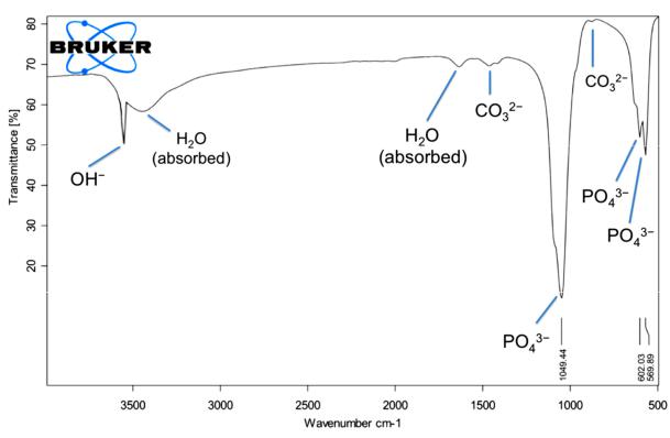
tion of the lattice OH− ions. The characteristic bands for PO43− appear at 569, 602, 1049 and 1093 cm−1. The observation of the asymmetric P−O schetching peak, together with the sharp peaks at 633, 602, 568 cm−1 correspond to the triply degenerate bending vibrrations of PO43−in hydroxyapatite.CO32− group forms weak peaks between 870 and 880 cm−1 and more intensive peaks between 1460 and 1530 cm−1. From the result, it was known that the producing hydroxide group and phosphate ion was observed while an insignificant amount of carbonate ion also was produced.
Figure 4. FTIR spectrum of hydroxyapatite from bovine bone
Conclusions. HA particles were successfully prepared from bovine bone. The morphology and particle surface of the as-prepared products, the crystallographic properties and the thermal stability on heating were determined. HA with high purity can further be used for potential applications such as bone tissue engineering scaffold materials and biomedical membranes.
148

Химия, физика и механика материалов. Выпуск № 4 (23), 2019
References
1.Abidi S.S.A. and Murtaza Q. Synthesis and Characterization of Nanohydroxyapatite Powder Using Wet Chemical Precipitation Reaction // J. Mater. Sci. Technol., 2014. vol. 30, no. 4, P. 307-310.
2.Kantharia N., Naik S., Apte S.K., Kheur M.G., Kheur S.M. and Kale B.B. Nano hydroxyapatite and its contemporary applications // Journal of Dental Research and Scientific Development. 2014. vol. 1, p.15-19
3.Muhammad A., Rashid A.,. Imran S, et al. Extracting hydroxyapatite and its precursors from natural resources // J. Mater. Sci., 2013.vol. 49, no. 4, P. 1461-1475,
4.Zhou H. and Lee J. Nanoscale hydroxyapatite particles for bone tissue engineering // Acta Biomater., 2011. vol. 7, no. 7, P. 2769-2781.
5.Ding T., Xue Y., Lu H. et al. Effect of Particle Size of Hydroxyapatite Nanoparticles on its Biocompatibility // IEEE Trans Nanobioscience, 2012. vol. 11, no. 4, P. 336-340.
6.Khoo W., Nor F.M., Ardhyananta H., and Kurniawan D., Preparation of Natural Hydroxyapatite from Bovine Femur Bones Using Calcination at Various Temperatures // Procedia Manuf., 2015.vol. 2, P. 196-201
7.Yousif, A. E., and Kareem, M. M. Extraction Of Hydroxyapatite From Bovine Femur Bone By Thermal Decomposition Method // i-manager’s Journal on Future Engineering and Technology, 2012. 7(2), P. 13-18.
8.Jamarun N. and Arief S. Characterization of Hydroxyapatite from Bovine Bone by Mechanical Combination Method // International Journal of Engineering and Techniques. 2018. vol. 4, no. 1, P. 6,
9.K. Tõnsuaadu, K.A. Gross, L. Plūduma, and M. Veiderma «A review on the thermal stability of calcium apatites» // J. Therm. Anal. Calorim, 2012.vol. 110, no. 2, P. 647-659.
10.S. Waheed, M. Sultan, T. Jamil, and T. Hussain «Comparative Analysis of Hydroxyapatite Synthesized by Sol-gel, Ultrasonication and Microwave Assisted Technique», Mater. Today Proc., 2015. vol. 2, no. 10, Part B, P. 5477-5484.
149