

Биоинженерия / ТИ_печень(органы_ЖКТ) / nihms667904
.pdfManuscript Author
Manuscript Author
Manuscript Author
Manuscript Author
Bhatia et al. |
Page 41 |
294.Wakita T, Pietschmann T, Kato T, Date T, Miyamoto M, Zhao ZJ, Murthy K, Habermann A, Krausslich HG, Mizokami M, Bartenschlager R, Liang TJ. Production of infectious hepatitis C virus in tissue culture from a cloned viral genome. Nat Med. 2005; 11:791–796. [PubMed: 15951748]
295.Zhong J, Gastaminza P, Cheng GF, Kapadia S, Kato T, Burton DR, Wieland SF, Uprichard SL, Wakita T, Chisari FV. Robust hepatitis C virus infection in vitro. Proc Natl Acad Sci U S A. 2005; 102:9294–9299. [PubMed: 15939869]
296.Buck M. Direct infection and replication of naturally occurring hepatitis C virus genotypes 1, 2, 3 and 4 in normal human hepatocyte cultures. PloS one. 2008; 3:e2660. [PubMed: 18628977]
297.Molina S, Castet V, Pichard-Garcia L, Wychowski C, Meurs E, Pascussi JM, Sureau C, Fabre JM, SaCunha A, Larrey D, Dubuisson J, Coste J, McKeating J, Maurel P, Fournier-Wirth C. Serum-derived hepatitis C virus infection of primary human hepatocytes is tetraspanin CD81 dependent. Journal of Virology. 2008; 82:569–574. [PubMed: 17942559]
298.Ploss A, Khetani SR, Jones CT, Syder AJ, Trehan K, Gaysinskaya VA, Mu K, Ritola K, Rice CM, Bhatia SN. Persistent hepatitis C virus infection in microscale primary human hepatocyte cultures. Proc Natl Acad Sci U S A. 2010; 107:3141–3145. [PubMed: 20133632]
299.Lindenbach BD, Rice CM. The ins and outs of hepatitis C virus entry and assembly. Nature reviews Microbiology. 2013; 11:688–700.
300.Kwong AD, Kauffman RS, Hurter P, Mueller P. Discovery and development of telaprevir: an NS3-4A protease inhibitor for treating genotype 1 chronic hepatitis C virus. Nature biotechnology. 2011; 29:993–1003.
301.Poordad F, McCone J Jr, Bacon BR, Bruno S, Manns MP, Sulkowski MS, Jacobson IM, Reddy KR, Goodman ZD, Boparai N, DiNubile MJ, Sniukiene V, Brass CA, Albrecht JK, Bronowicki JP. Boceprevir for untreated chronic HCV genotype 1 infection. N Engl J Med. 2011; 364:1195– 1206. [PubMed: 21449783]
302.Fried MW, Buti M, Dore GJ, Flisiak R, Ferenci P, Jacobson I, Marcellin P, Manns M, Nikitin I, Poordad F, Sherman M, Zeuzem S, Scott J, Gilles L, Lenz O, Peeters M, Sekar V, De Smedt G, Beumont-Mauviel M. Once-daily simeprevir (TMC435) with pegylated interferon and ribavirin in treatment-naive genotype 1 hepatitis C: the randomized PILLAR study. Hepatology. 2013; 58:1918–1929. [PubMed: 23907700]
303.Lawitz E, Mangia A, Wyles D, Rodriguez-Torres M, Hassanein T, Gordon SC, Schultz M, Davis MN, Kayali Z, Reddy KR, Jacobson IM, Kowdley KV, Nyberg L, Subramanian GM, Hyland RH, Arterburn S, Jiang D, McNally J, Brainard D, Symonds WT, McHutchison JG, Sheikh AM, Younossi Z, Gane EJ. Sofosbuvir for previously untreated chronic hepatitis C infection. N Engl J Med. 2013; 368:1878–1887. [PubMed: 23607594]
304.Jacobson IM, Gordon SC, Kowdley KV, Yoshida EM, Rodriguez-Torres M, Sulkowski MS, Shiffman ML, Lawitz E, Everson G, Bennett M, Schiff E, Al-Assi MT, Subramanian GM, An D, Lin M, McNally J, Brainard D, Symonds WT, McHutchison JG, Patel K, Feld J, Pianko S, Nelson DR. Sofosbuvir for hepatitis C genotype 2 or 3 in patients without treatment options. N Engl J Med. 2013; 368:1867–1877. [PubMed: 23607593]
305.Schwartz RE, Trehan K, Andrus L, Sheahan TP, Ploss A, Duncan SA, Rice CM, Bhatia SN. Modeling hepatitis C virus infection using human induced pluripotent stem cells. Proc Natl Acad Sci U S A. 2012
306.Wu X, Robotham JM, Lee E, Dalton S, Kneteman NM, Gilbert DM, Tang H. Productive hepatitis C virus infection of stem cell-derived hepatocytes reveals a critical transition to viral permissiveness during differentiation. PLoS pathogens. 2012; 8:e1002617. [PubMed: 22496645]
307.Yalaoui S, Huby T, Franetich JF, Gego A, Rametti A, Moreau M, Collet X, Siau A, van Gemert GJ, Sauerwein RW, Luty AJF, Vaillant JC, Hannoun L, Chapman J, Mazier D, Froissard P. Scavenger receptor BI boosts hepatocyte permissiveness to Plasmodium infection. Cell host & microbe. 2008; 4:283–292. [PubMed: 18779054]
308.van Schaijk BCL, Janse CJ, van Gemert G-J, van Dijk MR, Gego A, Franetich J-F, van de VegteBolmer M, Yalaoui S, Silvie O, Hoffman SL, Waters AP, Mazier D, Sauerwein RW, Khan SM. Gene disruption of Plasmodium falciparum p52 results in attenuation of malaria liver stage development in cultured primary human hepatocytes. PloS one. 2008; 3:e3549. [PubMed: 18958160]
Sci Transl Med. Author manuscript; available in PMC 2015 July 16.

Manuscript Author
Manuscript Author
Manuscript Author
Manuscript Author
Bhatia et al. |
Page 42 |
309.March S, Ng S, Velmurugan S, Galstian A, Shan J, Logan David J, Carpenter Anne E, Thomas D, Kim L, Sim B, Mota Maria M, Hoffman Stephen L, Bhatia Sangeeta N. A Microscale Human Liver Platform that Supports the Hepatic Stages of Plasmodium falciparum and vivax. Cell host & microbe. 2013; 14:104–115. [PubMed: 23870318]
310.Hickey RD, Lillegard JB, Fisher JE, McKenzie TJ, Hofherr SE, Finegold MJ, Nyberg SL, Grompe M. Efficient production of Fah-null heterozygote pigs by chimeric adeno-associated virus-mediated gene knockout and somatic cell nuclear transfer. Hepatology. 2011; 54:1351– 1359. [PubMed: 21674562]
311.Schuldiner M, Itskovitz-Eldor J, Benvenisty N. Selective ablation of human embryonic stem cells expressing a “suicide” gene. Stem cells. 2003; 21:257–265. [PubMed: 12743320]
312.Kiuru M, Boyer JL, O’Connor TP, Crystal RG. Genetic control of wayward pluripotent stem cells and their progeny after transplantation. Cell stem cell. 2009; 4:289–300. [PubMed: 19341619]
Sci Transl Med. Author manuscript; available in PMC 2015 July 16.

Manuscript Author
Manuscript Author
Manuscript Author
Manuscript Author
Bhatia et al. |
Page 43 |
Figure 1. Structure of the Liver
The liver is the largest internal organ in the body and performs over 500 functions, including numerous metabolic, synthetic, immunologic, and detoxification processes. (A) The liver exhibits a hierarchical structure consisting of repeated functional tissue units (liver lobules). Within a lobule, oxygenated blood enters through branches of the hepatic artery and portal vein, and flows in specialized sinusoidal vessels towards the central vein. Bile, that is produced and excreted by hepatocytes, flows in the counter direction towards the intrahepatic bile duct. (B) Hepatocytes are polarized epithelial cells that interact closely with a number of non-parenchymal cells types along the sinusoidal tracts of the liver lobule. Collectively, these cellular components and multiscale tissue structures contribute to the diverse functional roles of the liver.
Sci Transl Med. Author manuscript; available in PMC 2015 July 16.

Manuscript Author
Manuscript Author
Manuscript Author
Manuscript Author
Bhatia et al. |
Page 44 |
Figure 2. The Liver in Health and Disease
Mechanisms that lead to hepatocyte damage and reduce liver function include drugmediated toxicity, alcohol-induced and nonalcoholic fatty liver disease, hepatotrophic infections, and hereditary disorders. Fatty liver disease, resulting from both chronic alcohol exposure as well as nonalcoholic mechanisms, is increasingly common and leads to the chronic accumulation of fat droplets within the liver. Liver cirrhosis can be caused by hepatitis virus infection, autoimmune processes, chronic alcohol abuse, as well as chronic inflammation and fat accumulation. Cirrhosis is characterized by alterations in the sinusoidal structure and function of the liver and the accumulation of extracellular matrix, which is commonly referred to as scarring. These alterations lead to a reduction in hepatic function that can progress to hepatic failure and increased risk of hepatocellular carcinoma.
Sci Transl Med. Author manuscript; available in PMC 2015 July 16.
Manuscript Author
Manuscript Author
Manuscript Author
Manuscript Author
Bhatia et al. |
Page 45 |
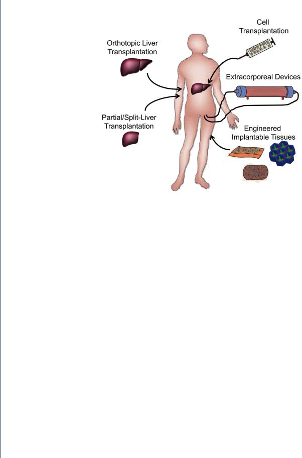
Figure 3. Organ transplantation and cell-based therapies
Currently, liver transplantation is the primary treatment for liver failure and the only therapy shown to improve survival in patients with liver failure. Due to the limited number of livers suitable for transplantation, advanced surgical procedures including split liver and partial donor transplants have been pursued clinically. Additionally, a diverse range of cell-based therapies are currently being explored to treat liver disease and liver failure. These include the transplantation of various adult and stem cell-derived cell populations, the development of extracorporeal bioartificial liver devices, and the implantation of engineered tissues.
Sci Transl Med. Author manuscript; available in PMC 2015 July 16.

Manuscript Author
Manuscript Author
Manuscript Author
Manuscript Author
Bhatia et al. |
Page 46 |
Figure 4. Extracorporeal bioartificial liver devices
Extracorporeal bioartificial liver devices incorporate functional liver cells and aim to provide an array of important liver functions (detoxification, metabolic, synthetic) for a patient by processing the patient’s blood/plasma outside of the body. This approach could serve as a temporary support and bridge until a liver becomes available for transplantation. Currently, such devices are either in the clinical trial or exploratory research stage. Liver cell-based bioreactor designs primarily fall into four general categories based on device configuration. These include hollow fiber devices, packed beds, flat plate systems, and encapsulation-based reactors. The majority of current clinical trials utilize a hollow fiber design in which cells are positioned outside the fibers and the patient’s blood/plasma is perfused through the fiber lumen. Cell sources include three categories; currently existing tumor cell lines, porcine and human primary hepatocytes, and hepatic cells derived from embryonic stem cells (ESC), induced pluripotent stem cells (iPSC), or reprogrammed from other cell types. Due to their increased availability compared to primary human hepatocytes, primary porcine hepatocytes are the most common cellular component of current bioartificial liver devices. Device design characteristics have been shown to affect the functional stability of the cellular components. Many design challenges exist including the balanced delivery of oxygen and nutrients to the cells, preventing mechanical shear forces from damaging the cells, and clinically relevant scale-up.
Sci Transl Med. Author manuscript; available in PMC 2015 July 16.

Manuscript Author
Manuscript Author
Manuscript Author
Manuscript Author
Bhatia et al. |
Page 47 |
Figure 5. In vitro culture systems for hepatocytes
Improved in vitro systems have been developed for culturing primary human hepatocytes. Elucidating the roles of many microenvironmental signals in governing hepatocellular processes has enabled optimization of in vitro culture systems. These optimized systems include co-cultures to provide specific cell-cell interactions as well as culture conditions with defined concentrations of soluble factors and extracellular matrix molecules (left). The application of microfabrication technologies to in vitro hepatic tissue engineering has facilitated the control of culture platforms down to the microscale, such as the patterning of co-culture configurations (middle). Further, natural and synthetic biomaterial systems have been applied towards the optimization of three-dimensional in vitro culture platforms (right). Collectively, these engineering approaches have further advanced the understanding of how combinations of microenvironmental cues influence cell functions, and provided important insights into the temporal and spatial dynamics of hepatic cell and tissue function.
Sci Transl Med. Author manuscript; available in PMC 2015 July 16.

Manuscript Author
Manuscript Author
Manuscript Author
Manuscript Author
Bhatia et al. |
Page 48 |
Figure 6. Sources of Hepatocytes
Obtaining appropriate sources of hepatocytes is a major limitation for developing cell-based therapies for treating liver disease. Many different approaches are under investigation including methods for improving the expansion of primary human hepatocytes in vitro, the directed differentiation of pluripotent stem cells (both embryonic stem cells and induced pluripotent stem cells), the differentiation of either intrahepatic or extrahepatic adult progenitor cells, as well as new methods for the direct reprogramming of hepatic cells from adult somatic cells.
Sci Transl Med. Author manuscript; available in PMC 2015 July 16.

Manuscript Author
Manuscript Author
Manuscript Author
Manuscript Author
Bhatia et al. |
Page 49 |
Figure 7. Liver cell and tissue engineering
Progress in the field of liver cell and tissue engineering serves a bidirectional role as both a means for establishing robust model systems for investigating the human liver in health and disease, as well as the foundation for the development of new cell-based therapies.
Consequently, applications exist on a continuum ranging from fundamental in vitro studies (left), to engineered approaches for interfacing with animal models (middle), and finally to translational clinical applications (right). In order to further understand human liver function and disease processes, engineered culture platforms can serve to complement animal models. Concurrently, the foundation of novel cell-based therapies is based on advances in cell and tissue engineering and the progression of these technologies from relevant animal disease models to clinical settings.
Sci Transl Med. Author manuscript; available in PMC 2015 July 16.