

Биоинженерия / ТИ_ССС / nm1391
.pdf© 2006 Nature Publishing Group http://www.nature.com/naturemedicine
T E C H N I C A L R E P O R T S
Monolayered mesenchymal stem cells repair scarred myocardium after myocardial infarction
Yoshinori Miyahara1,9, Noritoshi Nagaya1,9, Masaharu Kataoka1, Bobby Yanagawa1, Koichi Tanaka1, Hiroyuki Hao2, Kozo Ishino3, Hideyuki Ishida4, Tatsuya Shimizu5, Kenji Kangawa6, Shunji Sano3, Teruo Okano5, Soichiro Kitamura7 & Hidezo Mori8
Mesenchymal stem cells are multipotent cells that can differentiate into cardiomyocytes and vascular endothelial cells. Here we show, using cell sheet technology, that monolayered mesenchymal stem cells have multipotent and self-propagating properties after transplantation into infarcted rat hearts. We cultured adipose tissue–derived mesenchymal stem cells characterized by flow cytometry using temperature-responsive culture dishes. Four weeks after coronary ligation, we transplanted the monolayered mesenchymal stem cells onto the scarred myocardium. After transplantation, the engrafted sheet gradually grew to form a thick stratum that included newly formed vessels, undifferentiated cells and few cardiomyocytes. The mesenchymal stem cell sheet also acted through paracrine pathways to trigger angiogenesis. Unlike a fibroblast cell sheet, the monolayered mesenchymal stem cells reversed wall thinning in the scar area and improved cardiac function in
rats with myocardial infarction. Thus, transplantation of monolayered mesenchymal stem cells may be a new therapeutic strategy for cardiac tissue regeneration.
Myocardial infarction, a main cause of heart failure, leads to loss of cardiac tissue and impairment of left ventricular function. Therefore, restoring the scarred myocardium is desirable for the treatment of heart failure. Although needle injections of bone marrow cells into the myocardium have been performed for cardiac regeneration1–5, it is difficult to reconstruct sufficient cardiac mass in the thinned scar area after myocardial infarction.
Recently, our colleagues have developed cell sheets using tempera- ture-responsive culture dishes6. These cell sheets allow for cell-to-cell connections and maintain the presence of adhesion proteins because enzymatic digestion is not needed7–10. Therefore, cell sheet transplantation may be a promising strategy for partial cardiac tissue reconstruction. Skeletal myoblasts, fetal cardiomyocytes and embryonic stem cells have been considered as candidates for an implantable cell
source11–13. It is difficult, however, to produce a multilayered construct requiring a vascular network. Thus, autologous somatic stem cells with self-propagating properties that can induce angiogenesis are a desirable cell source for a transplantable sheet.
Mesenchymal stem cells (MSCs) are multipotent adult stem cells that reside within the bone marrow microenvironment14,15. MSCs can differentiate not only into osteoblasts, chondrocytes, neurons and skeletal muscle cells, but also into vascular endothelial cells16 and cardiomyocytes17–20. In contrast to their hematopoietic counterparts, MSCs are adherent and can expand in culture. Recently, MSCs have been isolated from adipose tissue21–24, which is typically abundant in individuals with cardiovascular disease. Here, we investigated the therapeutic potency of monolayered MSCs derived from adipose tissue using cell sheet technology.
RESULTS
Characteristics of adipose tissue–derived MSCs
We isolated MSCs from subcutaneous adipose tissue of male SpragueDawley rats on the basis of the adherent properties of these cells. We obtained 1.7 105 ± 0.2 105 cells from 1 g adipose tissue in a 12-h culture. By day 4 of culture of the minced adipose tissue, spindleshaped adherent cells were apparent and formed symmetric colonies. After approximately three to four passages, most adherent cells expressed CD29 and CD90 (Supplementary Fig. 1 online). In contrast, the majority of adherent cells were negative for CD34 and CD45. They were also negative for CD31, a marker for vascular endothelial cells, and negative for a smooth muscle actin (aSMA), a marker for smooth muscle cells. A small fraction of adherent cells expressed CD71, CD106 and CD117. These results were similar to those from bone marrow–derived MSCs15,22,25 (Supplementary Fig. 1 online). Using previously described methods16,22,26, we confirmed that these adipose-derived adherent cells, like bone marrow–derived MSCs, were multipotent, as judged by their ability to differentiate into adipocytes, osteoblasts and vascular endothelial cells. Thus, we
1Department of Regenerative Medicine and Tissue Engineering, National Cardiovascular Center Research Institute and 2Department of Pathology, National Cardiovascular Center, 5-7-1 Fujishirodai, Suita, Osaka, 565-8565, Japan. 3Department of Cardiovascular Surgery, Okayama University Graduate School of Medicine, Dentistry and Pharmaceutical Sciences, 2-5-1 Shikata-cho, Okayama, 700-8555, Japan. 4Department of Physiology, School of Medicine, Tokai University, Bohseidai, Isehara, Kanagawa, 259-1193, Japan. 5Institute of Advanced Biomedical Engineering and Science, Tokyo Woman’s Medical University, 8-1 Kawada-cho, Shinjuku-ku, Tokyo, 162-8666, Japan. 6Department of Biochemistry, National Cardiovascular Center Research Institute and 7Department of Cardiovascular Surgery, National Cardiovascular Center and 8Department of Cardiac Physiology, National Cardiovascular Center Research Institute, 5-7-1 Fujishirodai, Suita, Osaka, 565-8565, Japan. 9These authors contributed equally to this work. Correspondence should be addressed to N.N. (nnagaya@ri.ncvc.go.jp) or H.M. (hidemori@ri.ncvc.go.jp).
Received 9 August 2005; accepted 3 March 2006; published online 2 April 2006; doi:10.1038/nm1391
NATURE MEDICINE VOLUME 12 [ NUMBER 4 [ APRIL 2006 |
4 5 9 |
© 2006 Nature Publishing Group http://www.nature.com/naturemedicine
T E C H N I C A L R E P O R T S
a |
b |
c |
d |
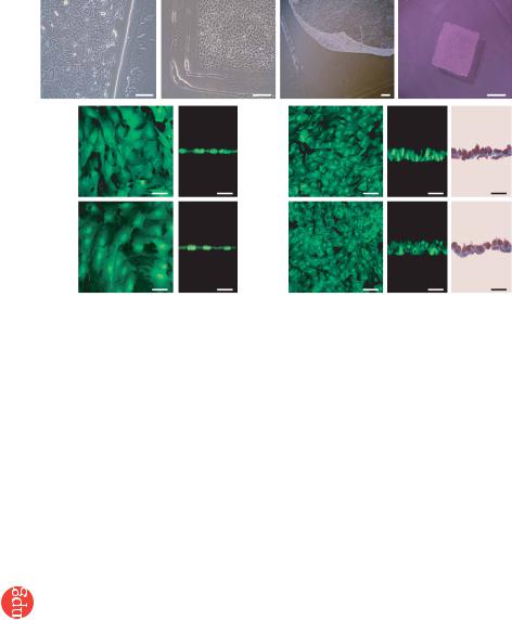
Figure 1 Preparation of monolayered MSCs. |
|
(a) MSCs 2 d after seeding on a temperature- |
|||||
|
|
|
|
||
|
|
|
|
responsive dish. (b) Cultured MSCs expanded |
|
|
|
|
|
to confluence within the square area of the dish |
|
|
|
|
|
by day 3. (c) The monolayered MSCs detached |
|
|
|
|
|
easily from the culture dish at 20 1C. (d) The |
|
|
|
|
|
completely detached monolayered MSCs were |
|
e |
|
f |
|
identified as a 12 12 mm square sheet. |
|
|
|
(e–h) Cross-sectional analysis of GFP-expressing |
|||
|
|
|
|
monolayered MSCs and DFBs before detachment |
|
|
|
|
|
(e and g, confocal images) and after detachment |
|
MSC |
|
MSC |
|
(f and h, left and center, confocal images; |
|
|
|
|
|
right, Masson trichrome). The thickness of |
|
|
|
|
|
both monolayers was 3.5-fold greater than the |
|
g |
|
h |
|
thickness before detachment, and constituent |
|
|
|
cells were compacted. Scale bars in a–c, |
|||
|
|
|
|
100 mm; in d, 5 mm; in e–h, 20 mm. |
DFB |
DFB |
confirmed that the majority of adherent cells isolated from adipose tissue were MSCs.
Preparation and transplantation of monolayered MSCs
We cultured adipose tissue–derived MSCs (5 105 cells) on tem- perature-responsive dishes for 3 d until confluent. MSCs were attached on the poly-N-isopropylacrylamide (PIPAAm)-grafted area (24 24 mm; Fig. 1a,b). As the culture temperature was decreased from 37 1C to 20 1C, MSCs detached spontaneously and floated up into the culture medium as a monolayer of MSCs within 40 min (Fig. 1c,d). As a control, we prepared dermal fibroblasts (DFBs) by the skin explant technique27. DFBs (8 105 cells) were cultured on the temperature-responsive dishes, and monolayered DFBs were fabricated as described above. The final cell counts for monolayered MSCs and DFBs before transplantation were 9.4 ± 0.6 105 and 8.6 ± 0.6 105 cells, respectively (n ¼ 6 each). To identify the thickness of
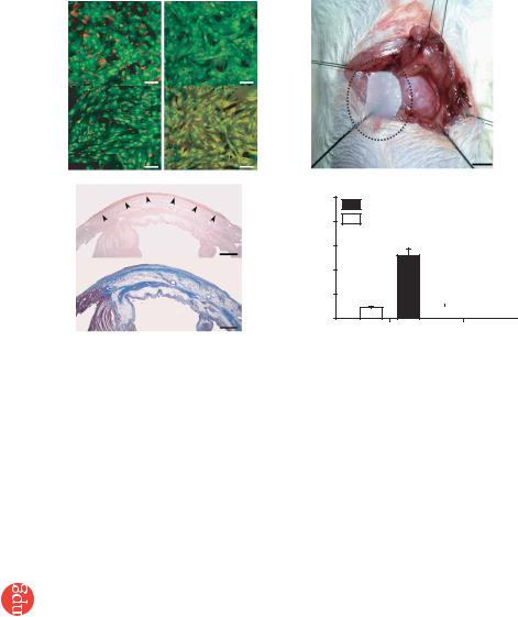
monolayered MSCs, we used green fluorescent protein (GFP)-expres- sing cell grafts derived from the GFP-transgenic Sprague-Dawley rats. Immediately after detachment, cells became compacted, possibly owing to cytoskeletal tensile reorganization, and the thickness of monolayered MSCs and DFBs was approximately 3.5-fold greater than the thickness before detachment (MSCs, 6.2 ± 0.3 to 21.5 ± 0.8 mm; DFBs, 6.5 ± 0.4 to 22.4 ± 1.1 mm; Fig. 1e–h). MSCs on the temperature-responsive dishes were positive for vimentin and slightly positive for collagen type 1, whereas DFBs were positive for both markers (Fig. 2a). We transferred detached monolayered MSCs above the myocardial scar (Fig. 2b) and then attached them to the surface of the anterior scar (Fig. 2c).
Secretion of angiogenic factors from monolayered MSCs
We measured secretion of angiogenic factors from MSCs 24 h after monolayers had formed, equivalent to day 4 after initial cell seeding. The monolayered MSCs secreted significantly larger amounts of angiogenic and antiapoptotic factors such as vascular endothelial growth factor (VEGF) and hepatocyte growth factor (HGF) than did the monolayered DFBs (P o 0.01; Fig. 2d). The control medium supplemented with 10% fetal calf serum contained less than 5 pg/ml of VEGF or HGF. These results suggest that the paracrine effects of monolayered MSCs on host myocardium are greater than those of monolayered DFBs.
Engraftment and growth of monolayered MSCs
To identify the transplanted cells in myocardial sections, we used GFP-expressing cell grafts derived from the GFP-transgenic Sprague-Dawley rats. We
grafted monolayered MSCs or DFBs onto the scar area of the anterior wall (Fig. 3). Fluorescence microscopy showed that GFP-expressing monolayered MSCs gradually grew in situ and developed into a thick stratum, up to B600 mm thick over the native tissue at 4 weeks (Fig. 3a–f). The engrafted MSC tissue tapered off toward the healthy myocardium (Fig. 3d,e), although most of the monolayered MSCs were attached only to the scar area in the anterior wall because of the large infarct. We rarely detected TUNEL-positive MSCs in the sheet (o1%) 48 h after transplantation (Fig. 3g), implying that cell viability in the sheet was maintained. In contrast, we frequently detected TUNEL-positive cells (15% ± 2%) in the DFB sheet, which was observed as a thin layer above the scar. Subsequently, the DFB sheet was undetectable 1 week later. Masson trichrome staining showed increased thickness of the anterior wall and attenuation of left ventricle enlargement after transplantation of monolayered MSCs (Fig. 3h), although the infarct size did not differ significantly among the untreated, DFB and MSC groups (Supplementary Table 1 online).
Reconstruction of cardiac mass
After growth in situ, GFP-expressing MSC tissue contained a number of mature vascular structures that had positive staining for von Willebrand factor (vWF) and aSMA (Fig. 4a,b). A small fraction of the MSC tissue had positive staining for cardiac troponin T and desmin (Fig. 4c,d). On the other hand, a large proportion of the MSC tissue was positive for vimentin, a marker for mesenchymal lineage cells (Fig. 4e). The percentages of graft-derived cells that expressed endothelial (vWF), smooth muscle (aSMA), cardiac (troponin T) and mesenchymal (vimentin) markers were 12.2% ± 0.6%, 5.0% ± 0.3%, 5.3% ± 0.3% and 57.8% ± 2.2%, respectively. Notably, based on expression of these markers, two-thirds of vascular endothelial cells, four-fifths of smooth muscle cells and one-twentieth of cardiomyocytes within the MSC tissue were GFP– and hence were derived from the host. The MSC tissue stained modestly for collagen type 1 (Fig. 4f). Picrosirius red staining showed that collagen deposition was found mainly in the extracellular matrix and the epicardial margin of the MSC tissue (Fig. 4g). Excluding staining in blood vessels, the MSC tissue was also negative for aSMA, a marker for myofibroblasts (Fig. 4b). This phenotype was consistent with properties of MSCs
4 6 0 |
VOLUME 12 [ NUMBER 4 [ APRIL 2006 NATURE MEDICINE |
© 2006 Nature Publishing Group http://www.nature.com/naturemedicine
a |
Vimentin |
Collagen type 1 |
b |
|
|
||
MSC |
|
|
|
DFB
c |
|
AW |
d |
|
|
|
cells) |
5,000 |
|
|
|
|
|
|
|
|
|
6 |
4,000 |
|
|
|
(pg/10 |
|
RV |
IVS |
LV |
3,000 |
|
secreted |
|
|||
|
|
|
2,000 |
|
|
|
|
Amount |
|
|
|
|
0 |
|
|
|
|
|
1,000 |
before transplantation (Fig. 2a and Supplementary Fig. 1 online), suggesting that the MSC tissue includes a number of undifferentiated MSCs. Taken together, the grown MSC tissue was composed of newly formed blood vessels, undifferentiated MSCs and few cardiomyocytes.
Fluorescence in situ hybridization analysis
We performed fluorescence in situ hybridization (FISH) to detect X and Y chromosomes after sex-mismatched transplantation of monolayered MSCs. We transplanted GFP-expressing monolayered MSCs derived from male rats to female Sprague-Dawley rats that had suffered an infarct. Four weeks later, newly formed cardiomyocytes that were positive for GFP had only one set of X and Y chromosomes, whereas we detected two X chromosomes exclusively in GFP– hostderived cells (Fig. 4h). We counted the X and Y chromosomes in male and female control rats and in the MSC sheet–transplanted rats (Supplementary Table 2 online), and we did not detect extra copies of the X or Y chromosome in graft-derived GFP+ cardiomyocytes. When we compared the frequencies of the occurrence of zero, one, two and more than two X chromosomes in the GFP+ cardiomyocytes with the frequencies in male control cardiomyocytes, the GFP+ cardiomyocytes did not show an increased proportion of X chromosomes (0.25 4 P 4 0.10, w2 test).
Effects of monolayered MSCs on cardiac function
Heart failure developed 8 weeks after coronary ligation, as indicated by an increase in left ventricle end-diastolic pressure (LVEDP) and attenuation of maximum and minimum rate of change in left ventricular pressure (dP/dt). Autologous transplantation of monolayered MSCs, however, resulted in decreased LVEDP (Fig. 5a). Left ventricle maximum and minimum dP/dt were significantly improved in the MSC group (Fig. 5b,c). We did not observe these hemodynamic improvements in the DFB group. The MSC group also had significantly lower right ventricular weight and lung weight than the DFB and untreated groups 4 weeks after transplantation (Supplementary Table 1 online). These results suggest that transplantation of monolayered MSCs has beneficial hemodynamic effects in rats with chronic heart failure.
T E C H N I C A L R E P O R T S
Figure 2 Characteristics of monolayered MSCs. (a) Properties of constituent cells in the
monolayered grafts. Compared with DFBs (green), MSCs (green) are positive for vimentin (red)
and slightly positive for collagen type 1 (red). (b) Monolayered MSCs (in the dotted circle) transferred to the infarcted heart. (c) Extent of monolayered MSCs 48 h after transplantation (arrows). AW, anterior wall; LV, left ventricle; RV right ventricle; IVS, interventricular septum. (d) Comparison of secretion of growth factors between monolayered MSCs and DFBs.
**P o 0.01 versus DFBs. Scale bar in a, 20 mm; in b, 5 mm; in c, 100 mm.
|
|
|
|
** |
|
|
|
|
|
|
Echocardiographic analysis showed that |
|
|
|
|
|
|
|
|
|
|
|
transplantation of monolayered MSCs signi- |
|
|
|
|
|
ficantly increased diastolic thickness of the |
|
|
|
|
|
infarcted anterior wall (Fig. 5d). Left ventricle |
|
|
|
|
|
end-diastolic dimension at 8 weeks was sig- |
|
|
|
|
|
nificantly smaller in the MSC group than in |
|
|
|
|
|
the DFB and untreated groups (Fig. 5e). |
|
|
|
|
||
|
|
HGF |
|
Transplantation of the monolayered MSCs |
|
|
|
|
|
|
significantly increased left ventricle fractional |
|
|
|
|
|
shortening (Fig. 5f). Left ventricle wall stress |
in diastole was markedly lower in the MSC group than in the DFB and untreated groups (Supplementary Table 3 online). Plasma atrial natriuretic peptide (ANP) in the DFB and untreated groups was markedly elevated 8 weeks after myocardial infarction (Fig. 5g). Transplantation of the monolayered MSCs inhibited the increase in plasma ANP.
Survival analysis
The Kaplan-Meier survival curve showed that 4-week survival after coronary ligation did not differ significantly between the untreated and MSC groups before transplantation (Fig. 5h). Notably, however, no rats died after transplantation of monolayered MSCs. Therefore, the survival rate after transplantation was markedly higher in the MSC group than in the untreated group (4-week survival after transplantation was 100% for the MSC group versus 71% for the untreated group, log-rank test, P o 0.05).
DISCUSSION
There are several advantages to monolayered MSC transplantation. First, the self-propagating property of MSCs in situ leads to the formation of a thick stratum on the surface of the scarred myocardium. Second, the multipotency of MSCs and their ability to supply angiogenic cytokines allows neovascularization in the MSC tissue. Third, the reconstruction of thick myocardial tissue reduces left ventricle wall stress and results in improvement of cardiac function after myocardial infarction. Finally, a substantial part of the transplanted tissue is composed of undifferentiated MSCs, and it is tempting to speculate that such cells may act against future progressive left ventricle remodeling.
Cellular cardiomyoplasty using needle injections is emerging as a treatment option for individuals with chronic heart failure, but it may be limited by failure to regenerate cardiac mass. The cell sheet allows for cell-to-cell connections owing to the lack a need for enzymatic digestion6–10. Thus, the cell sheet has attracted considerable interest as a tool for tissue engineering28. Here, we used adipose tissue–derived MSCs as a cellular source for the cell sheet, which resulted in successful autologous transplantation in heterogenic rats without immunological
NATURE MEDICINE VOLUME 12 [ NUMBER 4 [ APRIL 2006 |
4 6 1 |

T E C H N I C A L R E P O R T S
© 2006 Nature Publishing Group http://www.nature.com/naturemedicine
a |
b |
c |
d |
e |
|
f |
m) |
700 |
|
|
|
|
* |
|
|
|
|
||||||||||
|
( |
600 |
|
|
|
* |
|||||||
|
|
|
|
|
|
|
tissue |
|
|
|
|
||
|
|
|
|
|
|
|
500 |
|
|
* |
|
|
|
|
|
|
|
|
|
|
MSC |
|
|
|
|
||
|
|
|
|
|
|
|
400 |
|
|
|
|
||
|
|
|
|
|
|
|
|
|
|
|
|
||
|
|
|
|
|
|
|
of |
300 |
|
|
|
|
|
|
|
|
|
|
|
|
|
|
|
|
|
|
|
|
|
|
|
|
|
|
Thickness |
200 |
|
|
|
|
|
|
|
|
|
|
|
|
0 |
|
|
|
|
|
|
|
|
|
|
|
|
|
|
100 |
|
|
|
|
|
|
|
|
|
|
|
|
|
1 |
2 |
3 |
4 |
||
|
|
|
|
|
|
|
|
|
|
|
Time (weeks) |
|
|
|
|
|
|
|
|
g |
|
|
|
|
|
|
|
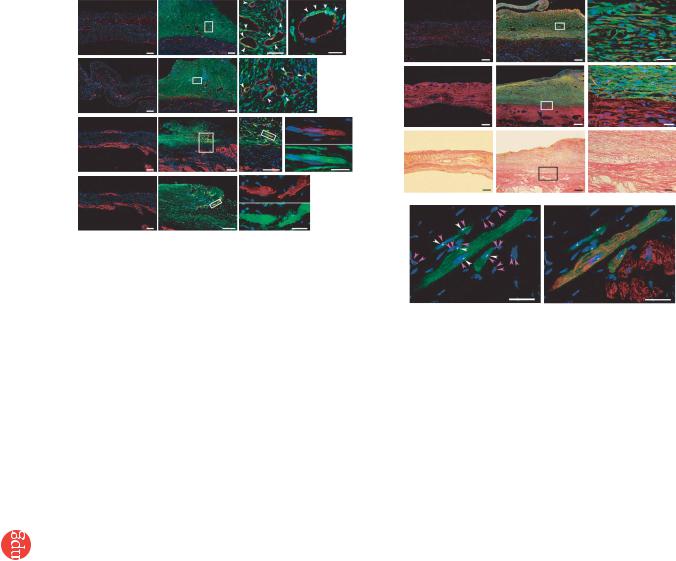
Figure 3 Engraftment, survival and growth of monolayered MSCs. To identify |
|
|
|
|
|
|
|
|
|
|
|||
the transplanted cells within myocardial sections, we used GFP-expressing cells |
|
|
|
|
|
|
|
|
|
|
|||
derived from GFP-transgenic Sprague-Dawley rats. (a–c) MSC tissue above |
h |
Sham |
|
Untreated |
DFB |
|
MSC |
||||||
the anterior scar at day 2 (a), day 14 (b) and day 28 (c). GFP-expressing |
|
|
|||||||||||
|
|
|
|
|
|
|
|
|
|
||||
monolayered MSCs were grafted to the native myocardium and grew gradually in situ, resulting in increased wall thickness. (d,e) MSC tissue above the surrounding area of the scar at day 2 (d) and day 28 (e). In a–e, upper row shows immunofluorescent staining of MSC tissue (green); nuclei are stained with DAPI (blue). Middle row shows identical sections stained with antibody to GFP and DAB by bright-field microscopy. Lower row shows hematoxylin and eosin–stained sections. (f) Time course of growth of MSCs expressing GFP.
*P o 0.05 versus thickness of GFP+ MSC tissue at 1 week. (g) TUNEL staining of transplanted MSCs (green, left) and DFBs (green, right) 48 h after transplantation. Nuclei are stained with DAPI (blue). Arrows indicate TUNELpositive nuclei (red). (h) Photomicrographs show two cases of representative myocardial sections stained with Masson trichrome in the individual groups. Left ventricle enlargement was attenuated by the transplantation of monolayered MSC. Scale bars in a–e and upper panels of h, 100 mm;
in g, 20 mm; in lower panels of h, 1 mm.
rejection. Using flow cytometry, we did not find any substantial differences between adipose tissue–derived MSCs and bone mar- row–derived MSCs, consistent with results from previous studies22,25. Adipose-derived MSCs readily attached to and propagated on the temperature-responsive dish. Abdominal subcutaneous adipose tissue is clinically redundant and easily accessible by rapid and minimally invasive surgery such as liposuction. Thus, adipose tissue may serve as a source of stem cells for therapeutic cell sheets.
Here, monolayered MSCs could readily be transferred and grafted to the scarred myocardium without additives or suturing. This may be attributable to cell-to-cell connections as well as extracellular matrix deposits on the basal surface of the monolayered MSCs. Regeneration of myocardial mass is thought to require multilayered constructs of the cell sheet. Unfortunately, however, the lack of a vascular network has limited the formation of a thick construct10,29. The transplanted monolayered MSCs thickened gradually, developing into a stratum of up to 600 mm in thickness over the native tissue 4 weeks after transplantation, suggesting that monolayered MSCs have an ability to grow in situ. As a result, the transplanted MSC tissue reversed wall thinning of the infarcted myocardium. On the other hand, the fibroblast sheet did not grow in situ. It should be noted that the MSC tissue included a large number of newly formed blood vessels. These vessels were composed of graft-derived cells, host-derived cells or both. The MSC sheet secreted a large amount of angiogenic and antiapoptotic cytokines, including VEGF and HGF, as compared with the fibroblast sheet. These results suggest that MSCs induce neovascularization within the sheet not only through their ability to differentiate into vascular cells but also through growth factor–mediated paracrine
regulation. Thus, we believe that the angiogenic action of MSCs is important for reconstruction of cardiac mass by the MSC tissue.
Four weeks after transplantation, a small fraction of the engrafted MSCs were positive for cardiac proteins such as cardiac troponin T and desmin, suggesting the presence of cardiomyocytes within the MSC tissue. FISH analysis suggested that the most cardiomyocytes within the MSC tissue were not derived from cell fusion, but we are unable to exclude the possibility that some were. Further studies are necessary to investigate the mechanisms by which MSCs within the MSC tissue regenerate cardiomyocytes. The majority of the MSC tissue was positive for vimentin, a marker for undifferentiated MSCs and fibroblasts. In addition, the majority of MSCs within the graft were negative for collagen type 1 and aSMA, a marker for myofibroblasts. These results suggest that the grown-up MSC tissue is composed of newly formed blood vessels, undifferentiated MSCs and few cardiomyocytes.
We have also shown that transplantation of the monolayered MSCs significantly increased left ventricle maximum dP/dt, decreased LVEDP and inhibited the development of left ventricle enlargement in rats with chronic heart failure secondary to myocardial infarction. These results suggest that transplantation of monolayered MSCs improves cardiac function. But the presence of cardiomyocytes within the MSC tissue seemed to be rare. Thus, this improvement may be explained mainly by growth factor–mediated paracrine effects of the MSC sheet and a decrease in left ventricle wall stress resulting from the thick MSC tissue. Furthermore, no rats treated with the monolayered MSCs died during the study period, although untreated rats died frequently. These results indicate that fatal arrythmogenic problems were not caused by integration of the MSC tissue.
4 6 2 |
VOLUME 12 [ NUMBER 4 [ APRIL 2006 NATURE MEDICINE |
© 2006 Nature Publishing Group http://www.nature.com/naturemedicine
T E C H N I C A L R E P O R T S
a |
Untreated |
MSC |
e |
Untreated |
MSC |
|
|
|
|||||
|
E |
E |
|
E |
E |
|
|
I |
I |
I |
|
I |
|
b |
E |
E |
|
|||
f |
E |
E |
||||
|
|
|||||
c |
I |
I |
|
|
|
|
E |
E |
I |
|
I |
||
|
|
|
g |
E |
E |
|
d |
I |
I |
|
|
|
|
E |
E |
I |
|
I |
||
|
|
|
h |
|
|
|
|
I |
I |
|
|
|
Figure 4 Differentiation of MSCs within the MSC tissue after growth in situ. (a,b) GFP-expressing MSCs (green) were identified as a thick stratum at the epicardial side of the myocardium. The MSC tissue contained a number of vascular structures positive for vWF (red, a) and aSMA (red, b). MSCs that did not participate in blood vessel formation were only rarely positive for aSMA, a marker for myofibroblasts. Arrows indicate transplanted MSCs positive for vWF or aSMA. (c,d) Some MSCs within the MSC tissue were positive for cardiac markers cardiac troponin T (red, c) and desmin (red, d). (e) Most of the MSC tissue was positive for vimentin (red). (f) The MSC tissue modestly stained for collagen type 1 (red). (g) Collagen deposition was also detected by picrosirius red staining. (h) FISH analysis. Newly formed cardiomyocytes (desmin, red) that were positive for GFP (green) had only one set of X (purple) and Y chromosomes (white), whereas two X chromosomes were detected exclusively in GFP– host-derived cells. Nuclei are stained with DAPI (blue, a–f and h). Scale bars in left three panels of a and c and in two left panels of b and d–g, 100 mm; in h and far right panels of a–g, 20 mm. E, epicardial side; I, intimal side.
In summary, adipose tissue–derived monolayered MSCs can be readily engrafted to the scarred myocardium, grow gradually in situ and become a thick stratum that includes newly formed vessels, cardiomyocytes and undifferentiated MSCs. The engrafted MSCs reversed wall thinning in the scar area and improved cardiac function and survival in rats with myocardial infarction. Thus, transplantation of monolayered MSCs may be a new therapeutic strategy for cardiac tissue regeneration.
METHODS
Model of heart failure. All protocols were performed in accordance with the guidelines of the Animal Care Ethics Committee of the Japanese National Cardiovascular Center Research Institute. We used male Sprague-Dawley rats (Japan SLC) weighing 187–215 g. A myocardial infarction model was produced by ligation of the left coronary artery, as described previously30. Briefly, we anesthetized rats with sodium pentobarbital (30 mg/kg) and ventilated them with a volume-regulated respirator. We exposed hearts by left thoracotomy, and ligated the left coronary artery 2–3 mm from its origin between the pulmonary artery conus and the left atrium with a 6-0 Prolene suture. The sham group underwent thoracotomy and cardiac exposure without coronary ligation. The surviving rats were maintained on standard rat chow.
Study protocol. We randomly placed rats into four groups: rats with chronic heart failure that underwent transplantation of monolayered MSCs (MSC group; n ¼ 12), rats with chronic heart failure given monolayered DFBs (DFB group; n ¼ 12), rats with chronic heart failure without transplantation (untreated group; n ¼ 12) and sham-operated rats without transplantation (sham group; n ¼ 10). Four weeks after coronary ligation, the MSC and DFB groups underwent autologous transplantation of each monolayered cell graft onto the anterior wall, including the scar area (Supplementary Methods online). The other two groups underwent the same operative procedures
without transplantation. We performed hemodynamic studies, echocardiography and histological assessments 4 and 8 weeks after coronary ligation (Supplementary Methods). Upon killing at 8 weeks after coronary ligation, only those rats with infarct size 425% of the left ventricle area were included in this study. Therefore, the variation in infarct size between the experimental rats was relatively low (28–41%, average 33.9% ± 1.9%).
Isolation and culture of MSCs from adipose tissue. Immediately after coronary ligation, we acquired subcutaneous adipose tissue (1.1 ± 0.1 g) from the right inguinal region of each rat. We minced adipose tissue with scissors and digested it with 10 ml of type 1 collagenase solution (0.1 mg/ml, Worthington Biochemical) for 1 h in a 37 1C water bath shaker. After filtration with mesh filter (Costar 3480, Corning) and centrifugation at 780g for 8 min, we suspended isolated cells in a-MEM supplemented with 10% FCS and antibiotics, plated them onto a 100-mm dish and incubated them at 37 1C with 5% CO2. A small number of spindle-shaped cells were apparent in visible symmetric colonies by days 5–7.
Preparation of temperature-responsive dishes. Specific procedures for preparation of square-designed PIPAAm-grafted dishes have been previously described9. Briefly, we spread IPAAm monomer (Kohjin) in 2-propanol solution onto 60-mm polystyrene culture dishes (Corning). We then subjected the dishes to irradiation (0.25-MGy electron beam dose) using an Area Beam Electron Processing system (Nisshin High-Voltage) to immobilize IPAAm on the dish surface; we then rinsed dishes with cold distilled water and dried them in nitrogen gas. In the second step, we masked the PIPAAm-grafted surface with a square glass coverslip (24 24 mm, Matsunami Glass). We spread acrylamide (AAm) monomer solution in 2-propanol onto the masked dish surface. We then irradiated the dish surface with an electron beam and washed it. As a result, the central square area of each dish was PIPAAm grafted (temperature responsive), and the surrounding border was poly-AAm grafted (non–cell adhesive). This PIPAAm-grafted surface is hydrophobic under culture
NATURE MEDICINE VOLUME 12 [ NUMBER 4 [ APRIL 2006 |
4 6 3 |

|
T E C H N I C A L R E P O R T S |
|
|
|
|
|
|
|
|
|
|
|
|
|
|
|
|
|
|
|
|
||||||||||
|
a 20 |
|
|
|
|
b 6,000 |
|
|
|
c |
|
–6,000 |
|
|
|
|
d 2.0 |
|
|
|
|
|
|
|
|
||||||
|
|
|
|
* |
|
|
|
|
|
|
|
|
*†‡ |
|
|
|
|
|
|
†‡ |
AWT diastole (mm) |
1.6 |
|
|
|
|
|
|
|
|
|
|
|
15 |
|
|
* |
|
Max dP/dt (mmHg/s) |
|
|
|
|
* |
Min dP/dt (mmHg/s) |
|
|
|
|
|
|
|
|
|
|
|
|
|
|
|
|||
|
LVEDP (mmHg) |
|
|
|
4,000 |
|
* |
|
–4,000 |
|
|
* |
|
|
|
|
|
|
|
|
|
|
|||||||||
|
|
|
|
|
|
|
|
|
|
* |
|
1.2 |
|
|
*†‡§ |
|
|
|
Sham |
||||||||||||
|
|
|
|
|
|
|
|
|
|
|
|
|
|
|
|
|
|
|
|
|
|
|
Untreated |
||||||||
|
10 |
|
|
|
|
|
|
|
|
|
|
|
|
|
|
|
|
|
|
|
|
|
|
|
|||||||
|
|
|
|
|
|
|
|
|
|
|
|
|
|
|
|
|
|
|
|
|
|
|
|
DFB |
|
||||||
|
|
|
|
|
*†‡ |
|
|
|
|
|
|
|
|
|
|
|
|
0.8 |
* |
|
* |
|
|
|
|
||||||
|
|
|
|
|
|
|
|
|
|
|
|
|
|
|
|
|
|
|
|
|
MSC |
||||||||||
|
|
|
|
|
2,000 |
|
|
|
|
–2,000 |
|
|
|
|
|
|
|
|
|
||||||||||||
|
5 |
|
|
|
|
|
|
|
|
|
|
|
|
|
|
|
|
|
|
|
|
|
|||||||||
|
|
|
|
|
|
|
|
|
|
|
|
|
|
|
|
|
0.4 |
* * |
|
* |
|
|
|
|
|
||||||
|
|
|
|
|
|
|
|
|
|
|
|
|
|
|
|
|
|
|
|
|
|
|
|
||||||||
|
|
0 |
|
|
|
|
|
|
0 |
|
|
|
|
|
|
0 |
|
|
|
|
|
0 |
|
|
|
|
|
|
|
|
|
http://www.nature.com/naturemedicine |
|
|
Sham |
ntreated |
DFB |
MSC |
|
|
|
Sham |
ntreated |
DFB |
MSC |
|
|
|
Sham |
ntreated |
DFB |
MSC |
|
Baseline |
After treatment |
|
|
|
|
||||
|
|
|
|
|
|
|
|
|
|
|
|
|
|
|
|
|
|
||||||||||||||
|
|
|
|
|
|
|
|
|
|
|
|
|
|
|
|
|
|
|
|
|
|
|
|
||||||||
|
|
U |
|
|
|
|
|
U |
|
|
|
|
|
U |
|
|
|
|
|
|
|
|
|
|
|
|
|||||
|
|
|
|
|
|
|
|
|
|
|
|
|
|
|
|
|
|
|
|
|
|
|
|
|
|
|
|||||
e |
11 |
|
|
|
*§ |
f |
50 |
|
|
|
g |
|
|
2,000 |
|
|
|
*§ |
h |
Coronary ligation |
Transplantation |
|
|
||||||||
|
|
|
|
|
|
|
|
|
|
|
|
|
|
||||||||||||||||||
|
|
|
|
|
|
|
|
|
|
|
|
|
|
|
|
100 |
|
|
|
|
|
|
|
|
|||||||
|
10 |
* |
|
*§ |
|
|
|
|
|
|
|
|
|
|
|
|
|
|
|
|
|
|
|
|
|
|
|
||||
|
|
|
40 |
|
|
|
|
(pg/ml) |
1,500 |
|
|
|
|
|
80 |
|
|
|
|
|
|
|
|
||||||||
|
|
* * |
|
|
|
|
|
|
|
|
|
|
|
|
|
|
|
|
|
|
|
MSC group |
|||||||||
|
9 |
|
*†‡ |
|
|
|
|
|
|
|
|
|
|
|
|
|
|
|
|
|
|
||||||||||
(mm) |
|
|
|
|
|
|
|
|
|
|
|
|
|
|
|
|
|
|
|
|
|
(n = 20) |
|||||||||
|
|
30 |
|
|
|
|
level |
|
|
|
|
|
|
|
|
|
|
|
|
|
|||||||||||
|
|
|
|
|
|
|
|
|
|
|
|
|
|
|
|
60 |
|
|
|
|
|
|
|
|
|||||||
8 |
|
|
|
|
|
|
|
|
|
|
*†‡ |
1,000 |
* |
|
|
*§ |
|
|
|
|
|
|
|
|
|
||||||
|
|
|
|
|
|
|
|
|
|
|
|
|
|
|
|
|
|
|
|
|
|
||||||||||
LVDD |
|
|
|
|
|
20 |
|
* |
|
Plasma ANP |
|
|
|
40 |
|
|
|
|
|
|
|
|
|||||||||
7 |
|
|
|
|
|
|
|
* |
|
|
|
|
|
|
|
|
|
|
|
Untreated group |
|||||||||||
|
|
|
|
|
|
|
|
|
|
500 |
* * |
|
|
*†‡ |
|
|
|
|
|
|
|
||||||||||
6 |
|
|
|
§ |
|
10 |
* * |
|
* |
|
|
|
20 |
|
|
|
|
|
|
(n = 20) |
|||||||||||
|
|
|
|
|
|
|
|
|
|
|
|
|
|
|
|
|
|
||||||||||||||
|
5 |
|
|
|
|
|
0 |
|
|
|
|
|
|
0 |
|
|
|
|
|
0 |
|
|
|
|
|
|
|
|
|||
|
Baseline |
After treatment |
|
Baseline |
After treatment |
|
|
Baseline |
After treatment |
|
0 |
1 |
2 |
3 |
4 |
5 |
6 |
7 |
8 |
||||||||||||
|
|
|
|
|
|
|
|
|
|||||||||||||||||||||||
Figure 5 Cardiac structure and function afterPercentageFS |
transplantation of monolayered MSCs. (a–c) Hemodynamic parametersSurvivalrate(percent) |
|
|
Time (weeks) |
|
|
|
||||||||||||||||||||||||
obtained by catheterization. LVEDP, left |
|||||||||||||||||||||||||||||||
Group |
ventricle end-diastolic pressure. (d–f) Echocardiographic findings. AWT, anterior wall thickness; LVDD, left ventricle end-diastolic dimension; FS, fractional |
||||||||||||||||||||||||||||||
shortening. (g) Plasma atrial natriuretic peptide (ANP) level. Baseline represents measurements 4 weeks after coronary ligation; ’after treatment’ represents |
|||||||||||||||||||||||||||||||
measurements taken 4 weeks after transplantation (8 weeks after coronary ligation). Data are mean ± s.e.m. *P o 0.05 versus sham group; wP o 0.05 |
|||||||||||||||||||||||||||||||
versus untreated group; zP o 0.05 versus DFB group; yP o 0.05 versus baseline. (h) Survival of rats with chronic heart failure with or without monolayered |
|||||||||||||||||||||||||||||||
Publishing |
|||||||||||||||||||||||||||||||
MSC transplantation. The Kaplan-Meier survival curve demonstrates an 8-week survival rate of 65% for the MSC group versus 45% for the untreated group. |
|||||||||||||||||||||||||||||||
Survival rate after transplantation was significantly higher in the MSC group than in the untreated group (100% versus 71% 4-week survival rate after |
|||||||||||||||||||||||||||||||
transplantation, log-rank test, P o 0.05). |
|
|
|
|
|
|
|
|
|
|
|
|
|
|
|
|
|
|
|
|
|
|
|
||||||||
|
|
|
|
|
|
|
|
|
|
|
|
|
|
|
|
|
|
|
|
|
|
|
|
|
|
|
|
|
|
||
Nature |
conditions at 37 1C and becomes reversibly hydrophilic below 32 1C. Therefore, |
|
COMPETING INTERESTS STATEMENT |
|
|
|
|
|
|
|
|||||||||||||||||||||
cultured cells that adhere to the dish surface spontaneously detach from the |
|
The authors declare competing financial interests (see the Nature Medicine website |
|||||||||||||||||||||||||||||
grafted surface without enzymatic digestion. |
|
|
|
|
|
|
|
for details). |
|
|
|
|
|
|
|
|
|
|
|
|
|||||||||||
2006 |
|
|
|
|
|
|
|
|
|
|
|
|
|
|
|
|
|
|
|
||||||||||||
Preparation of monolayered cell grafts. We suspended MSCs at the third or |
|
Published online at http://www.nature.com/naturemedicine/ |
|
|
|
|
|||||||||||||||||||||||||
© |
fourth |
passage from adipose |
tissue or DFBs at the second passage by |
|
Reprints and permissions information is available online at http://npg.nature.com/ |
||||||||||||||||||||||||||
|
trypsinization, and plated the cell suspension containing 3 ml of complete |
|
reprintsandpermissions/ |
|
|
|
|
|
|
|
|
|
|
||||||||||||||||||
|
medium onto a 60-mm temperature-responsive dish at 5 105 cells per dish |
|
|
|
|
|
|
|
|
|
|
|
|
|
|
|
|
||||||||||||||
|
(MSCs) or 8 105 cells per dish (DFBs) and cultured cells at 37 1C. After 3 d of |
|
1. |
Liu, J. et al. Autologous stem cell transplantation for myocardial repair. Am. J. Physiol. |
|||||||||||||||||||||||||||
|
culture, confluently cultured MSCs or DFBs on the temperature-responsive |
|
|
Heart Circ. Physiol. 287, H501–H511 (2004). |
|
|
|
|
|
|
|||||||||||||||||||||
|
dishes were incubated at 20 1C. By 40 min, both MSCs and DFBs detached |
|
2. |
Reinlib, L. & Field, L. Cell transplantation as future therapy for cardiovascular disease?: |
|||||||||||||||||||||||||||
|
spontaneously and floated up into the medium as monolayered cell grafts. |
|
|
A workshop of the National Heart, Lung, and Blood Institute. Circulation 101, E182– |
|||||||||||||||||||||||||||
|
Immediately after detachment, we gently aspirated the monolayered cell grafts |
|
|
E187 (2000). |
|
|
|
|
|
|
|
|
|
|
|||||||||||||||||
|
|
3. Schuster, M.D. et al. Myocardial neovascularization by bone marrow angioblasts results |
|||||||||||||||||||||||||||||
|
using a 1,000 ml pipette tip and transferred them onto an elastic plastic sheet. |
|
|||||||||||||||||||||||||||||
|
|
|
in cardiomyocyte regeneration. Am. J. Physiol. Heart Circ. Physiol. 287, H525–H532 |
||||||||||||||||||||||||||||
|
Statistical analysis. Numerical values are expressed as mean ± s.e.m. There are |
|
|
(2004). |
|
|
|
|
|
|
|
|
|
|
|
|
|||||||||||||||
|
|
4. |
Kocher, A.A. et al. Neovascularization of ischemic myocardium by human bone- |
||||||||||||||||||||||||||||
|
four groups of continuous variables in this study. Therefore, for multiple |
|
|
marrow-derived angioblasts prevents cardiomyocyte apoptosis, reduces remodeling |
|||||||||||||||||||||||||||
|
comparisons of more than two groups, we performed one-way analysis of |
|
|
and improves cardiac function. Nat. Med. 7, 430–436 (2001). |
|
|
|
||||||||||||||||||||||||
|
variance (ANOVA). If the ANOVA was significant, we used the Newman-Keul |
|
5. Bel, A. et al. Transplantation of autologous fresh bone marrow into infarcted myocar- |
||||||||||||||||||||||||||||
|
|
|
dium: a word of caution. Circulation 108, II247–II252 (2003). |
|
|
|
|||||||||||||||||||||||||
|
procedure as a post hoc test. For repeated measurement such as echocardio- |
|
|
|
|
|
|||||||||||||||||||||||||
|
|
6. |
Yamada, N. et al. Thermo-responsive polymeric surface: control of attachment and |
||||||||||||||||||||||||||||
|
graphic parameters, we performed two-way repeated ANOVA with the |
|
|
detachment of cultured cells. Makromol. Chem. Rapid Commun. 11, 571–576 |
|||||||||||||||||||||||||||
|
Newman-Keul test. Comparisons of parameters between two groups were made |
|
|
(1990). |
|
|
|
|
|
|
|
|
|
|
|
|
|||||||||||||||
|
by unpaired Student t-test. A value of P o 0.05 was considered significant. |
|
|
7. Okano, T., Yamada, H., Sakai, H. & Sakurai, Y. A novel recovery system for cultured |
|||||||||||||||||||||||||||
|
|
|
|
cells using plasma-treated polystyrene dishes grafted with poly (N-isopropylacryla- |
|||||||||||||||||||||||||||
|
|
|
|
|
|
|
|
|
|
|
|
|
|
|
|
|
|||||||||||||||
|
Note: Supplementary information is available on the Nature Medicine website. |
|
|
|
mide). J. Biomed. Mater. Res. 27, 1243–1251 (1993). |
|
|
|
|
|
|||||||||||||||||||||
|
|
|
8. |
Shimizu, T. et al. Fabrication of pulsatile cardiac tissue grafts using a novel 3- |
|||||||||||||||||||||||||||
|
|
|
|
|
|
|
|
|
|
|
|
|
|
|
|
|
dimensional cell sheet manipulation technique and temperature-responsive cell |
||||||||||||||
|
ACKNOWLEDGMENTS |
|
|
|
|
|
|
|
|
|
|
|
culture surfaces. Circ. Res. 90, e40–e48 (2002). |
|
|
|
|
|
|
||||||||||||
|
We thank J.I. Hoffman for his statistical advice. We thank T. Iwase, T. Ito, S. |
|
|
9. |
Hirose, M., Kwon, O.H., Yamato, M., Kikuchi, A. & Okano, T. Creation of designed |
||||||||||||||||||||||||||
|
Murakami, N. Sakata and Y. Isono for their technical support. We thank Y. Tsuboi |
|
|
shape cell sheets that are noninvasively harvested and moved onto another surface. |
|||||||||||||||||||||||||||
|
|
|
Biomacromolecules 1, 377–381 (2000). |
|
|
|
|
|
|
|
|||||||||||||||||||||
|
and H. Sonoda for their assistance with microscopic analysis of monolayered cell |
|
|
|
|
|
|
|
|
|
|
||||||||||||||||||||
|
|
|
10. Kushida, A. et al. Decrease in culture temperature releases monolayer endothelial cell |
||||||||||||||||||||||||||||
|
grafts. We also thank Y. Sawa for his suggestions on this study. This work was |
|
|
||||||||||||||||||||||||||||
|
|
|
|
sheets together with deposited fibronectin matrix from temperature-responsive culture |
|||||||||||||||||||||||||||
|
supported by research grants for Cardiovascular Disease (16C-6) and Human |
|
|
|
|||||||||||||||||||||||||||
|
|
|
|
surfaces. J. Biomed. Mater. Res. 45, 355–362 (1999). |
|
|
|
|
|
||||||||||||||||||||||
|
Genome Tissue Engineering 005 and 009 from the Japanese Ministry of Health, |
|
|
|
|
|
|
|
|
||||||||||||||||||||||
|
|
|
11. Herreros, J. et al. Autologous intramyocardial injection of cultured skeletal muscle- |
||||||||||||||||||||||||||||
|
Labor and Welfare, and the Program for Promotion of Fundamental Studies in |
|
|
|
derived stem cells in patients with non-acute myocardial infarction. Eur. Heart J. 24, |
||||||||||||||||||||||||||
|
Health Science of the Japanese National Institute of Biomedical Innovation. |
|
|
|
2012–2020 (2003). |
|
|
|
|
|
|
|
|
|
|
||||||||||||||||
|
4 6 4 |
|
|
|
|
|
|
|
|
|
|
|
|
|
|
|
|
|
VOLUME 12 [ NUMBER 4 [ |
APRIL 2006 |
NATURE MEDICINE |
||||||||||

T E C H N I C A L R E P O R T S
© 2006 Nature Publishing Group http://www.nature.com/naturemedicine
12.Skobel, E. et al. Transplantation of fetal cardiomyocytes into infarcted rat hearts results in long-term functional improvement. Tissue Eng. 10, 849–864 (2004).
13.Hodgson, D.M. et al. Stable benefit of embryonic stem cell therapy in myocardial infarction. Am. J. Physiol. Heart Circ. Physiol. 287, H471–H479 (2004).
14.Makino, S. et al. Cardiomyocytes can be generated from marrow stromal cells in vitro.
J.Clin. Invest. 103, 697–705 (1999).
15.Pittenger, M.F. et al. Multilineage potential of adult human mesenchymal stem cells. Science 284, 143–147 (1999).
16.Reyes, M. et al. Origin of endothelial progenitors in human postnatal bone marrow.
J.Clin. Invest. 109, 337–346 (2002).
17.Toma, C., Pittenger, M.F., Cahill, K.S., Byrne, B.J. & Kessler, P.D. Human mesenchymal stem cells differentiate to a cardiomyocyte phenotype in the adult murine heart. Circulation 105, 93–98 (2002).
18.Wang, J.S. et al. Marrow stromal cells for cellular cardiomyoplasty: feasibility and potential clinical advantages. J. Thorac. Cardiovasc. Surg. 120, 999–1005 (2000).
19.Jiang, Y. et al. Pluripotency of mesenchymal stem cells derived from adult marrow. Nature 418, 41–49 (2002).
20.Nagaya, N. et al. Transplantation of mesenchymal stem cells improves cardiac function in a rat model of dilated cardiomyopathy. Circulation 112, 1128–1135 (2005).
21.Rangappa, S., Fen, C., Lee, E.H., Bongso, A. & Wei, E.S. Transformation of adult mesenchymal stem cells isolated from the fatty tissue into cardiomyocytes. Ann. Thorac. Surg. 75, 775–779 (2003).
22.Zuk, P.A. et al. Human adipose tissue is a source of multipotent stem cells. Mol. Biol. Cell 13, 4279–4295 (2002).
23.Gaustad, K.G., Boquest, A.C., Anderson, B.E., Gerdes, A.M. & Collas, P. Differentiation of human adipose tissue stem cells using extracts of rat cardiomyocytes. Biochem. Biophys. Res. Commun. 314, 420–427 (2004).
24.Planat-Benard, V. et al. Plasticity of human adipose lineage cells toward endothelial cells: physiological and therapeutic perspectives. Circulation 109, 656–663 (2004).
25.Lee, R.H. et al. Characterization and expression analysis of mesenchymal stem cells from human bone marrow and adipose tissue. Cell. Physiol. Biochem. 14, 311–324 (2004).
26.Li, J., Takaishi, K., Cook, W., McCorkle, S.K. & Unger, R.H. Insig-1 ‘‘brakes’’ lipogenesis in adipocytes and inhibits differentiation of preadipocytes. Proc. Natl. Acad. Sci. USA 100, 9476–9481 (2003).
27.Vande Berg, J.S., Rudolph, R. & Woodward, M. Comparative growth dynamics and morphology between cultured myofibroblasts from granulating wounds and dermal fibroblasts. Am. J. Pathol. 114, 187–200 (1984).
28.Nishida, K. et al. Corneal reconstruction with tissue-engineered cell sheets composed of autologous oral mucosal epithelium. N. Engl. J. Med. 351, 1187–1196 (2004).
29.Shimizu, T., Yamato, M., Kikuchi, A. & Okano, T. Cell sheet engineering for myocardial tissue reconstruction. Biomaterials 24, 2309–2316 (2003).
30.Nishikimi, T., Uchino, K. & Frohlich, E.D. Effects of a1-adrenergic blockade on intrarenal hemodynamics in heart failure rats. Am. J. Physiol. Regul. Integr. Comp. Physiol. 262, R198–R203 (1998).
NATURE MEDICINE VOLUME 12 [ NUMBER 4 [ APRIL 2006 |
4 6 5 |