

163. INFARCTION OF
LUNG DUE TO
TUBERCULOSIS
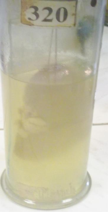
320.SCARS AFTER THE INFARCTION OF KIDNEY.
•The retraction of the lesions under capsule of the kidney with sizes from 0.5-2.5 cm in a diameter are visble.
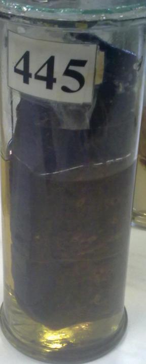
455.THROMBOEMBOLIUM OF BLOOD VESSELS IN LUNGS WITH HEMORRHAGIC INFARCTION.
•In dissection of lungs in the vessels we see red obturated thrombosis. In the lower part of lungs having lesion dark-reddish color, dim, escaping under pleura.
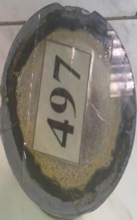
497.MICROFOCI OF CARDIOSCLEROSIS
•There are whitish colored lucid foci in the section of the myocardium

234.NECROSIS OF LYMPH NODE
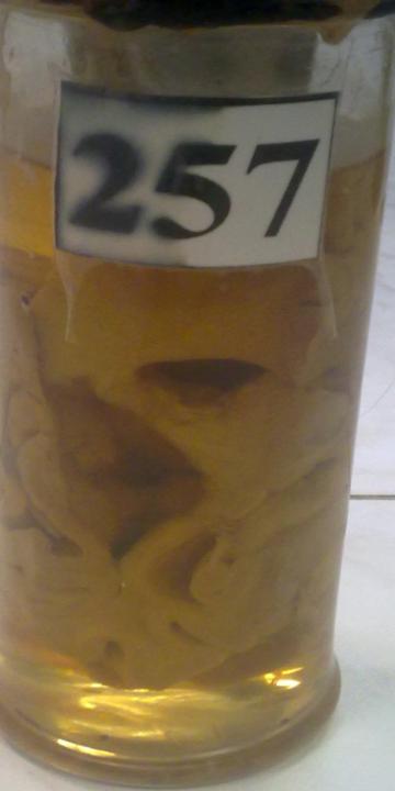
257.CYST IN THE
BRAIN

21.INFARCTION OF MYOCARDIUM WITH PARIETAL THROMBOSIS.
•In a zone of infarctionof the muscle fibers are disposed of nucleikaryolysis. Between necrotic muscle fiber and in border with normal muscles we see aggregation of leucocytes-demarcation inflammation.
•Indicate in the figure: 1-necrosis of muscle fiber,2-aggregation of leucocytes 3-normal cardiac fibers 4- thrombosis.
16. KARYOLYSIS,
KARYORRHEXIS, AND A
KARYOPYKNOSIS
•In the lesions of necrosis in lungs during tuberculosis on the background of homogenic parts, we see separate cells with changed nucleuses: a karyorrhexisfragmentation into many granular clumps; a karyopyknosis-condensation of nuclear chromatin in the form of dark ball; a karyolysis-dissolution of nucleus, cells without a nucleus or with its “shadow”
•Indicate in the figure: 1-normal cell, 2- karyolysis, 3-karyopyknosis, 4- karyorrhexis.