

Bovine Viral Diarrhea Virus Diagnosis, Management, and Control
.pdf66 |
BVDV: Diagnosis, Management, and Control |
sequence of viruses classified as BVDV led to the conclusion that there were actually two genotypes of BVDV, BVDV 1, and BVDV 2 (Pellerin et al., 1994; Ridpath et al., 1994a). The degree of sequence identity between pestiviruses in the 5’ UTR region is the most frequently used parameter for differentiation pestivirus species (Ridpath et al., 1994a; Harasawa and Mizusawa, 1995; Harpin et al., 1995; Harasawa, 1996; Baule et al., 1997; Becher et al., 1997; Giangaspero et al., 1997; Ridpath and Bolin, 1997; Sandvik et al., 1997; Vilcek et al., 1997; Wolfmeyer et al., 1997; Harasawa and Giangaspero, 1998; Ridpath and Bolin, 1998; Shimazaki et al., 1998; Letellier et al., 1999; Sakoda et al., 1999; Flores et al., 2000; Ridpath et al., 2000; Falcone et al., 2001; Tajima et al., 2001; Tajima et al., 2001; Vilcek et al., 2001b; Beer et al., 2002; Couvreur et al., 2002; Evermann and Ridpath, 2002; Flores et al., 2002). However, differences between BVDV 1 and BVDV 2 strains are found throughout the genome (Ridpath and Bolin, 1995b, 1997).
All pestiviruses are antigenically cross-reactive. Although antigenic differences do exist between pestivirus species, they are not extensive enough for pestivirus species to be recognized as serotypes. Convelescent sera generated against a virus from a particular pestivirus species will generally have a higher titer against other viruses from that species as opposed to viruses from the other pestivirus species (Ridpath, 2003). However, animal-to-animal variation and divergence among viruses from the same pestivirus species can make it difficult to reproducibly and reliably differentiate pestivirus species based on serology alone (Muller et al., 1997; Bolin and Ridpath, 1998). Mabs have been developed that differentiate between the pestivirus species (Wensvoort et al., 1989a; Zhou et al., 1989; Dahle et al., 1991; Edwards et al., 1991; Kosmidou et al., 1995). However, cross-reactivity between pestivirus species and variation within any one species make it difficult to generate a Mab that simultaneously differentiates between species and still recognizes all the viruses within one species. Antigenic variation is particularly pronounced among BVDV 1 and BVDV 2 strains (Ridpath et al., 1994a) and impacts on both detection and control.
In addition to the four recognized pestivirus species, three putative species have been suggested. These putative species are based on viruses isolated from a giraffe, a reindeer, and a pronghorn antelope (Becher et al., 1997; van Rijn et al., 1997; Harasawa et al., 2000; Vilcek et al., 2001b). Only one virus has been isolated for each of these putative species,
and no correlation with clinical disease has been reported.
THE PESTIVIRUS VIRION
Pestivirus virions are enveloped, spherical particles that are 40 to 60 nm in diameter. The virions are made up of a central capsid, composed of the virally encoded C protein and the genome RNA, surrounded by a lipid bilayer. The capsid appears as an electron-dense inner core with a diameter of approximately 30 nm (Horzinek et al., 1971). The structure and symmetry of the core has not been determined. Three virus encoded proteins—Erns, E1, and E2—are associated with the lipid bilayer envelope. The envelope surrounding the virion is pleomorphic, which impedes purification of infectious particles by banding in sucrose gradients and identification by electron microscopy. There appear to be 10–12 nm ring-like subunits on the surface of the virus envelope (Heinz et al., 2000). The Mr of the virion is estimated as 6.0 107, and the buoyant density in sucrose is 1.10–1.15 gm/cm3 (Heinz et al., 2000).
Virions are stable within a pH range of 5.7 to 9.3 (Hafez and Liess, 1972). Infectivity is not affected by freezing but decreases at temperatures above 40°C (Heinz et al., 2000). Like other enveloped viruses, BVDV are inactivated by organic solvents and detergents. Other methods of inactivation include Trypsin treatment (0.5 mg/ml, 37°C, 60 min) (Liess, 1990), ethylenimine (reduction of 5 log10 units using 10 mM at 37°C for 2 h) (Preuss et al., 1997), electron beam irradiation (4.9 and 2.5 kGy needed to reduce virus infectivity 1 log10 unit for frozen and liquid samples respectively) (Preuss et al., 1997), and gamma irradiation (20–30 kGy) (Miekka et al., 1998).
THE PESTIVIRUS GENOME
The pestivirus genome, in the absence of insertions, is approximately 12.3 Kb in length (Collett et al., 1988a, b; Moormann et al., 1990; Deng and Brock, 1992; Ridpath and Bolin, 1995b, 1997). The long open reading frame (approximately 4000 codons) is bracketed by relatively large 5’ (360–390 bases) and 3’ (200–240 bases) untranslated regions (UTR). The 5’ terminus does not contain a cap structure (Brock et al., 1992; Deng and Brock, 1993), and no poly(A) tract is present at the 3’ end. All pestivirus genomes terminate at the 3’ end with a short poly(C) tract. The highest nucleic acid sequence identity among pestiviruses is found in the 5’ UTR (Ridpath and Bolin,
Classification and Molecular Biology |
67 |
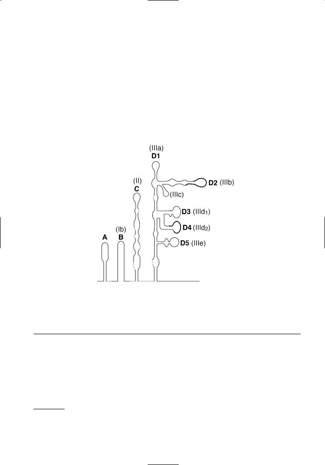
Figure 3.1. Conservation and predicted pseudoknots in pestivirus 5’ UTR sequences. Top. The alignment and comparison of 5’UTR sequences from recognized pestivirus species is shown. Hyperviable regions are boxed. Conserved sequences used in RT-PCR tests to detect pestiviruses are underlined. The first ATG of the open reading frame is in bold type. Bottom. A schematic of predicted tertiary structures in the 5’ UTR is shown. Deng and Brock (1993) domain designations are shown in bold, and Pestova and Hellen (1999) domain designations are shown in parentheses. Note that domain D5 of Deng and Brock is just a portion of the domain IIIe of Pestova and Hellen. The location of hypervariable regions shown in Figure 3.1a are denoted by a thickened line in the schematic.
1997). Although sequence identity is high among pestiviruses in the 5’ UTR, there are two short regions that are notable for their variability (Figure 3.1A). These are located between nucleotides 208– 223 and nucleotides 294–323 (nucleotide position numbers based on the sequence of BVDV 1-SD-11). Sequence variations in these regions have been ex-
ploited in PCR-based tests designed to differentiate BVDV genotypes (el-Kholy et al., 1998; Ridpath and Bolin, 1998). High conservation of 5’ UTR sequences is related to formation of tertiary structures required for internal ribosomal entry mediated initiation of translation (Deng and Brock, 1993; Pestova and Hellen, 1999) (Figure 3.1B).
1Although BVDV 1a-NADL and BVDV 2-890 are the type viruses for genotypes BVDV 1 and BVDV 2, respectively, both genomes have inserted sequences. Insertions can cause confusion when indicating genomic location based on nucleotide number. For this reason BVDV 1a-SD-1 is used as the reference for nucleotide position. It was the first noncytopathic BVDV1 sequenced and does not have an insertion. The accession number for BVDV 1a-SD-1 is M96751.

68 |
BVDV: Diagnosis, Management, and Control |
ENTRY OF THE VIRUS INTO CELLS, TRANSLATION, AND REPLICATION
The binding and entry of BVDV to susceptible cells has not been extensively studied. Based on the behavior of other flaviviruses and pestiviruses, it is hypothisized that binding and entry is a multistep process initiated by receptor-mediated endocytosis involving cell surface molecules and the viral proteins Erns and E2 (Schelp et al., 1995; Xue et al., 1997; Iqbal et al., 2000; Schelp et al., 2000; Iqbal and McCauley, 2002). The genomic RNA is uncoated following endocytosis and serves as the mRNA. No subgenomic mRNA molecules have been detected. Initiation of translation is mediated by a cap-independent internal initiation mechanism that requires an internal ribosome entry site (IRES) located within the 5’ UTR. A secondary structure model for the pestivirus 5’ UTR was first proposed by Deng and Brock (1993). Further refinements of a BVDV specific model have been developed (Poole et al., 1995; Pestova et al., 1998; Pestova and Hellen, 1999). Unfortunately a universal terminology system was not adopted. The different names used for the same structural domains by different research groups can be confusing. The proposed 5’ UTR secondary structures shown in Figure 3.1B are identified by both the domain names given by Deng and Brock and those given by Pestova and Hellen. It should be noted that domain D5 of Deng and Brock is just a portion of the IIIe domain of Pestova and Hellen. The IIIe domain contains the D5 domain plus the nucleotides between D5 and nucletide 380 (numbering based on BVDV 1a-SD-1). Domains C (II) and D (III) contain structures critical for the attachment of the initiation complex. A pseudoknot formed just upstream of the start codon is necessary for IRES function. The hypervariable regions used in PCR based tests to differentiate BVDV genotypes are located in domains D2 (IIIb) and D4 (IIId2).
VIRAL PROTEINS
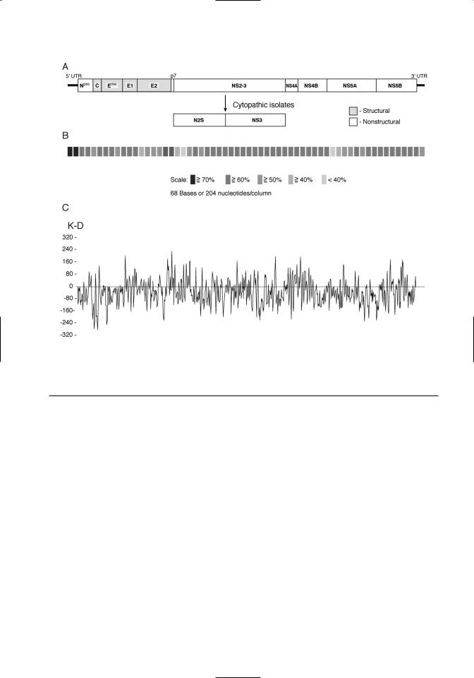
The large open reading frame is translated as a polyprotein. The order of the individual viral proteins within the polyprotein is as follows: Npro- C-Erns-E1-E2-p7-NS2/3-NS4A-NS4B-NS5A- NS5B (Figure 3.2A). The polyprotein is processed cotranslationally and posttranslationally by host and viral proteases. The size, function, and characteristics of the viral proteins are summarized in Table 3.1. Conservation of the polyprotein coding sequences among pestiviruses is shown in Figure 3.2B. The predicted hydrophobicity profile of the
polyprotein is shown in Figure 3.2C. The hydrophobicity profile of the polyprotein is conserved within the pestivirus genus.
Viral structural proteins
The proteins associated with the mature virion are C, Erns, E1, and E2. C is the virion nucleocapsid protein. It is highly basic and relatively conserved among the pestivirus species. It is postulated that the C terminus of the nucleocapsid protein contains an internal signal sequence that directs translocation of structural glycoproteins to the endoplasmic reticulum (Heinz et al., 2000). The Erns protein is glycosylated and forms disulfide-linked homodimers (Weiland et al., 1990). In tissue culture systems Erns may be found both associated with released virus and free in the culture medium. This protein has an unusual ribonuclease activity. Although the function of this ribonuclease activity in the viral life cycle is unknown, antibodies that inhibit ribonuclease activity neutralize virus infectivity of classical swine fever viruses (CSFV) (Windisch et al., 1996). The ribonuclease activity does not require dimer formation or glycosylation (Windisch et al., 1996). The Erns of CSFV possesses epitopes that induce neutralizing antibodies (hereafter referred to as neutralizing epitopes) and CSFV Erns subunit vaccines induce neutralizing antibodies (Konig et al., 1995). It is not known whether the Erns of BVDV possesses neutralizing epitopes that are important in disease control. However, protection afforded by killed BVDV vaccines does not appear to be dependent upon Erns specific antibodies (Bolin and Ridpath, 1990).
Both the E1 and E2 proteins possess potential membrane-spanning domains and are glycosylated. They are predicted to be integral membrane proteins and interact to form heterodimers (Weiland et al., 1990). The E2 protein is the immunodominant structural protein and possesses neutralizing epitopes (Donis et al., 1988; Bolin and Ridpath, 1989). Mabs produced against the E2 have been used to differentiate pestivirus species (Peters et al., 1986; Wensvoort et al., 1986; Bolin et al., 1988; Hess et al., 1988; Wensvoort et al., 1989a; Corapi et al., 1990a; Edwards and Sands, 1990; Dahle et al., 1991; Deregt et al., 1994; Paton et al., 1994; Ridpath et al., 1994a; Ridpath and Bolin, 1997). The coding region for E2 neutralizing epitopes are found in a hypervariable region located in the N-terminal portion of the E2 protein (Paton et al., 1992; Deregt et al., 1998) (refer to Figure 3.2B). E2 antigenic variation is particularly pronounced among BVDV strains and contributes to vaccine failure (Bolin et al., 1988; Bolin and Rid-
Classification and Molecular Biology |
69 |
Figure 3.2. The pestivirus genome. A. Organization of the pestivirus genome is shown. Structural proteins are denoted by shading. B. Sequence conservation among pestivirus species is shown. These results are a composite of comparisons between the four recognized pestivirus species. Sequence similarity is indicated by different levels of shading. C. A hydrophobicity plot of a pestivirus consensus sequence is shown. A consensus sequence was derived by comparison of representatives of noncytopathic strains from each of the four recognized pestivirus species.
path, 1989, 1990; Bolin et al., 1991a; Bolin et al., 1994; Ridpath et al., 1994a; Van Campen et al., 1997; Bolin and Ridpath, 1998; Van Campen et al., 1998; Ridpath et al., 2000; Van Campen et al., 2000).
Viral nonstructural proteins
The first viral protein encoded by the BVDV ORF is the nonstructural protein, Npro. This protein, which is unique to the pestivirus genus, is an autoprotease whose only known function is to cleave itself from the polyprotein. The next nonstructural protein, p7, follows the structural protein E2 in the polyprotein (Elbers et al., 1996). It consists of a central charged region flanked by hydrophobic termini. The role of this cell-associated protein is unknown. It is hypothesized that p7 is required for production of infectious virus but not for RNA replication (Harada et al., 2000). It is inefficiently cleaved from the E2, leading to two intracellular forms of E2 with different C termini (E2 and E2-p7) (Elbers et al., 1996).
Neither p7 nor E2-p7 are found associated with infectious virus.
Following p7 the next nonstructural protein in the polyprotein is the serine protease, NS2-3. In BVDV strains from the cytopathic biotype (see discussion of biotype below), the NS2-3 is cleaved to NS2 and NS3. The serine protease activity of the NS2-3 resides in the NS3 portion of the protein. The function of the NS2 is unknown. It is not required for RNA replication, and its cleavage from the NS2-3 does not affect serine protease activity (Behrens et al., 1998). Sequence analysis reveals regions in the NS2 with homology to zinc-finger motifs present in DNA binding proteins (De Moerlooze et al., 1991). However, a function for the putative DNA binding activity of NS2 in viral replication has not been demonstrated.
Both the uncleaved NS2-3 and the cleaved NS3 act as serine proteases (Tautz et al., 1997) that cleave the remaining nonstructural proteins from the

70 |
|
BVDV: Diagnosis, Management, and Control |
||
Table 3.1. Pestivirus proteins |
|
|
||
|
Estimated |
|
|
|
Viral |
Size |
|
Neutralizing |
|
Protein |
(K Daltons) |
Attributes |
Epitope(s) |
Function |
|
|
|
|
|
Npro |
020 |
Nonstructural |
N |
Autoproteolysis |
|
|
|
|
Not required for RNA replication |
C |
014 |
Structural |
N |
Forms nucleocapsid of virion |
|
|
Conserved |
|
|
Erns |
|
Highly basic |
|
|
048 |
Structural |
Y |
Envelope-associated glycoprotein |
|
|
|
7–9 glycosylation sites |
|
Ribonuclease activity |
E1 |
025 |
Structural |
N |
Envelope-associated glycoprotein |
|
|
2–3 glycosylation sites |
|
Integral membrane protein |
E2 |
053 |
Structural |
Y |
Envelope-associated glycoprotein |
|
|
4–6 glycosylation sites |
|
Integral membrane protein |
|
|
Least conserved of structural proteins |
Immunodominant structural protein |
|
p7 |
007 |
Nonstructural |
N |
Function unknown |
|
|
Central charged region flanked |
|
Required for production of |
|
|
by hydrophobic termini |
|
infectious virus but not RNA |
|
|
|
|
replication |
NS 2/3 |
125 |
Nonstructural |
N |
NS2 has a zinc-finger–like domain |
NS2 |
054 |
In cytopathic biotype NS 2/3 cleaved |
NS2/3 and NS3 contain RNA |
|
NS3 |
080 |
to NS2 and NS3 |
|
helicase and N-terminal serine |
|
|
Conserved |
|
protease domains; cleaves itself |
|
|
|
|
and remaining nonstructural pro- |
|
|
|
|
teins from viral polyprotein |
|
|
|
|
Immunodominant nonstructural |
|
|
|
|
protein |
NS4A |
7.2 |
Nonstructural |
N |
Serine protease cofactor |
|
|
Hydrophobic |
|
|
NS4B |
38–39 |
Nonstructural |
N |
Replicase component |
|
|
Hydrophobic |
|
|
NS5A |
55–56 |
Nonstructural |
N |
Replicase component |
|
|
Phosphorylated |
|
|
NS5B |
81–82 |
Nonstructural |
N |
RNA-dependent RNA polymerase |
polyprotein. Purified BVDV NS3 also possesses RNA helicase and RNA-stimulated NTPase activities (Tamura et al., 1993; Warrener and Collett, 1995). All three activities (serine protease, RNA helicase, and RNA-stimulated NTPase) are essential to virus viability (Grassmann et al., 1999; Gu et al., 2000). Antibodies to the NS2-3 and NS3 do not neutralize infectivity. However, the NS2-3 and NS3 (but not the NS2) are strongly recognized by polyclonal convalescent sera (Donis et al., 1991). Animals vaccinated with modified live vaccines also have a strong antibody response to the NS2-3 and/or NS3 protein (Bolin and Ridpath, 1989). In contrast, animals vaccinated with inactivated (killed) vaccines
primarily react with structural proteins and not the NS2-3 or NS3 (Bolin and Ridpath, 1990). The difference in recognition of NS2-3 or NS3 may be useful in differentiating between immune responses to inactivated vaccines and immune responses to natural infection.
The NS4A and NS4B proteins are similar in size, composition and hydrophobicity to the NS4A and NS4B proteins of other flaviviruses (Lindenbach and Rice, 2001). NS4A acts as a cofactor for the NS2-3 and NS3 serine protease activity. NS4B and NS5A probably are replicase complex components. RNA polymerase activity has been demonstrated for the NS5B protein (Lai et al., 1999).

Classification and Molecular Biology |
71 |
Nomenclature for proteins
The names of the viral proteins have changed over time and these variations in terminology can be confusing as one reads past literature. The names used above and in Table 3.1 are those designated in the Seventh Report of the International Committee on Taxonomy of Viruses (van Regenmortel et al., 2000). The first studies of BVDV reported two glycosylated structural polypeptides, with molecular weights of 55K and 45K, which were termed VP1 and VP2, respectively (Matthaeus, 1979). Later reviews referred to these proteins as E1 and E2 (Westaway et al., 1985). When Collett et al. first derived the genomic map for BVDV, gene products were designated by their molecular weights and glycosylation status (Collett et al., 1988b). Thus, the viral protein now known as E2, may be referred to as VP1, E1, or gp53 in the literature. Similarly, Erns may be reported as VP2, E0, E2, or gp48.
TRANSLATION AND PROCESSING OF THE
POLYPROTEIN
As stated above, the first viral protein in the ORF is a nonstructural protein unique to the pestivirus genus, Npro. The Npro is an autoprotease whose only known function is to cleave itself from the polyprotein. Cellular signal peptidases are responsible for the cleavages between C and Erns, E1, and E2, and E2 and p7 (Rumenapf et al., 1993; Stark et al., 1993; Elbers et al., 1996). The mechanism that produces the cleavage between Erns and E2 is unknown. As stated above, the cleavage between E2 and p7 may not be complete. Depending on the biotype of the virus (see section on biotypes below), the NS2/3 protein may be further processed to the NS2 and the NS3 proteins. Both NS2/3 and NS3 have serine protease activity that cleaves the remaining downstream proteins from the polyprotein. The NS4A viral protein facilitates the protease activity of the NS2/3 (and/or NS3) at the cleavage between NS4B and NS5A and between NS5A and NS5B.
VIRAL REPLICATION
Viral proteins, generated by processing the translated polyprotein, participate in viral replication. It has been proposed that a secondary structure motif in the 5’ UTR enables the switch of viral RNA from a template for translation to a template for replication (Yu et al., 2000). RNA replication occurs via a replication complex, composed of viral RNA and viral nonstructural proteins, in association with intracytoplasmic membranes. The replication of BVDV RNA is similar to that of other flaviviruses.
After translation, RNA replication begins with the synthesis of complementary negative strands. Using these negative strands as templates, genome-length positive strands are synthesized by a semiconservative mechanism involving replicative intermediates and replicative forms (Gong et al., 1996; Gong et al., 1998). Because viral proteins are not detected on the surface of infected cells, it is thought that virions mature in intracellular vesicles and are released by exocytosis (Heinz et al., 2000). A substantial fraction of the infectious virus remains cell associated (Lindenbach and Rice, 2001).
BVDV GENOTYPES AND PHENOTYPES
As stated above, BVDV strains may belong to one of two different genotypes, BVDV 1 and BVDV 2. Further, viruses from either genotype may exist as one of two biotypes, cytopathic and noncytopathic.
BVDV GENOTYPES
Genotyping is grouping based on comparison of genomic sequences. There are no hard and fast taxonomic rules governing how different two genomes have to be before they are designated different genotypes. However, for genotypic designations to be meaningful, some practical considerations should be taken into account. Genotypes should be distinct, discrete, and stable. That is, there should be a clear defined break between groups (discreet groups as opposed to a continuum), and isolates should not be able to easily switch back and forth between groups. Ideally genotypes would be associated with practical observations such as geographic distribution, antigenic variation, or variations in virulence. The differentiation between the BVDV 1 and the BVDV 2 genotypes meets these practical considerations.
The first reported division of BVDV strains into two different genotypes was based on comparison of the 5’ UTR (Pellerin et al., 1994; Ridpath et al., 1994a). Previous studies of vaccination cross protection and monoclonal antibody binding revealed that BVDV strains were quite variable (Moennig et al., 1987; Bolin et al., 1988; Corapi et al., 1990a; Bolin et al., 1991a; Donis et al., 1991; Edwards et al., 1991). Although these studies indicated that there was considerable variation among BVDV strains, no standard means of grouping viruses based on these variations was generated. Meanwhile, hybridization analysis and sequence comparison suggested that the 5’ UTR was highly conserved compared to other portions of the genome (Lewis et al., 1991; Ridpath and Bolin, 1991; Ridpath et al.,

72 |
BVDV: Diagnosis, Management, and Control |
1993). The highly conserved sequences in the 5’ UTR were evaluated by several groups as targets for polymerase chain reaction (PCR) tests designed to detect the wide range of BVDV strains or to differentiate BVDV from CSFV or BDV (Hofmann et al., 1994; Vilcek, 1994). Concurrent to this research a highly virulent form of BVD, termed hemorrhagic syndrome, was reported in Canada and the United States (Corapi et al., 1989; Corapi et al., 1990b; Carman, 1995; Carman et al., 1998). Phylogenetic analysis of the BVDV strains, isolated from animals suffering from hemorrhagic syndrome, grouped them separately from the BVDV strains commonly used, at that time, in vaccine production, diagnostic tests, and research (Pellerin et al., 1994; Ridpath et al., 1994a). The newly recognized group of BVDV was designated BVDV genotype II, and the group containing the strains used in vaccines, detection, and research was termed BVDV genotype I. The names of these two genotypes were later modified to BVDV 1 and BVDV 2 in keeping with taxonomic conventions in use with other viruses (Heinz et al., 2000). It was further noted that viruses from the BVDV 2 genotype were also isolated from persistently infected calves born to dams that had been vaccinated with vaccines based on BVDV 1 isolates (Ridpath et al., 1994a). Comparison of monoclonal antibody binding profiles revealed clear differences between BVDV 1 and BVDV 2 strains (Ridpath et al., 1994a). Although initial phylogenetic differentiation was based on comparison of 5’ UTR sequences, comparison of complete genomic sequences showed that differences between BVDV 1 and BVDV 2 strains were found throughout the genome (Ridpath and Bolin, 1995b).
There are two common misconceptions relating to the BVDV 2 genotype. One relates to origin of the BVDV 2 genotype and the other relates to the virulence of BVDV 2 strains. In the late 1990s there was some speculation that BVDV 2 strains represented newly emerging viruses that originated in the United States as a result of use of vaccines by U.S. producers and were then transferred to Europe. Although BVDV 2 strains were first recognized in 1994, retrospective characterization of strains collected from BVDV outbreaks in Ontario that occurred between 1981 and 1994 demonstrated that BVDV 2 were present in North America at least since the the early 1980s. However, the first BVDV 2 strain described in the literature was isolated in Europe prior to 1979 (Wensvoort et al., 1989b). Interestingly, this strain was isolated from a pig and was referred to as an atypical CSFV. Subsequent comparison of this iso-
late to BVDV 2 strains isolated from North America led to the segregation of this virus to the BVDV 2 genotype (Paton et al., 1995b). Further, a phylogenetic survey of BVDV 1 and BVDV 2 strains showed similar levels of sequence variation in the 5’ UTR (Ridpath et al., 2000). If one assumes that the evolutionary clock ticks at the same rate for both genotypes, similar rates of variation in the 5’ UTR suggests that the two genotypes have been evolving for approximately the same time span. This suggests that viruses from the BVDV 2 genotype are not newly emerging viruses, but newly recognized ones.
Initially the BVDV 2 genotype was associated with outbreaks of hemorrhagic syndrome (Corapi et al., 1989; Corapi et al., 1990b; Pellerin et al., 1994; Carman et al., 1998), a clinically severe form of acute BVDV infection. This led to the misconception that all BVDV 2 strains cause severe acute disease. In an initial survey of BVDV 2 strains, only 32 out of 76 strains were associated with clinically severe disease (Ridpath et al., 1994a). Since then, animal studies have shown that there is a continuum of virulence seen in acute infections with type 2 BVDV (Flores et al., 2000; Fulton et al., 2000a; Hamers et al., 2000; Ridpath et al., 2000; Tajima et al., 2001; Evermann and Ridpath, 2002; Kelling et al., 2002; LieblerTenorio et al., 2002; Liebler-Tenorio et al., 2003). Thus, it appears that only a minority of BVDV 2 strains cause severe acute clinical disease. The majority of BVDV 2 strains present in North America appear to be no more virulent than BVDV 1 strains.
Prevalence of BVDV genotypes
Both BVDV genotypes have been reported in North America (Pellerin et al., 1994; Ridpath and Neill, 1998), Europe (Becher et al., 1995; Edwards and Paton, 1995; Paton, 1995a, b; Sandvik et al., 1997; van Rijn et al., 1997; Giangaspero et al., 2001; Luzzago et al., 2001; Pratelli et al., 2001; Couvreur et al., 2002; Vilcek et al., 2002), and South America (Canal et al., 1998; Jones et al., 2001), although the reported prevalence of BVDV 2 is higher in North America than in Europe or South America. Reports of the prevalence of the BVDV 2 genotype in commercial cattle herds in North America range from 24–47% (Carman, 1995; Bolin and Ridpath, 1998; Carman et al., 1998; Fulton et al., 1998; Fulton et al., 2000b; Evermann and Ridpath, 2002).
Similarities between viruses from the BVDV 1 and the BVDV 2 genotype
Viruses from both genotypes may exist as one of two biotypes, cytopathic and noncytopathic (see

Classification and Molecular Biology |
73 |
below). Noncytopathic viruses from either genotype can cross the placenta and establish persistent infections in the fetus.
Aside from severe acute BVDV, it is difficult to distinguish BVDV 1 infections from BVDV 2 infections based on clinical signs. There is considerable heterogeneity among strains from both genotypes. Although virulence is strain-dependent, clinical presentation may also be effected by immune status, reproductive status, stress, and the presence of secondary pathogens.
Heterogeneity, among strains within each genotype, also affects antigenicity. Although strains from the BVDV 1 and BVDV 2 genotypes may be distinguished by monoclonal antibody binding, there are smaller, but still significant, differences among strains from the same genotype (Ridpath et al., 1994a; Ridpath et al., 2000).
Differences between viruses from the BVDV 1 and the BVDV 2 genotype
To date only viruses from the BVDV 2 genotype are associated with severe acute BVDV (Corapi et al., 1989; Corapi et al., 1990b; Bolin and Ridpath, 1992; Pellerin et al., 1994; Ridpath et al., 1994a; Carman et al., 1998; Liebler-Tenorio et al., 2002). BVDV 1 and BVDV 2 strains are antigenically distinct, as demonstrated by serum neutralization using polyclonal sera and monoclonal antibody binding (Ridpath et al., 2000). The practical significance of antigenic differences is indicated by the birth of BVDV 2 persistently infected animals to dams that had been vaccinated against BVDV 1 strains (Bolin et al., 1991a; Ridpath et al., 1994a). Although modified live BVDV 1 vaccines may induce antibodies against BVDV 2 strains, the titers average one log less than titers against heterologous BVDV 1 strains (Cortese et al., 1998).
Subgenotypes of BVDV 1 and BVDV 2
Subgenotypes of both BVDV 1 and BVDV 2 have been described, although the biological significance of these subgroupings is a matter of debate. BVDV 1 strains from North America can be segregated to two subgenotypes, BVDV 1a and BVDV 1b (Pellerin et al., 1994). These two subgenotypes can be distinguished by monoclonal antibody binding (Bolin and Ridpath, 1998) and RT-PCR (Ridpath and Bolin, 1998). Epidemiological surveys suggest that BVDV 1b strains may predominate in respiratory cases (Fulton et al., 2002), and BVDV 1a strains may predominate in fetal infections occurring late in gestation (>100 days gestation) (Evermann and Rid-
path, 2002). While the reported incidence of BVDV 2 strains is lower, European BVDV 1 strains appear to be more variable than North American BVDV 1 strains. The European BVDV 1 strains have been separated into at least 11 subgroups (Vilcek et al., 2001a). The biological significance of these 11 groups has yet to be examined.
Similarly, BVDV 2 strains from North and South American have been segregated into two subgroups, BVDV 2a and BVDV 2b (Flores et al., 2002). In North America BVDV 2b strains are relatively rare; in South America the prevalence of BVDV 2a strains and BVDV 2b strains are similar.
BVDV BIOTYPES
As stated above, both BVDV 1 strains and BVDV 2 strains may exist as one of two biotypes, cytopathic and noncytopathic. The division into biotypes is based on the activity of the strain when propagated in cultured epithelial cells (Lee and Gillespie, 1957; Gillespie et al., 1960). Recall that both noncytopathic and cytopathic strains code for the nonstructural protein NS2-3. Among cytopathic strains the NS2-3 is processed to an NS2 and an NS3 protein. Processing of the NS2-3 protein in cytopathic strains may occur by several different strategies depending on the viral strain (Meyers et al., 1991a, b; Meyers et al., 1992; Qi et al., 1992; Ridpath et al., 1994b; Kupfermann et al., 1996; Tautz et al., 1996; Becher et al., 1998; Mendez et al., 1998; Meyers et al., 1998; Qi et al., 1998; Becher et al., 1999; Baroth et al., 2000; Kummerer and Meyers, 2000; Ridpath and Neill, 2000; Vilcek et al., 2000; Neill and Ridpath, 2001; Becher et al., 2002; Ridpath and Bolin, 1995a). Most commonly, the generation of the NS3 is associated with insertion of sequences into the viral genome. Generally the amino acid sequence flanking the carboxy-terminus of the insertion corresponds to either amino acid position 1535 or position 1589 (numbering based on the noncytopathic BVDV 1a strain SD-1, acc. # M96751, see footnote a). These two positions are referred to as positions A and B, respectively (Meyers et al., 1998). Position B corresponds to the cleavage site that frees the N-terminus of the NS3 from the NS2- 3 precursor. It is thought that insertions at the B position result in the processing of the NS2-3, either by introducing a new cleavage site at carboxy terminus of the insertion or by introducing sequences with autocatalytic activity that act at the carboxy terminus of the insertion. The end result is that cleavage occurs at position 1589, making Gly1589 the first amino acid of NS3. Insertions at position A, al-

74 |
BVDV: Diagnosis, Management, and Control |
though upstream of the cleavage site, also produce cleavage at amino acid position 1589. It has been proposed that insertions upstream of the cleavage site induce conformational changes that allow cleav-
age via a cryptic mechanism at Gly1589 (Meyers and Thiel, 1996). The most frequently described inser-
tions into cytopathic BVDV 1 strains have been at position B and consist of duplicated viral genomic sequence and/or ubiquitin coding sequences. Insertions in cytopathic BVDV 2 strains are more frequently reported in the vicinity of position A and consist of portions of a gene coding for a DnaJ-like protein (Ridpath and Neill, 2000; Neill and Ridpath, 2001). Included among the possible explanations for these differences in relative frequency of position and source of inserted sequences are (i) differences in recombination frequencies due to sequence variation; (ii) differences in the stability of recombinations; and (iii) differences in the relative amounts of mRNA coding for ubiquitin and DnaJ-like proteins in BVDV 1 and BVDV 2 infected cells.
Practical significance of biotypes
Only noncytopathic BVDV have been reported to establish persistent infections. This has led to some speculation that cytopathic BVDV are not able to cross the placenta and thus do not infect the fetus. However, the detection of cytopathic BVDV strains in fetal calf serum and fetal sero conversion in utero after exposure to cytopathic BVDV call into question this assumption (Brownlie et al., 1989; Bolin et al., 1991b; Bolin and Ridpath, 1998). Because cytopathic BVDV do not establish persistent infections in vivo, they are frequently the biotype of choice for modified live vaccines. Superinfection of an animal, persistently infected with a noncytopathic BVDV, with a cytopathic BVDV can lead to the development of a highly fatal form of BVDV called mucosal disease (Brownlie et al., 1984; Bolin et al., 1985). Mucosal disease is a comparatively rare form of BVDV and does not represent a major source of loss for producers (Houe, 1995, 1999). The majority of losses incurred by producers due to BVDV infections are associated with acute infections with noncytopathic BVDV (Houe, 1995; Fulton et al., 1998; Houe, 1999; Fulton et al., 2000a, b; Evermann and Ridpath, 2002; Fulton et al., 2002).
Noncytopathic BVDV strains predominate over cytopathic BVDV in nature. In the field, cytopathic BVDV strains are typically isolated from mucosal disease outbreaks or post-vaccinal disease outbreaks. Cytopathology in cultured epithelial cells does not correlate with virulence in acute infections
in vivo. All highly virulent viruses studied to date have been noncytopathic (Corapi et al., 1990b; Bolin and Ridpath, 1992; Carman et al., 1998; Ellis et al., 1998; Ridpath et al., 2000; Liebler-Tenorio et al., 2002). In short, from a management standpoint, determining the genotype of a strain is generally more important than determining the biotype.
Importance of variation among BVDV
Heterogeneity is the defining characteristic of BVDV viruses. Variation between BVDV genotype 1 and BVDV genotype 2 has practical significance. In particular, variation in antigenicity impacts on detection and management programs. The impact of variation within each genotype is less well defined. Understanding and compensating for variation among BVDV is essential to developing a successful control program.
REFERENCES
Baroth M, Orlich M, Thiel HJ, Becher P: 2000, Insertion of cellular NEDD8 coding sequences in a pestivirus. Virology 278:456–466.
Baule C, van Vuuren M, Lowings JP, Belak S: 1997, Genetic heterogeneity of bovine viral diarrhoea viruses isolated in Southern Africa. Virus Res 52:205–220.
Becher P, Konig M, Paton DJ, Thiel HJ: 1995, Further characterization of border disease virus isolates: Evidence for the presence of more than three species within the genus pestivirus. Virology 209:200–206.
Becher P, Orlich M, Konig M, Thiel HJ: 1999, Nonhomologous RNA recombination in bovine viral diarrhea virus: Molecular characterization of a variety of subgenomic RNAs isolated during an outbreak of fatal mucosal disease. J Virol 73:5646–5653.
Becher P, Orlich M, Shannon AD, et al.: 1997, Phylogenetic analysis of pestiviruses from domestic and wild ruminants. J Gen Virol 78:1357–1366.
Becher P, Orlich M, Thiel HJ: 1998, Ribosomal S27a coding sequences upstream of ubiquitin coding sequences in the genome of a pestivirus. J Virol 72:8697–8704.
Becher P, Thiel HJ, Collins M, et al.: 2002, Cellular sequences in pestivirus genomes encoding gammaaminobutyric acid (A) receptor-associated protein and Golgi-associated ATPase enhancer of 16 kilodaltons. J Virol 76:13069–13076.
Beer M, Wolf G, Kaaden OR: 2002, Phylogenetic analysis of the 5’-untranslated region of german BVDV type II isolates. J Vet Med B Infect Dis Vet Public Health 49:43–47.

Classification and Molecular Biology |
75 |
Behrens SE, Grassmann CW, Thiel HJ, et al.: 1998, Characterization of an autonomous subgenomic pestivirus RNA replicon. J Virol 72:2364–2372. Bolin S, Moennig V, Kelso Gourley NE, Ridpath J:
1988, Monoclonal antibodies with neutralizing activity segregate isolates of bovine viral diarrhea virus into groups. Brief report. Arch Virol 99:117–123.
Bolin SR, Littledike ET, Ridpath JF: 1991a, Serologic detection and practical consequences of antigenic diversity among bovine viral diarrhea viruses in a vaccinated herd. Am J Vet Res 52:1033–1037.
Bolin SR, Matthews PJ, Ridpath JF: 1991b, Methods for detection and frequency of contamination of fetal calf serum with bovine viral diarrhea virus and antibodies against bovine viral diarrhea virus. J Vet Diagn Invest 3:199–203.
Bolin SR, McClurkin AW, Cutlip RC, Coria MF: 1985, Severe clinical disease induced in cattle persistently infected with noncytopathic bovine viral diarrhea virus by superinfection with cytopathic bovine viral diarrhea virus. Am J Vet Res 46:573–576.
Bolin SR, Ridpath JF: 1989, Specificity of neutralizing and precipitating antibodies induced in healthy calves by monovalent modified-live bovine viral diarrhea virus vaccines. Am J Vet Res 50:817–821.
Bolin SR, Ridpath JF: 1990, Range of viral neutralizing activity and molecular specificity of antibodies induced in cattle by inactivated bovine viral diarrhea virus vaccines. Am J Vet Res 51:703–707.
Bolin SR, Ridpath JF: 1992, Differences in virulence between two noncytopathic bovine viral diarrhea viruses in calves. Am J Vet Res 53:2157–2163.
Bolin SR, Ridpath JF: 1998, Prevalence of bovine viral diarrhea virus genotypes and antibody against those viral genotypes in fetal bovine serum. J Vet Diagn Invest 10:135–139.
Bolin SR, Ridpath JF, Black J, et al.: 1994, Survey of cell lines in the American Type Culture Collection for bovine viral diarrhea virus. J Virol Methods 48:211–221.
Brock KV, Deng R, Riblet SM: 1992, Nucleotide sequencing of 5’ and 3’ termini of bovine viral diarrhea virus by RNA ligation and PCR. J Virol Methods 38:39–46.
Brownlie J, Clarke MC, Howard CJ: 1984, Experimental production of fatal mucosal disease in cattle. Vet Rec 114:535–536.
Brownlie J, Clarke MC, Howard CJ: 1989, Experimental infection of cattle in early pregnancy with a cytopathic strain of bovine virus diarrhoea virus. Res Vet Sci 46:307–311.
Canal CW, Strasser M, Hertig C, et al.: 1998, Detection of antibodies to bovine viral diarrhoea
virus (BVDV) and characterization of genomes of BVDV from Brazil. Vet Microbiol 63:85–97.
Carman S: 1995, Typing of Ontario BVD Virus Isolates from 1981 to 1994. Ontario Ministry of Agriculture, Food, and Rural Affairs, Guelph.
Carman S, van Dreumel T, Ridpath J, et al.: 1998, Severe acute bovine viral diarrhea in Ontario, 1993–1995. J Vet Diagn Invest 10:27–35.
Collett MS, Anderson DK, Retzel E: 1988a, Comparisons of the pestivirus bovine viral diarrhoea virus with members of the flaviviridae. J Gen Virol 69:2637–2643.
Collett MS, Larson R, Belzer SK, Retzel E: 1988b, Proteins encoded by bovine viral diarrhea virus: the genomic organization of a pestivirus. Virology 165:200–208.
Corapi WV, Donis RO, Dubovi EJ: 1990a, Characterization of a panel of monoclonal antibodies and their use in the study of the antigenic diversity of bovine viral diarrhea virus. Am J Vet Res 51:1388–1394.
Corapi WV, Elliott RD, French TW, et al.: 1990b, Thrombocytopenia and hemorrhages in veal calves infected with bovine viral diarrhea virus. J Am Vet Med Assoc 196:590–596.
Corapi WV, French TW, Dubovi EJ: 1989, Severe thrombocytopenia in young calves experimentally infected with noncytopathic bovine viral diarrhea virus. J Virol 63:3934–3943.
Cortese VS, Whittaker R, Ellis J, et al.: 1998, Specificity and duration of neutralizing antibodies induced in healthy cattle after administration of a modified-live virus vaccine against bovine viral diarrhea. Am J Vet Res 59:848–850.
Couvreur B, Letellier C, Collard A, et al.: 2002, Genetic and antigenic variability in bovine viral diarrhea virus (BVDV) isolates from Belgium. Virus Res 85:17–28.
Dahle J, Liess B, Moennig V, Coulibaly CO: 1991, Use of monoclonal antibodies for the differential diagnosis of pestivirus infections in swine. Tierarztl Prax 19:151–155.
De Moerlooze L, Renard A, Lecomte C, Martial JA: 1991, A “zinc finger-like” domain in the 54KDA protein of several pestiviruses. Arch Virol Suppl 3:41–46.
Deng R, Brock KV: 1992, Molecular cloning and nucleotide sequence of a pestivirus genome, noncytopathic bovine viral diarrhea virus strain SD-1.
Virology 191:867–869.
Deng R, Brock KV: 1993, 5’ and 3’ untranslated regions of pestivirus genome: primary and secondary structure analyses. Nucleic Acids Res 21:1949–1957.
Deregt D, Bolin SR, Heckert RA, Loewen KG: 1994, Monoclonal antibodies to bovine viral diarrhea