

- •At a Glance
- •Preface
- •Contents
- •Cells
- •Epithelium
- •Exocrine Glands
- •Connective and Supportive Tissue
- •Muscular Tissue
- •Nerve Tissue
- •Blood Vessels, Blood and Immune System
- •Endocrine Glands
- •Digestive System
- •Respiratory System
- •Urinary Organs
- •Male Reproductive Organs
- •Female Reproductive Organs
- •Integumentary System, Skin
- •Somatosensory Receptors
- •Sensory Organs
- •Central Nervous System
- •Tables
- •Index
- •Cells
- •Epithelial Tissue
- •Exocrine Glandular Epithelium
- •Connective and Supportive Tissue
- •Muscular Tissue
- •Nerve Tissue
- •Blood Vessels, Blood and Immune System
- •Endocrine Glands
- •Digestive System
- •Respiratory System
- •Urinary Organs
- •Male Sexual Organs
- •Female Sexual Organs
- •Integumentary System, Skin
- •Somatosensory Receptors
- •Sensory Organs
- •Central Nervous System
- •Tables
- •Photographic Credits
- •Index

Sensory Organs
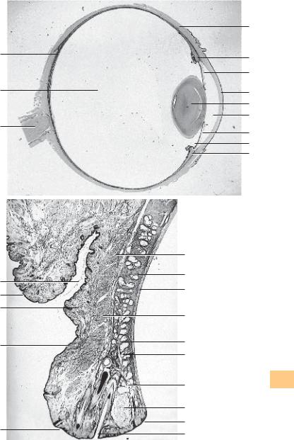
624 Eyeball—Bulbus Oculi
Horizontal center section of the left eyeball (bulbus oculi).
1 Cornea
2 Anterior camera oculi, anterior chamber of the eye
3 Iris
4 Lens
5 Posterior camera oculi, posterior chamber of the eye
6 Corpus ciliare, ciliary body
7 Sclera, tunica fibrosa bulbi
8 Corpus vitreum, vitreous body
9 Retina
10 Optic nerve
Stain: hematoxylin-eosin; magnifying glass
625 Eyelids—Palpebrae
Eyelids are skin folds, which can be actively moved. They consist of a tough connective tissue skeleton 1 (tarsus superior and tarsus inferior). Toward the outside, it is covered by the musculus orbicularis oculi (pars palpebralis) 2 . The surface covering of the eyelid is a multilayered keratinizing squamous epithelium with only a few velum hairs. The outer lid is about 2 mm wide and consists of a dull anterior 4 and a sharp-edged posterior palpebral limb 5 . This tissue continues in the multilayered nonkeratinizing squamous epithelium of the palpebral part of the conjunctiva (conjunctiva tarsi) 6 . A multilayered columnar epithelium with goblet cells is only found beyond the level of the fornix of the conjunctiva. Long cilia (eyelashes) 7 protrude from the anterior rim of the lid. They are rooted in the lid plate (see Fig. 626). The sebaceous glands (Zeis glands), apocrine scent glands and the sweat glands of the cilia (Moll glands) end in the hair follicle of the eyelashes. The right side of the figure shows numerous tarsal holocrine sebaceous glands (Meibomian glands) 8 with long secretory ducts that end on the anterior edge of the lid (posterior limbus). The tight tarsal fiber meshwork envelops the lobed glands. Smooth muscle cells run both before and behind the Meibomian glands at the rim of the lid. The palpebral part of the musculus orbicularis oculi 2 is located in front of the tarsus. The subcutaneous tissue of the lid consists of loosely structured, cell-rich connective tissue 9 , which is usually free of adipose tissue. The epithelium on the anterior eyelid is thin. The skin of the lids contains many melanocytes. This explains the dark pigmentation.
1 Tarsus
2 Palpebral part of the orbicularis oculi muscle
3 Skin
4 Anterior palpebral limb
5 Posterior palpebral limb
6 Palpebral part of the conjunctiva
7 Eyelashes
8 Tarsal glands, Meibomian glands
9 Loosely structured subcutaneous connective tissue
10Deep skin fold of the upper lid
11Chalazion (infection of the Meibomian gland) Stain: hematoxylin-eosin; magnifying glass
Kuehnel, Color Atlas of Cytology, Histology, and Microscopic Anatomy © 2003 Thieme All rights reserved. Usage subject to terms and conditions of license.
9
8
10
10
3
9
3
4
2
1
6
2
1
8
8
11
5
7
7
6
3
1
4
2
3
5
6
Sensory Organs
Kuehnel, Color Atlas of Cytology, Histology, and Microscopic Anatomy © 2003 Thieme All rights reserved. Usage subject to terms and conditions of license.

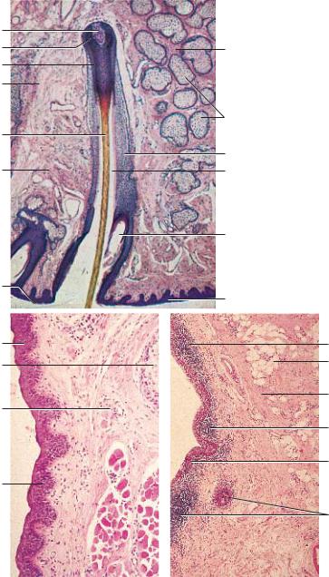
626 Eyelids—Palpebrae
Detail magnification of an upper eyelid, with the rim of the lid and eyelashes. The following structures are shown:
1 Rim (edge) of the lid
2 Hair funnel
3 Hair shaft, scapus pili of the eyelash
4 Outer root sheath
5 Rim of the eyelid, multilayered keratinizing squamous epithelium
6 The terminal portions of a Meibomian gland end in the hair follicle clearance 8 Tarsus superior
9 Hair bulb
10Hair papilla
11Subcutaneous tissue of the lids
Compare with Figs. 625–629. Heinrich J. Meibom (1638–1700).
Stain: alum hematoxylin-eosin; magnification: × 10
Sensory Organs
627 Eyelids—Palpebrae
Detail magnification of a sagittal section through the eyelid of an adult human (cf. Fig. 625). The skin on the outside of the lids is very thin. There is usually no adipose tissue 1 . It is malleable and can move laterally. Keratinization of the multilayered squamous epithelium of the epidermis is marginal. Dermis and subcutaneous connective tissue are thin. The dermis of the skin on the lid contains branched chromatophores. The palpebral part of the striated musculus orbicularis oculi 3 is shown in the lower right corner of the microphotograph. Part of the sebaceous follicle of a holocrine Meibomian gland 4 is visible in the upper right of the figure 4 .
1 Skin of the eyelid
2 Subcutaneous tissue
3 Palpebral part of the orbicularis oculi muscle
4 Meibomian gland
Stain: alum hematoxylin-eosin; magnification: × 20
628 Eyelids—Palpebrae
The inner face of the eyelids is lined by the moist mucous membrane of the palpebral part of the tunica conjunctiva, which is part of the conjunctival sac. The keratinizing squamous epithelium of the skin of the lid continues in the nonkeratinizing multilayered squamous epithelium of the palpebral part of the conjunctiva at the posterior palpebral limb (see Fig. 625). It is only at the fornix conjunctivae that a multilayered columnar epithelium is found.
The figure shows a sagittal section of the eyelid close to the fornix conjunctivae with conjunctival epithelium 1 and an underlying accumulation of lymphocytes 2 . The tunica propria 3 contains adipocytes 4 .
1 Conjunctival epithelium
2 Accumulation of lymphocytes
3 Lamina propria
4 Adipocytes
Stain: alum hematoxylin-eosin; magnification: × 20
Kuehnel, Color Atlas of Cytology, Histology, and Microscopic Anatomy © 2003 Thieme All rights reserved. Usage subject to terms and conditions of license.
10
9
4
11
3
11
1
1
4
2
1
3 
8
7
4
2
6
5
1
4
3 |
Organs |
|
2 |
||
Sensory |
||
1 |
||
|
||
2 |
|
|
|
||
|
Kuehnel, Color Atlas of Cytology, Histology, and Microscopic Anatomy © 2003 Thieme All rights reserved. Usage subject to terms and conditions of license.
629 Eyelids—Palpebrae
Detail magnification of a sagittal section through the eyelid close to the rim of the lid. It shows the apocrine glands of Moll (ciliary glands) 1 . They are located in the vicinity of the roots of the eyelashes and end at the rim of the lid or in the hair follicles. The glands of Moll are classified as sweat glands.
There are striated muscle fiber bundles of the musculus orbicularis oculi 2 close to the glands. They can pull the rim of the lid to the eyeball (cf. Fig. 625).
1 Glands of Moll, apocrine sweat glands
2 Orbicularis oculi muscle
Stain: alum hematoxylin-eosin; magnification: × 20
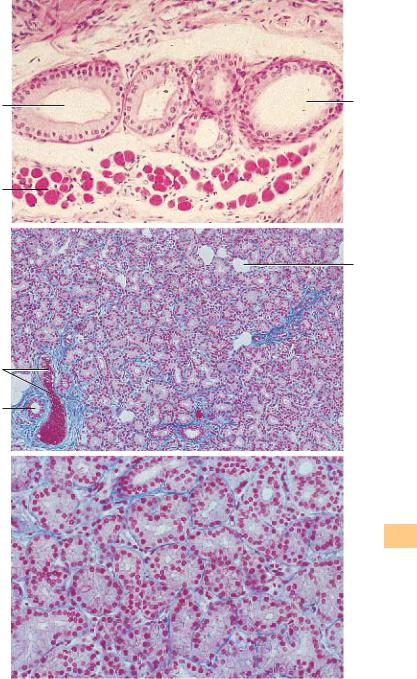
630 Lacrimal Gland
The lacrimal gland has approximately the shape of an almond. It consists of lobes that are intersected by connective tissue septae. The lacrimal gland is a tubular, purely serous gland without intercalated ducts. The gland tubules end in intralobular ducts 1 , which merge to larger ducts. About 8–12 ducts channel the tears to the fornix conjunctivae. In cross-sections, the acini of the lacrimal gland resemble those of the parotid glands. They show all the morphological criteria of serous glands (cf. Figs. 129, 379–381). The secretory cells often show a fine cytoplasmic granulation (see Fig. 632). The interstitial connective tissue is sometimes rich in lymphocytes and plasma cells. With increasing age, there are often also adipocytes 2 .
1 Intralobular ducts
2 Adipocytes
3 Artery
Stain: azan; magnification: × 80
Sensory Organs
631 Lacrimal Gland
The tubules of the lacrimal gland often have wide lumina. They are therefore often referred to as tubuloalveolar glands. The irregularly shaped tubules can be clearly assessed at higher magnification. Note the shape of the gland cells (secretory cells) (cf. Fig. 630). The round nuclei are in basal position. The cytoplasm appears light, and cell borders can be clearly recognized in some places. Myoepithelial cells are found between the gland epithelium and the basal membrane. Note the delicate connective tissue between the tubules (here stained blue).
The secretory product of the lacrimal glands (tears) moisturizes the cornea and the conjunctiva of the eyeball as well as the eyelids.
Stain: azan; magnification: × 200
Kuehnel, Color Atlas of Cytology, Histology, and Microscopic Anatomy © 2003 Thieme All rights reserved. Usage subject to terms and conditions of license.
1
2
3
1
1
2
Sensory Organs
Kuehnel, Color Atlas of Cytology, Histology, and Microscopic Anatomy © 2003 Thieme All rights reserved. Usage subject to terms and conditions of license.

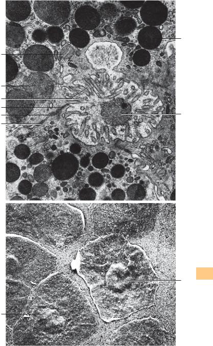
632 Lacrimal Gland
The varieties of secretory products of the exocrine and endocrine gland cells are stored in the cells as secretory granules or secretory droplets. Intracellularly stored secretory granules may display a variety of appearances. The figure shows three acinar cells of the lacrimal gland. The secretory granules appear either homogeneous 1 or show low electron microscopic densities. They contain finely dispersed secretory particles. As shown in the figure, the secretory granules are released individually in response to stimuli. Microvilli 3 protrude into the acinar lumen. Note the granule in the lumen 7 . It has already lost part of its membrane. The lateral surfaces of the secretory cells show apical junctional complexes (terminal bars) 456 .
1 Secretory granules
2 Mitochondrion
3 Microvilli
4 Zonula occludens
5 Zonula adherens
6 Desmosome
7 Secretory granule in the acinar lumen Electron microscopy; magnification: × 25 000
Sensory Organs
633 Cornea
View of the surface of the corneal epithelium (surface cells, superficial cells). The polygonal cells are flattened and about 5 μm thick, with diameters up to 50 μm. Their nuclei protrude slightly 1 . The surface cells are sloughed off continuously and replaced. The large cell in the center of the figure is about to detach. The cells of the next lower layer contain fine, dense surface plicae  , which serve the intercellular attachment. Compare the surface view of this tissue with the sections in figures 117, 634–636).
The cornea consists of five layers:
1 Epithelium
2 Lamina limitans anterior (Bowman’s membrane)
3 Stroma
4 Lamina limitans posterior (Descemet’s membrane)
5 Endothelium (see Figs. 634–636)
Scanning electron microscopy; magnification: × 2000
Kuehnel, Color Atlas of Cytology, Histology, and Microscopic Anatomy © 2003 Thieme All rights reserved. Usage subject to terms and conditions of license.
1
2
1
3
4
5 7
6
Sensory Organs
1
1
Kuehnel, Color Atlas of Cytology, Histology, and Microscopic Anatomy © 2003 Thieme All rights reserved. Usage subject to terms and conditions of license.
634 Cornea
This vertical section through the cornea provides a clear image of the layered structure. The outer covering consists of five or six layers of nonkeratinizing cells (multilayered nonkeratinizing squamous epithelium) 1 (cf. Figs. 117, 635, 636). It is about 70 μm high and is supported by a basal membrane. The limiting lamina (Bowman’s membrane) 2 follows as a relatively wide layer (see Figs. 635–637). The thick corneal stroma (substantia propria corneae) 3 features 200–250 densely stacked lamellae about 2 μm thick, with interleaved parallel oriented collagen fibrils (see Fig. 638). Fibrocytes (keratinocytes) with cytoplasmic processes (“branched fibrocytes”; see Fig. 639) are found between collagen fibrils. The corneal fibrocytes appear spindle-shaped in vertical sections (see Figs. 103, 117, 635, 636). The thinner posterior limiting lamella (Descemet’s membrane) separates the corneal stroma from the about 5 μm thick posterior single-layered corneal epithelium (corneal endothelium) (cf. Figs. 103, 104, 640).
1 Anterior corneal epithelium
2 Anterior limiting lamina, Bowman’s membrane
3 Corneal stroma, substantia propria corneae with fibrocytes (keratinocytes)
4 Posterior corneal epithelium (corneal endothelium) Stain: alum hematoxylin-eosin; magnification: × 50
Sensory Organs
635 Cornea
Multilayered nonkeratinizing squamous epithelium of the cornea. There are surface cells 1 , intermediary cells 2 and basal cells 3 . Compare with Fig. 636.
1 Surface cells
2 Intermediary cells
3 Basal cells
4 Anterior limiting lamina, Bowman’s membrane
5 Corneal stroma
6 Keratinocyte
Stain: hematoxylin-eosin; magnification: × 500
636 Cornea
This vertical section of the cornea shows the corneal epithelium, Bowman’s membrane and the corneal stroma. Note the different shapes of the basal cells 3 and compare them with those in Fig. 635. An intermediary cell often spans over two basal cells like an umbrella. The two uppermost layers consist of flattened, about 5 μm thick and up to 50 μm long flat surface cells 1 (see Fig. 633). The anterior limiting lamina (Bowman’s membrane) 4 lies under the epithelium. The lower part of the figure shows the corneal stroma 5 with long spindle-shaped fibrocytes (keratinocytes) (cf. Fig. 639).
1 Surface cells
2 Intermediary cells
3 Basal cells
4 Bowman’s membrane
5 Stroma corneae
Semi-thin section; stain: methylene blue-azure II; magnification: × 80
Kuehnel, Color Atlas of Cytology, Histology, and Microscopic Anatomy © 2003 Thieme All rights reserved. Usage subject to terms and conditions of license.
1
2
3
1
2
3
4
1
2
3
4
5
4
5
6
Sensory Organs
Kuehnel, Color Atlas of Cytology, Histology, and Microscopic Anatomy © 2003 Thieme All rights reserved. Usage subject to terms and conditions of license.

637 Cornea—Bowman’s Membrane
Bowman’s membrane (anterior limiting lamina) stretches underneath the subepithelial basal lamina. It appears homogeneous in light microscopic images and is about 8–14 μm thick. Bowman’s membrane represents a modified condensed form of the substantia propria corneae. It is free of cells and shows a weakly positive PAS reaction. With 14–17 μm, the collagen fibrils of Bowman’s membrane are thinner than the fibrils of the substantia propria. In addition to the collagen fibrils, the lamina contains a proteoglycan-rich basic substance (cf. Figs. 103, 117, 634, 636).
Electron microscopy; magnification: × 42 500
Sensory Organs
638 Cornea—Corneal Stroma
The sclera consists of a weave of tough undulating collagen fibers, which provide controlled plasticity and tensibility. In contrast, the collagen fibers of the corneal stroma (substantia propria corneae) are stretched (see Fig. 634). The about 500-μm thick corneal stroma is considered a modified connective tissue, which ascertains stability and translucency. Its fibrils contain collagen types I, III, V and VII and form parallel bundles with a cross-striation periodicity of 21 or 64 nm. The fibers form lamellae, which are layered parallel over the corneal surface. Lamellae are between 1 and 6 μm thick. There are therefore variable numbers of fibrils in each stack of lamellae. There are up to 250 lamellae. The direction of adjacent fibrils may be offset by an angle. However, the fibrils in each lamella are strictly parallel and stacked in equal distance. The basic substance predominantly consists of glycosaminoglycans. It fills the spaces between and inside the lamellae (interfibrillar substance). Fibrocytes (keratinocytes) and their branched cytoplasmic processes are embedded in the basic substance (not shown; see Figs. 636, 639). Sporadically, macrophages are found.
This figure shows a Vibratome section through the substantia propria corneae with its lamellar structure.
Scanning electron microscopy; magnification: × 700
639 Cornea
This parallel section through the corneal stroma shows the fibrocytes (keratinocytes) and their long, branched processes. The processes form a twodimensional fibrocyte web, which is layered between the collagen lamellae (see Fig. 638). The applied impregnation technique does not stain the collagen lamellae.
Stain: gold chloride impregnation; magnification: × 200
Kuehnel, Color Atlas of Cytology, Histology, and Microscopic Anatomy © 2003 Thieme All rights reserved. Usage subject to terms and conditions of license.
Sensory Organs
Kuehnel, Color Atlas of Cytology, Histology, and Microscopic Anatomy © 2003 Thieme All rights reserved. Usage subject to terms and conditions of license.

640 Cornea
Vertical section through the cornea. It shows the stroma corneae (substantia propriae corneae) 1 with spindle-shaped keratinocytes, Descemet’s membrane (posterior limiting lamina) 2 and posterior corneal epithelium 3 (cf. Figs. 103, 636, 639). Descemet’s membrane (basal membrane) is about 10 μm thick and shows a positive PAS reaction.
1 Corneal stroma with keratinocytes
2 Descemet’s membrane, posterior limiting lamina
3 Posterior corneal epithelium (corneal endothelium)
Semi-thin section; stain: methylene blue-azure II; magnification: × 800
641 Iris
Section of the iris (detail enlargement) with pigmented epithelium 1 , iris dilator 2 and pupillary sphincter (sphincter pupillae) 3 . The next layer is the stroma of the iris (stroma iridis) 4 . The border between iris and anterior chamber of the eye consists of a discontinuous mesothelium 6 .
1 Pigmented epithelium
2 Iris dilator
3 Pupillary sphincter
4 Stroma of the iris
5 Vessels
6 Mesothelium of the anterior face of the iris
7 Crypts of Fuchs
8 Anterior chamber of the eye
Stain: alum hematoxylin-eosin; magnification: × 50
Sensory Organs
642 Iris and Lens
Section of the pupillary zone of the iris and the adjacent lens 6 (detail enlargement). The stroma of the iris 4 consists of collagen fiber bundles.
1 Pigmented epithelium |
5 |
Ectropion |
|
2 |
Pupillary sphincter |
6 |
Lens |
3 |
Iris dilator |
7 |
Anterior face of the lens |
4 |
Stroma of the iris |
|
|
Stain: alum hematoxylin-eosin; magnification: × 50
643 Eye and Lens
The lens (see Fig. 624) consists of lens capsule (capsula lentis), the lens epithelium (epithelium lentis), subcapsular epithelium and the lens fibers (fibrae lentis ). The lens fibers arise from the lens epithelium (see textbooks of embryology). Lens fibers are thin cells, 7–10 μm long and about 2-μm thick, containing crystallin. The cells lose their nuclei during development. They form long bands that appear hexagonal in cross-sections. The bands consist of concentric layers of radial lamellae. The lamellae are connected via focal desmosomes (junctional complexes). Interdigitations are present at the corner structures of the hexagonal lamellae. Note the focal desmosomes between intercellular processes. There are about 2300 lamellae in the adult human.
Scanning electron microscopy; magnification: × 4000
Kuehnel, Color Atlas of Cytology, Histology, and Microscopic Anatomy © 2003 Thieme All rights reserved. Usage subject to terms and conditions of license.

1
2
3
1
2
4
5
3
7
5
6
4
8
1
2
4
3
7
5
6
Sensory Organs
Kuehnel, Color Atlas of Cytology, Histology, and Microscopic Anatomy © 2003 Thieme All rights reserved. Usage subject to terms and conditions of license.
644 Angle of the Eye—Iridocorneal Angle
The upper part of the figure shows the substantiae propriae sclerae 1 and the cornea 2 . The slits in the sclera represent the canal of Schlemm (sinus venosus sclerae). The ciliary body with the ciliary muscle 6 forms lamellashaped ciliary processes with a thin pars plana and a pars plicata. The processes are covered by a two-layered epithelium, which is thought to produce
the intraocular fluid. The pars plicata 8 |
features about 70 ciliary processes |
||
(see Fig. 645). |
|
|
|
1 |
Substantia propria sclerae |
5 |
Iris |
2 |
Substantia propria corneae |
6 |
Ciliary muscle |
3 |
Angle of the eye (Schwalbe’s line) |
7 |
Pigmented epithelium of the iris |
4 |
Anterior chamber of the eye |
8 |
Ciliary processus |
Stain: hematoxylin-eosin; magnification: × 40
645 Ciliary Processes and Zonula Filaments
The functions of the ciliary body are accommodation and the release of ocular fluid. Both parts of the ciliary body (see Fig. 644) are covered by a twolayered epithelium. The suspensor filaments for the lens (zonula filaments, zonula lentis, Zinn zonule) 2 extend from the surface of the ciliary processus 1 to the lens capsule. This establishes the connection between lens and ciliary muscle. There are thick suspensor fibers and thinner tensile fibers. The latter are not anchored in the ciliary epithelium but span the distance between ciliary fold and basal membrane of the nonpigmented epithelium. The thinner fibers may be considered part of the basal membrane.
The figure shows the attachment of the zonula filaments to the walls of the ciliary processus.
1 Ciliary processus
2 Lens zonula, zonula filaments
Scanning electron microscopy; magnification: × 660
Sensory Organs
646 Angle of the Eye—Corneoscleral Trabeculae
The anterior chamber of the eye is limited in the front by the cornea and the peripheral sclera, in the back by the anterior iris and the pupillary part of the lens. The border between anterior and posterior chamber of the eye is the angle of the eye or Schwalbe’s line (angulus iridocornealis) (see Fig. 644). At that line, the corneal epithelium continues in the epithelium of the conjunctiva and the lamellae of the corneal stroma interweave with the connective tissue of the sclera. From the corneal epithelium and Descemet’s membrane arise the corneoscleral trabeculae, which are a loosely arranged sponge-like connective tissue. The ocular fluid flows through the meshwork of this sponge-like tissue to the canal of Schlemm . Endothelial cells cover the trabecular surfaces. The intertrabecular space is the space of Fontana. It is the connection between Schwalbe’s line and the canal of Schlemm. This figure conveys an impression of the trabecular structure. The structure cannot be obtained in this form using light microscopy (cf. Fig. 644).
Scanning electron microscopy; magnification: × 800
Kuehnel, Color Atlas of Cytology, Histology, and Microscopic Anatomy © 2003 Thieme All rights reserved. Usage subject to terms and conditions of license.
1
3
6
8
1
1
 2 4
2 4
5
7
8
2
2
1
Sensory Organs
Kuehnel, Color Atlas of Cytology, Histology, and Microscopic Anatomy © 2003 Thieme All rights reserved. Usage subject to terms and conditions of license.

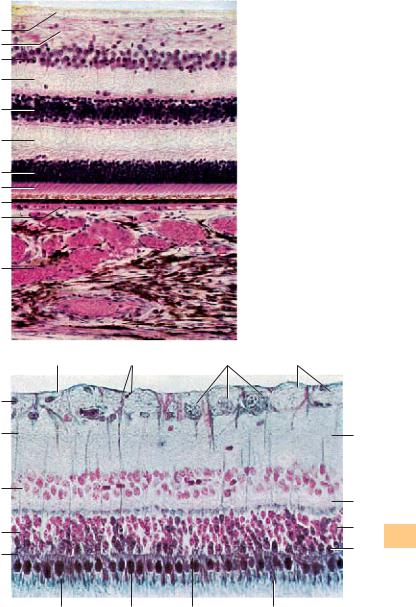
647 Retina and Choroid
Vertical section through the optic part of the retina and the choroidea. Note: the iris, the choroidea and the ciliary body appear combined as uvea. The choroid is the posterior part of the uvea.
1 Stratum limitans internum (internal limiting lamina)
2 Stratum neurofibrarum, optic nerve fiber layer with anchoring parts of Müller cells
3Stratum ganglionicum nervi optici (3rd neuron), the ganglion cell layer is single-layered, the multilayered appearance is an artifact due to the thickness of the section
4Stratum plexiforme internum, inner plexiform layer (IPL) (only lightly stained). The synapses between bipolar and multipolar ganglia cells of the ganglion cell layer are located here
5 Stratum nucleare internum, inner nuclear layer (INL) (2nd neuron) The blue-violet stained nuclei are part of bipolar ganglia cells (amacrine cells)
6 Stratum plexiform externum, outer plexiforme layer (OPL) (lightly stained). Synapses between receptor cells and bipolar ganglia cells are formed in this layer
7 Stratum neuroepitheliale, rods and cones, outer nuclear layer (1st neuron) 8 Inner and outer segments of rods and cones
9 Stratum pigmentosum retinae, pigment epithelium
10Lamina choroidocapillaris, choroidal capillary layer; its capillaries attach to the lower basal membrane on the pigmented epithelium
11The lower third of the figure represents the choroidea with the vascular layer and filled veins as well as pigmented epithelial cells (cf. Fig. 648)
Stain: alum hematoxylin-eosin; magnification: × 65
Sensory Organs
648 Retina
This microphotograph of a retina displays the separate layers particularly well (cf. Fig. 647).
1 External limiting lamina, (junctional complexes)
2 Rods and cones layer
3 Inner nuclear layer
4 Müller cells
5 Nucleus of a neuroglial cell
6 Internal limiting lamina
7 Anchor portion of a Müller cell
8 Ganglia cells of the optic nerve (3rd neuron)
9 Fiber bundle of the optic nerve
10Inner plexiform layer (IPL)
11Outer plexiform layer (OPL)
12Nucleus of a rod cell
13Nucleus of a cone cell
14Outer segment of a rod cell
15Inner segment of a rod cell
16Inner segment of a cone cell
17Outer segment of a cone cell
Stain: nuclear fast red-eosin-nigrosin (steel gray); magnification: × 320
Kuehnel, Color Atlas of Cytology, Histology, and Microscopic Anatomy © 2003 Thieme All rights reserved. Usage subject to terms and conditions of license.
1
2
3
4
5
6
7
8
9 
10 
11
|
6 |
7 |
8 |
9 |
5
4
3
2
1
17 |
16 |
15 |
14 |
10 |
Organs |
|
|
11 |
Sensory |
|
12
13
Kuehnel, Color Atlas of Cytology, Histology, and Microscopic Anatomy © 2003 Thieme All rights reserved. Usage subject to terms and conditions of license.

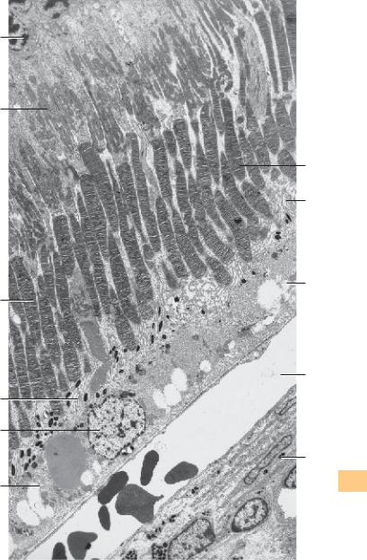
649 Retina
Incomplete section of a human retina. It shows the following layers:
1 External nuclear layer. This layer contains the nuclei of the rods and cones
2Inner segments of the rods and cones with a distal acidophilic part (ellipsoid) and a proximal basophilic part (myoid). The distal ellipsoid part is filled with mitochondria, the proximal myoid part contains smooth endoplasmic reticulum membranes, Golgi complexes, and free ribosomes
3Outer segments of the rods and cones. Their structures are in principal similar to the structure of the inner segments. Outer and inner segments of the rods are approximately of equal length. The cylindrical outer segments contain stacks of 600—1000 flat disks, which look like rolls of coins. They are enveloped by a plasma membrane. The outer segments of the cones are cone-shaped and shorter than the outer segments of rods. They are often described as flask-shaped. The membrane stacks of cones consist of equally spaced involutions of the plasma membrane, not of separate disks. The outer third of the
outer segments of the cones is surrounded by the microvilli of the pigmented epithelium
7
4Pigment epithelium. The single-layered cuboidal epithelium is interwoven with the choroidea. Microvilli protrude from the apical portion of the epithelium. They extend far into outer segments of the rods and cones of the bacillary layer. The basal membrane borders on Bruch’s membrane and shows many folds. Pigmented epithelial cells contain large round nuclei 8 . The apical cell region contains many melanosomes and phagosomes
5Choroidal capillary layer (lamina choroidocapillaris). This layer is found close to the pigment epithelium. It consists of a tight vascular network, which contains capillary lobes. Only one capillary is sectioned longitudinally in this figure. The capillaries contain ery-
throcytes
6Lamina vasculosa. The lower right corner of the figure shows an arteriole. There are three layers in the choroidea: the outer layer close to the sclera (Haller’s layer), the lamina vasculosa and the lamina choroidocapillaris close to the pigment epithelium. Compare with Figs. 647 and 648
Electron microscopy; magnification: × 3500
Sensory Organs
Kuehnel, Color Atlas of Cytology, Histology, and Microscopic Anatomy © 2003 Thieme All rights reserved. Usage subject to terms and conditions of license.
1
2
3
7
8
4
3
7
4
5
6
Sensory Organs
Kuehnel, Color Atlas of Cytology, Histology, and Microscopic Anatomy © 2003 Thieme All rights reserved. Usage subject to terms and conditions of license.

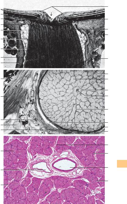
650 Optic Nerve—Papilla of the Optic Nerve
Exit point of the optic nerve, longitudinal section. The nerve fibers continue as optic nerve at the papilla of the optic nerve (blind spot).
1 External sheath of the optic nerve (dura mater)
2 Subdural space
3 Internal sheath of the optic nerve (pia mater)
4 Arachnoidea
5 Subarachnoidal space
6 Sclera
7 Choroidea
8 Retina
9 Lamina cribrosa sclera (perforated plate of collagen fibrils)
10Papilla of the optic nerve
11Unmyelinated nerve fiber bundle before pushing through the lamina cribrosa
12Myelinated nerve fiber bundle of the optic nerve behind the lamina cribrosa
13Lamina episcleralis
14Ciliary nerve
At the eyeball, the dura mater continues in the cornea; the arachnoidea and pia mater continue in the choroid.
Stain: alum hematoxylin-eosin; magnification: × 6
651 Optic Nerve
Section of the optic nerve behind the lamina cribrosa sclerae (see Fig. 653).
Sensory Organs
1 Ciliary nerve
2 Sclera
3 Pigment epithelium of the lamina suprachoroidea
4 Fiber bundles of the optic nerve
5 Central retinal artery
6 Septa of the pia mater
7 Central retinal vein
8 Internal sheath of the optic nerve (pia mater)
9 External sheath of the optic nerve (dura mater)
10Subdural space
11Arachnoidea
12Short posterior ciliary artery
Stain: hematoxylin-eosin; magnification: × 20
652 Optic Nerve
Cross-section of the optic nerve behind the lamina cribrosa sclerae. The axons of the ganglia cells are combined in bundles 1 . They are enveloped by a thin septum of the pia mater 2 . The optic nerve contains a pia mater sheath, an arachnoidea sheath and a dura mater sheath (see Figs. 650, 651). The pia mater sheath attaches directly to the optic nerve. Septa originate with the pia mater and guide blood vessels to the myelinated nerve fibers. The nerve fiber bundles contain astrocytes and oligodendrocytes. Arteries 3 and central retinal veins 4 are visible in the center of the section. The vessels are sheathed by the loose connective tissue of the pia mater.
1 Nerve fiber bundles
2 Pia mater septa
3 Central retinal artery
4 Central retinal vein
Stain: hematoxylin-eosin; magnification: × 40
Kuehnel, Color Atlas of Cytology, Histology, and Microscopic Anatomy © 2003 Thieme All rights reserved. Usage subject to terms and conditions of license.
8
7
6
9
5
4
3
2
1
3
2
12
1
11
1
4
2
10
11
12
13
14 |
|
|
6 |
|
|
4 |
|
|
7 |
|
|
5 |
|
|
6 |
|
|
10 |
|
|
8 |
|
|
9 |
Organs |
|
2 |
||
Sensory |
||
1 |
3
1
Kuehnel, Color Atlas of Cytology, Histology, and Microscopic Anatomy © 2003 Thieme All rights reserved. Usage subject to terms and conditions of license.

653 Optic Nerve—Lamina Cribrosa Sclerae
The optic nerve, a longitudinal fascicle, has an intraocular, orbital, intracanalicular and an intracranial segment. The about 2-mm long pars intraocularis corresponds to the papilla of the optic nerve (cf. Fig. 650). The nerve fibers in this intraocular segment are unmyelinated. Examination with the ophthalmoscope allows a view of the sieve-like, loosely structured lamina cribrosa sclerae, which shines through the unmyelinated nerve fibers at the point where the optic nerve traverses the lamina cribrosa. The lamina cribrosa is a continuation of the sclera. The axons of the optic nerve are myelinated only after traversing the lamina cribrosa.
This figure shows the lamina cribrosa sclerae. The viewer looks on the circular arrangements of collagen fibers around each traversing axon. The collagen fibers have been isolated by maceration with a 10% NaOH solution, which disintegrates the axons of the optic nerve. The center represents the space for the artery and the central retinal vein. The collagen fibers around the center are called scleral ring.
Lamina cribrosa sclerae of an 89-year-old woman.
Scanning electron microscopy; magnification: × 60
Sensory Organs
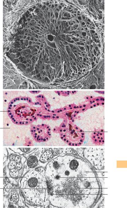
654 Choroid Plexus
The choroid plexus consists of a richly vascularized layer of leptomeninges (pia mater and arachnoidea), which is covered by a single-layered cuboidal epithelium 1 . The plexus structure is tree-like. In the ciliated connective tissue occur fibroblasts as well as plasma cells, mast cells and macrophages. This preparation contains many vessels with erythrocytes 2 . The cuboidal epithelium of the choroid plexus stains heavily with Eosin. Note the large, round central nuclei.
1 Epithelium of the choroid plexus
2 Capillaries with erythrocytes
3 Cerebrospinal fluid (CSF) with cells
Stain: alum hematoxylin-eosin; magnification: × 300
655 Axodendritic Synapse
This axodendritic synapse consists of the presynaptic membrane, the synaptic gap and the postsynaptic membrane 3 . The axolemma region appears denser in the area of the presynaptic membrane. The synaptic bouton of the axon 1 contains several synaptic vesicles 2 with diameters between 20 and 65 nm as well as mitochondria and a few small dense vesicles (dense bodies) 4 . The synaptic vesicles contain a transmitter (chemical synapse).
1 Synaptic bouton of an axon
2 Synaptic vesicle
3 Synaptic membrane complex
4 Dense granule
5 Dendrite
Electron microscopy; magnification: × 35 800
Kuehnel, Color Atlas of Cytology, Histology, and Microscopic Anatomy © 2003 Thieme All rights reserved. Usage subject to terms and conditions of license.
1
3
5
3
 2
2
2 OrgansSensory
1
4
2
4
Kuehnel, Color Atlas of Cytology, Histology, and Microscopic Anatomy © 2003 Thieme All rights reserved. Usage subject to terms and conditions of license.
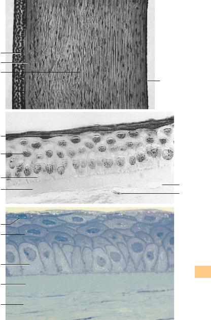
656 Inner Ear—Cochlea
Longitudinal section of a human cochlea.
1 Scala tympani (contains perilymph)
2 Cochlear duct (contains endolymph), triangular in cross-sections 3 Scala vestibuli (contains perilymph)
4 Osseous spiral lamina
5 Helicotrema (scala vestibuli and scala tympani join)
6 Cecum cupulare (copular blind sac, caecum cupulare)
7 Modiolus (end of the osseous spiral lamina)
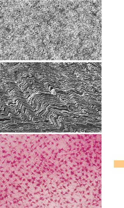
8 Cochlear cupola (azimuth of the apical cochlear turn)
9Longitudinal canals of the modioli (canales longitudinales modioli), central inner canals of the cochlea
10Spiral canal of the modioli (canalis spiralis modioli), canal in the outer wall of the osseous spiral lamina
11Facial nerve in the bony facial canal
12Vestibular membrane (Reissner’s membrane, membrana vestibularis, upper wall of the cochlea)
13Spiral crest, spiral ligament (ligamentum spirale cochlea)
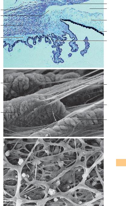
14Basilar membrane (connective tissue between cochlear duct and scala tympani)
15Osseous spiral lamina
16Area of the facial nerve of the internal acoustic meatus (fundus)
17Cochlear ganglion (cochlear nerve)
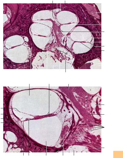
18Transverse crest
19Base of the modiolus (base of the cochlea)
20Cochlear nerve
21Internal acoustic meatus (fundus)
22Spiral canal of the modioli (see 10)
Stain: hematoxylin-eosin; magnification: × 10
Sensory Organs
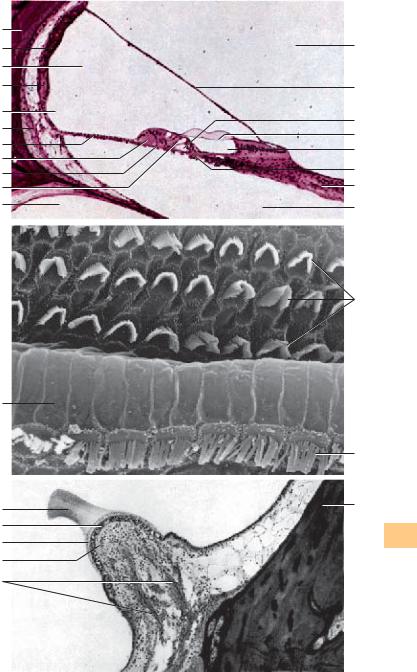
657 Cochlea—Inner Ear
Middle bend of the cochlea.
1 Cochlear duct (contains endolymph)
2 Spiral ligament, spiral crest
3 Stria vascularis, vascularized tissue over the spiral prominence
4 Reissner’s membrane, vestibular membrane
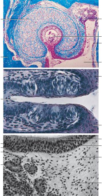
5 Scala vestibuli
6 Osseous spiral lamina
7 Cochlear ganglion
8 Spiral canal of the modioli (bony canal in the outer wall of the osseous spiral lamina) 9 Longitudinal canal of the modioli (central inner canal)
10Tractus spiralis foraminosus, a few spirally arranged openings around the inner cochlear canal, which permit access of cochlear nerve ganglia to the cochlea
11Cochlear nerve (branch of the vestibulocochlear nerve for the cochlear audiosensory organ)
12Internal acoustic meatus (fundus)
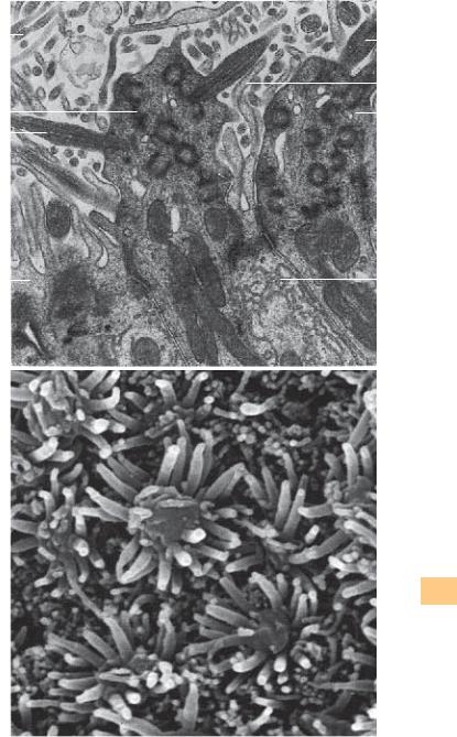
13Scala tympani (contains perilymph)
14Organ of Corti
15Basilar membrane (connective tissue layer between cochlear duct and scala tympani) Stain: hematoxylin-eosin; magnification: × 25
Kuehnel, Color Atlas of Cytology, Histology, and Microscopic Anatomy © 2003 Thieme All rights reserved. Usage subject to terms and conditions of license.
|
4 |
|
|
|
5 |
|
6 7 |
8 |
|
|
|
|
|
|
|
|
|
|||||||
|
|
|
|
|
|
|
|
|
|
|
|
|
|
|
|
|
|
|
|
|
|
|
|
11 |
|
|
|
|
|
|
|
|
|
|
|
|
|
|
|
|
|
|
|
|
|
|
|
|
|
|
|
|
|
|
|
|
|
|
|
|
|
|
|
|
|
|
|
|
|
|
|
|
|
|
|
|
|
|
|
|
|
|
|
|
|
|
|
|
|
|
|
|
|
|
|
|
|
|
|
|
|
|
|
|
|
|
|
|
|
|
|
|
|
|
|
|
|
|
|
|||||
|
|
|
|
|
|
|
|
|
|
|
|
|
|
|
|
|
|
|
10 |
|||||
|
|
|
|
|
|
|
|
|
|
|
|
|
|
|
|
|
|
|
||||||
|
|
|
|
|
|
|
|
|
|
|
|
|
|
|
|
|
|
9 |
||||||
3 |
|
|
|
|
|
|
|
|
|
|
|
|
|
|
|
|
|
|
|
|
|
|
12 |
|
|
|
|
|
|
|
|
|
|
|
|
|
|
|
|
|
|
|
|
|
|||||
|
|
|
|
|
|
|
|
|
|
|
|
|
|
|
|
|
|
|
|
|
13 |
|||
|
|
|
|
|
|
|
|
|
|
|
|
|
|
|
|
|
|
|
||||||
2 |
|
|
|
|
|
|
|
|
|
|
|
|
|
|
|
|
|
14 |
||||||
|
|
|
|
|
|
|
|
|
|
|
|
|
|
|
|
|
||||||||
|
|
|
|
|
|
|
|
|
|
|
|
|
|
|
|
|
||||||||
1 |
|
|
|
|
|
|
|
|
|
|
|
|
|
|
|
|
|
15 |
||||||
|
|
|
|
|
|
|
|
|
|
|
|
|
|
|
|
|
||||||||
|
|
|
|
|
|
|
|
|
|
|
|
|
|
|
|
|
|
|
|
|
|
|
16 |
|
|
|
|
|
|
|
|
|
|
|
|
|
|
|
|
|
|
|
|
|
|
|
|
||
|
|
|
|
|
|
|
|
|
|
|
|
|
|
|
|
|
|
|
|
|
|
|
||
|
|
|
|
|
|
|
|
|
|
|
|
|
|
|
|
|
|
|
|
|
||||
|
|
|
|
|
|
|
|
|
|
|
|
|
|
|
|
|
|
|
||||||
|
|
|
|
|
|
|
|
|
|
|
|
|
|
|
|
|
|
|
|
|
|
|||
|
|
|
|
|
|
|
|
|
|
|
|
|
|
|
|
|
|
|
|
|
|
|
|
|
|
|
|
|
|
|
|
|
|
|
|
|
|
|
|
|
|
|
|
|
|
|
|
|
|
|
22 |
17 |
21 |
20 |
19 |
17 |
18 |
|
|
|
|
|
||||||||||||
|
5 |
|
6 |
|
|
|
|
|
|
|
7 |
|
|
|
|
|
|
|
|
|||||
8
4
3
2
1
9
10 |
Organs |
|
|
11 |
Sensory |
|
15 |
14 |
13 |
7 |
12 |
Kuehnel, Color Atlas of Cytology, Histology, and Microscopic Anatomy © 2003 Thieme All rights reserved. Usage subject to terms and conditions of license.

658 Cochlea—Inner Ear
Section of the cochlear duct from the apical turn of the cochlea with the organ of Corti (organum spirale).
1 Spiral ligament, spiral crest
2 External spiral groove of the cochlear duct
3 Spiral prominence (border of the external spiral groove)
4 Stria vascularis
5 Bony wall of the cochlea
6 Cochlear duct
7 Vestibular membrane, Reissner’s membrane
8 Scala vestibuli
9 Cochlear nerve
10Scala tympani
11Tectorial membrane (located over the organ of Corti and the inner cochlear canal)
12Inner cochlear canal (canalis spiralis cochleae)
13Inner hair cells
14Inner tunnel, contains endolymph
15Outer hair cells
16Cells of Henson
17Cells of Claudius
18Basilar membrane
Stain: hematoxylin-eosin; magnification: × 80
Sensory Organs
659 Cochlea—Organ of Corti
The organ of Corti (organum spirale, papilla spiralis) rests on the basilar membrane. It represents the part of the cochlear duct, which has differentiated into a sensory epithelium. There are two groups of cells with different functions in this epithelial layer. 1. The sensory cells or hair cells. The fibers of the acoustic nerve end at the hair cells. 2. The phalangeal support cells 3 . Both cell types form rows. A surface view confirms the cell arrangement in rows. This figure shows the singular rows of inner sensory cells (hair cells) 1 and the triple or quadruple rows of the outer hair cells 2 . The hair consists of stereocilia (cochlear stereocilia). Inner and outer hair cells show numerous differences in their morphological attributes, and their functions are different. Both inner and outer hair are directed toward the endolymph.
1 Inner hair cells
2 Outer hair cells
3 Phalangeal cells
Scanning electron microscopy; magnification: × 2000
660 Osseous Semicircular Canals—Ampullary Crest
The ampullary crest consists of sensory organs, which extend into the ampullary spaces of the osseous semicircular canals. The laminae of the sensory organs are capped by the gelatinous cupula ampullaris 2 . A fiber bundle of the vestibular part of the ampullary nerve 3 is visible in the center of the figure. The bony capsule of the ampulla membranaceae 4 is shown on the right.
1 Sensory epithelium |
4 Bony capsule |
||
2 |
Cupula ampullaris, ampullary dome |
5 |
Sensory hair |
3 |
Ampullary nerve |
6 |
Lamina propria |
Stain: hematoxylin-eosin; magnification: × 25
Kuehnel, Color Atlas of Cytology, Histology, and Microscopic Anatomy © 2003 Thieme All rights reserved. Usage subject to terms and conditions of license.
5
4
6
3
2
1
18
17
16
15
8
3
2
1
5
6 
3
8
7
14
11
12
13
9
10
2
1
4
Sensory Organs
Kuehnel, Color Atlas of Cytology, Histology, and Microscopic Anatomy © 2003 Thieme All rights reserved. Usage subject to terms and conditions of license.
661 Eustachian Tube—Auditory Tube
The Eustachian tube 1 connects the nasal part of the pharynx and the middle ear. It is about 35–40 mm long. There are a bony part (pars ossea) 2 and a cartilaginous part (pars fibrocartilaginea ) 3 with different wall components. In the medium part of the cochlea, the human hyaline tube cartilage has the form of a hook. The lamina membranacea (basal plate) 4 covers the tube opening which has an oval shape in this segment. The part of the tube that is closed by the hook-shaped tube cartilage (stapes) and the basal plate takes the role of a pressure compensating tube 5 . The epithelium of the mucous membrane is a multilayered ciliated epithelium with goblet cells. The lamina propria 6 of the tubular mucous membrane contains seromucous glands. It also contains lymphocytes. Cross-section of the pars fibrocartilaginea tubae of the guinea pig.
1 Eustachian tube
2 Bone
3 Stapes, hyaline tube cartilage (ossicle)
4 Basal plate (closes the oval window), lamina membranacea
5Pressure compensation tube with parietal multilayered ciliated epithelium, which contains goblet cells
6 Lamina propria with lymphocytes
Stain: azan; preparation; magnification: × 10
Sensory Organs
662 Taste Buds
Sections of two neighboring foliate papillae of the rabbit tongue. There are six taste buds in the walls. They consist of sensory cells with taste receptors, support cells and basal cells. Taste buds are intraepithelial organs. Cross-sec- tions display their oval shape. Their circumference tapers off at the lamina propria and is even smaller at the epithelial surface. Each taste bud features 40–70 long, longitudinally oriented cells, which are straight in the center, but slightly arcuate, following the overall shape of the taste bud at the ends. The cells have the layered appearance of an onion. The sensory cells reach to the gustatory pores at the epithelial surface. A short duct ends at the epithelial surface. Routine staining leaves the cytoplasm of the sensory cells light.
1 Groove
2 Multilayered nonkeratinizing squamous epithelium Stain: Heidenhain iron hematoxylin; magnification: × 400
663 Olfactory Epithelium—Olfactory Region
The epithelium of the olfactory mucous lamina is multilayered and contains specific sensory cells 1 , support cells 2 and basal cells 3 . The cones of the bipolar olfactory cells 4 are visible in some areas of the epithelial surface. Olfactory glands 5 are present in the vascularized innervated lamina propria. The serous glands consist of winding tubes. They release mucus to dissolve and remove olfactory materials. Compare with Figs. 664 and 665.
1 Sensory cells |
5 |
Olfactory glands |
|
2 |
Support cells |
6 |
Glimpse of junctional complexes (terminal bars) |
3 |
Basal cells |
7 |
Plasma cell |
4 |
Conical olfactory cells |
8 |
Capillary |
Stain: alum hematoxylin-eosin; magnification: × 100
Kuehnel, Color Atlas of Cytology, Histology, and Microscopic Anatomy © 2003 Thieme All rights reserved. Usage subject to terms and conditions of license.
3
2
1
2
1
6
5
6
5
4
1
2
2 |
Organs |
|
|
||
4 |
Sensory |
|
3 |
||
|
||
7 |
|
|
|
8
Kuehnel, Color Atlas of Cytology, Histology, and Microscopic Anatomy © 2003 Thieme All rights reserved. Usage subject to terms and conditions of license.

664 Olfactory Epithelium—Olfactory Region
The multilayered columnar epithelium of the olfactory mucous lamina consists of basal cells, support cells and sensory olfactory cells. The nuclei of these cells occupy different levels (cf. Fig. 663). The support cells are the most numerous cells. Basal, support and sensory cells originate on the basal lamina, forming a pseudostratified epithelium. This figure shows the apical regions of two support cells and two sensory cells. The support cells are usually wider at their apical surfaces than at their bases. Long microvilli 4 protrude from the free surfaces of support cells. They contain many organelles, a large Golgi apparatus, extended smooth ER and secretory granules. The olfactory sensory cells are bipolar neurons. Their apex is wider than their basal part. Their olfactory bulbs extend over the epithelial surface. Six to eight long cilia 3 protrude from the olfactory cells, which contain the olfactory receptors. The base of each cilia shows the typical 9 × 2 + 2 microtubule organization. The microtubule continues in a thin nontubular process. The cilia protrude into a mucous film. Olfactory and support cells are connected via a network of terminal bars (tight junctions). Note the abundance of mitochondria in the sensory cells. The basal processes of the sensory cells are axons, which run to the base of the epithelium and traverse the basal membrane. Apposed plasmalemma Schwann cell processes envelop the axons only after they have traversed the basal lamina. The olfactory bulbs with their cilia make up the apical portion of the dendritic processes of these sensory cells.
1 Support cells with microvilli
2 Olfactory bulb with cilia
3 Kinocilia
4 Microvilli
Electron microscopy; magnification: × 24 000
Sensory Organs
665 Olfactory Epithelium—Olfactory Region
View of an area of the olfactory mucous lamina (cf. Fig. 664). The olfactory bulbs extend over the epithelial surface. Long cilia protrude from the olfactory bulbs. The sensory cells are interspersed with support cells with microvilli. The olfactory bulbs are about 4 μm high.
Scanning electron microscopy; magnification: × 17 800
Kuehnel, Color Atlas of Cytology, Histology, and Microscopic Anatomy © 2003 Thieme All rights reserved. Usage subject to terms and conditions of license.
|
|
|
|
|
||||
4 |
|
|
|
|
|
|
|
3 |
|
|
|
|
|
|
|
||
|
|
|
|
|
|
|
||
2 |
|
|
|
|
|
|
|
4 |
|
|
|
|
|
|
|
||
|
|
|
|
|
|
|
2 |
|
|
|
|
|
|
|
|||
3 |
|
|
|
|
|
|
|
|
|
|
|
|
|
|
|
|
|
1 |
|
|
|
1 |
|
|
Sensory Organs
Kuehnel, Color Atlas of Cytology, Histology, and Microscopic Anatomy © 2003 Thieme All rights reserved. Usage subject to terms and conditions of license.