

100_Cases_in_Clinical_Medicine
.pdfFigure 45.1 Ovarian ultrasound.
KEY POINTS
•True hirsutism is due to excessive androgens, whereas constitutional hirsutism is found in certain ethnic groups.
•The association of menstrual irregularities and obesity is suggestive of true hirsutism and polycystic ovary syndrome.
•In transplant patients, the anti-rejection drug ciclosporin causes marked hirsutism.
119
This page intentionally left blank

CASE 46: NAUSEA AND VERTIGO
History
A 17-year-old woman is admitted to the emergency department complaining of severe vertigo. This has developed over the past few hours and previously she was well. She has the sensation of her surroundings spinning around her. She feels nauseated and sleepy. She does not have a headache. She has not had any previous medical illnesses. She is a non-smoker, and says that she does not drink alcohol or take recreational drugs and she is taking no regular medication. She lives with her parents and is due to sit her A-levels in 3 weeks. Her father suffers from epilepsy and her mother has hypothyroidism.
Examination
She is drowsy and her speech is slurred. Her pulse rate is 64/min, blood pressure 90/70 mmHg and respiratory rate 12/min. Examination of her cardiovascular, respiratory and abdominal systems is otherwise normal. Her peripheral nervous system examination is normal apart from impaired co-ordination and a staggering gait. Funduscopy is normal. Her pupils are equal and reacting. There is a normal range of eye movements but she has multidirectional nystagmus. Her hearing is normal as is the rest of her cranial nerve examination.
Questions
•What is the diagnosis?
•What are the major differential diagnoses of vertigo?
•How would you manage this patient?
121

ANSWER 46
The acute onset of these symptoms and signs with drowsiness in a 17-year-old girl raise the possibility of a drug overdose. Her father is epileptic and is likely to be taking anticonvulsants. The most likely explanation is that this patient has taken a phenytoin overdose, tablets which her father uses to control his epilepsy. She has taken an overdose as a result of concern about her imminent exams. Excessive ingestion of barbiturates, alcohol and phenytoin all cause acute neurotoxicity manifested by vertigo, dysarthria, ataxia and nystagmus. In severe cases coma, respiratory depression and hypotension occur.
Vertigo is an awareness of disordered orientation of the body in space and takes the form of a sensation of rotation of the body or its surroundings.
! Causes of vertigo
Peripheral lesions |
Central lesions |
Benign positional vertigo |
Brainstem ischaemia |
Vestibular neuronitis |
Posterior fossa tumours |
Ménière’s disease |
Multiple sclerosis |
Middle-ear diseases |
Alcohol/drugs |
Aminoglycoside toxicity |
Migraine, epilepsy |
The duration of attacks is helpful in distinguishing some of these causes of vertigo. Benign positional vertigo lasts less than 1 min. Attacks of Ménière’s disease are recurrent and last up to 24 h. Vestibular neuronitis does not recur but lasts several days, whereas vertigo due to ototoxic drugs is usually permanent. Brainstem ischaemic attacks occur in patients with evidence of diffuse vascular disease, and long tract signs may be present. Multiple sclerosis may initially present with an acute attack of vertigo that lasts for 2–3 weeks. Posterior fossa tumours usually have symptoms and signs of space-occupying lesions. Acoustic neuromas often present with vertigo and deafness. Migrainous attacks are often accompanied by nausea and vomiting. Temporal lobe epilepsy may also produce rotational vertigo, often associated with auditory and visual hallucinations. Central lesions produce nystagmus which is multidirectional and may be vertical. Peripheral lesions induce a unilateral horizontal nystagmus.
The diagnosis in this case can be made by measuring plasma phenytoin levels and by asking the patient’s father to check if his tablets are missing. Gastric lavage should be carried out if it is within 12 h of ingestion of the tablets. Oral activated charcoal may be useful. National poisons information services are available to advise on treatment. Before discharge she should have counselling and treatment by adolescent psychiatrists.
KEY POINTS
•Vertigo can be caused by a variety of neurological disorders.
•A careful history and examination may reveal the cause of vertigo.
•Overdose should be considered in any patient presenting with decreased conscious level and respiratory depression.
122
CASE 47: CHEST PAIN
History
A 64-year-old woman has a 10-year history of retrosternal pain. The pain is often present in bed at night and may be precipitated by bending down. Occasionally, the pain comes on after eating and on some occasions it appears to have been precipitated by exercise. The pain has been described as having a burning and a tight quality to it. The pain is not otherwise exacerbated by respiratory movements or position.
Her husband has angina and on one occasion she took one of his glyceryl trinitrate tablets. She thinks that this probably helped her pain since it seemed to go off a little faster than usual. She has also bought some indigestion tablets from a local pharmacy and thinks that these probably helped also.
Examination
She is 1.62 m (5 ft 4 in) tall and weighs 82 kg, giving her a body mass index of 31.3 (recommended range 20–25) There are no abnormalities to find in the cardiovascular, respiratory or gastrointestinal systems.
INVESTIGATIONS
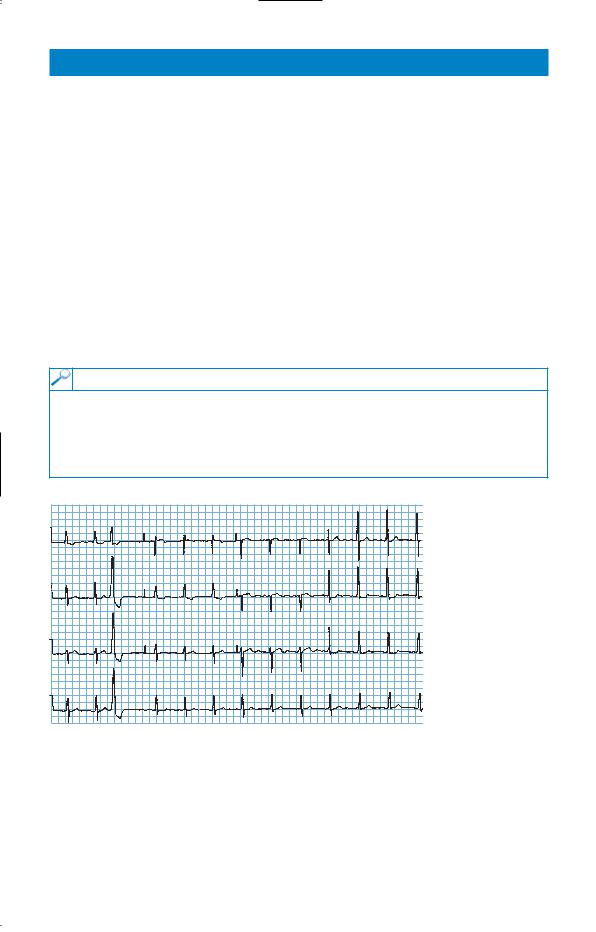
•Her chest X-ray is normal and the electrocardiogram is shown in Fig. 47.1.
•She had an exercise electrocardiogram (ECG) performed and she was able to perform 8-min exercise. Her heart rate went up to 130/min with no change in the ST segments on the ECG and normal heart and blood-pressure responses.
•The haemoglobin, renal and liver function are normal.
I |
|
V4 |
|
|
|
|
aVR |
V1 |
II |
aVL |
V5 |
|
|
V2 |
V6
aVF
V3
II
Figure 47.1 Electrocardiogram.
Questions
•What is the likely diagnosis?
•What would be appropriate management?
123

ANSWER 47
A number of features in the history make oesophageal reflux a likely diagnosis. The character and position of the pain and the relation to lying flat and to bending mean reflux is more likely. She is overweight, increasing the likelihood of reflux. The improvement with glyceryl trinitrate and with proprietary antacids is inconclusive. The ECG shows one ventricular ectopic and some T-wave changes in leads I, aVl, V5 and V6 which would be compatible with myocardial ischaemia but are not specific. The exercise ECG was negative which reduces the likelihood of ischaemic heart disease although it certainly does not rule it out. Other causes of chest pain are less likely with the length of history.
In view of the long history and the features suggesting oesophageal reflux, it would be reasonable to initiate a trial of therapy for oesophageal reflux with regular antacid therapy, H2-receptor blockers or a proton pump inhibitor (omeprazole or lansoprazole). If the pain responds to this form of therapy, then additional actions such as weight loss (she is well above ideal body weight) and raising the head of the bed at night should be added. If doubt remains, a barium swallow should show the tendency to reflux and a gastroscopy would show evidence of oesophagitis. There is a broad association between the presence of oesophageal reflux, evidence of oesophagitis at endoscopy and biopsy, and the symptoms of heart burn. However, each can occur independently of the others.
Recording of pH in the oesophagus over 24 h can provide additional useful information. It is achieved by passing a small pH-sensitive electrode into the oesophagus through the nose. This provides an objective measure of the amount of acid reaching the oesophagus and the times when this occurs.
This woman had an endoscopy which showed oesophagitis, and treatment with omeprazole and an alginate relieved her symptoms. Attempts at weight loss were not successful.
KEY POINTS
•In non-specific chest pain with a normal ECG, the oesophagus is a common source of the pain.
•24-h pH recording in the oesophagus provides further information on acid reflux.
124

CASE 48: HEADACHES
History
A 44-year-old woman presents to her general practitioner (GP) complaining of headaches. These headaches have been present in previous years but have now become more intense. She describes the headaches as severe and present on both sides of her head. They tend to worsen during the course of the day. There is no associated visual disturbance or vomiting. She also complains of loss of appetite and difficulty sleeping, with early morning waking. She has had eczema and irritable bowel syndrome diagnosed in the past but these are not giving her problems at the moment. She is divorced with two children aged 10 and 12 years, whom she looks after. She has a part-time job as an office cleaner. Her mother has recently died of a brain tumour. She smokes about 20 cigarettes per day and drinks 15 units of alcohol per week. She takes regular paracetamol or ibuprofen for her headaches.
Examination
She looks withdrawn. Her pulse is 74/min and regular, blood pressure is 118/76 mmHg. Examination of the cardiovascular, respiratory and gastrointestinal systems, breasts and reticuloendothelial system is normal. There are no abnormal neurological signs and funduscopy is normal.
Questions
•What is the diagnosis?
•What are the major differential diagnoses?
•How would you manage this patient?
125

ANSWER 48
This patient has a chronic tension headache. This is the commonest form of headache. It occurs mainly in patients under the age of 50 years. The headache is usually bilateral, often with diffuse radiation over the vertex of the skull, although it may be more localized. The pain is often characterized as a sense of pressure on the head. Visual symptoms and vomiting do not occur. The pain is often at its worst in the evening. Patients may show symptoms of depression (this woman has biological symptoms of loss of appetite and disturbed sleep pattern). Sufferers may reveal sources of stress such as bereavement or difficulty with work. There may be an element of suggestion as in this case, with concern that she may have inherited a brain tumour from her mother. She is looking after two children alone and working part-time. A normal neurological examination is important for reassurance.
!Major differential diagnoses of chronic headaches
•Classic migraine: characterized by visual symptoms followed within 30 min by the onset of severe hemicranial throbbing, headache, photophobia, nausea and vomiting lasting for several hours. The onset is usually in early adult life and a positive family history may be present.
•Cluster headaches: mainly affect men. The pain is unilateral, usually orbital and severe in nature. It characteristically occurs 1–2 h after sleeping, and lasts 1–2 h and recurs nightly for 6–8 weeks.
•Headache caused by a space-occupying lesion (such as tumour or abscess): Often the headache is initially mild but over a few weeks becomes severe and is exacerbated by coughing or sneezing. The headache is usually worse in the morning and is associated with vomiting. There will often be other signs, including personality change and focal neurological signs.
•Miscellaneous causes: sinusitis, dental disorders, cervical spondylosis, glaucoma, post-traumatic headache.
It is important to come to a clear diagnosis and to address the patient’s beliefs and concerns about the symptoms. In some circumstances it may be necessary to perform a computed tomography (CT) head scan for reassurance. The question of depression needs to be explored further and may need treating with antidepressants.
KEY POINTS
•Tension headaches occur mainly in those aged under 50, and patients often show features of depression.
•Tension headache should be diagnosed after other causes have been excluded.
126
CASE 49: HEADACHE AND CONFUSION
History
A 55-year-old man is admitted to hospital with headache and confusion. He has a cough and a temperature of 38.2°C. He does not complain of any other symptoms. Two months earlier he had been admitted with a productive cough and acid-fast bacilli had been found in the sputum on direct smear. He had lost weight and complained of occasional night sweats. He had a history of a head injury 10 years previously. He smoked 15 cigarettes a day and drank 40–60 units of alcohol each week. He was found a place in a local hostel for the homeless and sent out after 1 week in hospital on antituberculous treatment with rifampicin, isoniazid, ethambutol and pyrazinamide together with pyridoxine. His chest X-ray at the time was reported as showing probable infiltration in the right upper lobe.
Examination
He looked thin and unwell and he was slightly drowsy. His mini mental test score was 8/10. There were some crackles in the upper zones of the chest posteriorly. His respiratory rate was 22/min. There were no neurological signs.
INVESTIGATIONS
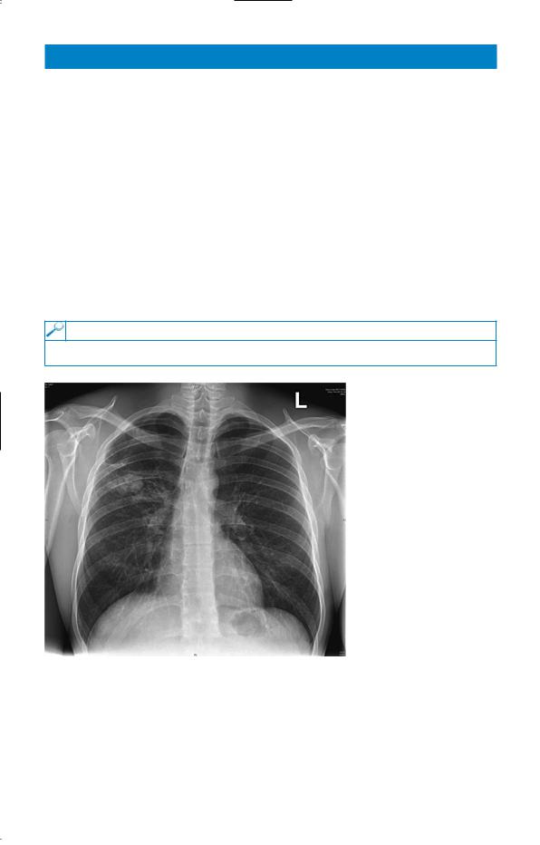
His chest X-ray is shown in Fig. 49.1.
Figure 49.1 Chest X-ray.
Question
• What might be the cause of his second admission?
127

ANSWER 49
The chest X-ray shows extensive changes in the right upper zone which seem as if they are likely to be more extensive than those described at the first admission 2 months earlier. It is likely that this is a worsening of his pulmonary tuberculosis. This might have occurred because he had a resistant organism or, more likely, because he had not taken his treatment as prescribed. Risk factors for development of tuberculosis are poor nutrition, high alcohol intake and immunosuppression (HIV, immunosuppressive therapy). Higher rates occur in those from the Indian subcontinent and parts of Africa.
The headache and confusion raise the possibility of tuberculous meningitis. Other possibilities would be liver damage from the antituberculous drugs and the alcohol, although clinical jaundice would be expected, or electrolyte imbalance. If these are not present a lumbar puncture would be indicated, provided that there is no sign to suggest raised intracranial pressure. It would be advisable to do a computed tomography (CT) scan of the brain first since a fall related to his high alcohol consumption might have led to a subdural haemorrhage to give him his headache and confusion.
It is now 2 months since the initial finding of acid-fast bacilli in the sputum and the cultures and sensitivities of the organism should now be available. These should be checked to be sure that the organism was Mycobacterium tuberculosis and that it was sensitive to the four antituberculous drugs which he was given. As a check on compliance, blood levels of antituberculous drugs can be measured. The urine will be coloured orangy-red by metabolites of rifampicin taken in the last 8 h or so.
Comparison with his old chest X-rays showed extension of the right upper-lobe shadowing. It is difficult to be sure about activity from a chest X-ray but extension of shadowing is obviously suspicious. ‘Softer’ more fluffy shadowing is more likely to be associated with active disease. A direct smear of the sputum showed that acid-fast bacilli were still present on direct smear. He confirmed that he was not taking his medication regularly. His headache and confusion resolved as he stopped his high alcohol intake. Subsequently the antituberculous therapy should be given as directly observed therapy (DOT) in a thriceweekly regime supervised at each administration by a district nurse or health visitor.
KEY POINTS
•Poor adherence to treatment regimes is the commonest cause of failure of antituberculous and other treatment.
•Directly observed therapy should be used when there is any doubt about adherence to treatment.
•Four drugs should be used (rifampicin, isoniazid, pyrazinamide and ethambutol) when there is a higher risk of resistant organisms, e.g. immigrants from Africa, Asia, previously treated patients, patients of no fixed abode.
128