

ethicon_wound_closure_manual
.pdfANUALM LOSUREC OUNDW
WOUND CLOSURE MANUAL

PREFACE


As a surgeon and medical professional, the complexities and importance of tissue repair and wound closure are critical to your success. From your first days in medical school to training in residency and as a practicing professional, you have recognized the need to continually learn and embrace the best practices to provide the best possible outcomes for your patients. At ETHICON, INC., we understand the importance of that and are pleased to provide you with the updated Wound Closure Manual to aid you in your quest for excellence.
This manual includes helpful information to assist you in critical decision making, and provides an overview of materials available today as well as basic principles of tissue healing and wound closure. Please use this as a guide, a reference, a training tool for your staff, or any other way you find most useful.
ETHICON, INC., a Johnson & Johnson company, is not only the world's leader in providing quality surgical sutures, but is also committed to advancing the standard of care by providing innovative products for suturing and topical skin closure. We are also committed to you and value your importance to the medical industry. We have been with you since the beginning and are committed to being with you in the future.

CONTRIBUTING
David L. Dunn, MD, PhD
Jay Phillips Professor and Chairman of
University of Minnesota
We thank Dr. Dunn for his contributions to the Wound Closure Manual. Dr. Dunn is currently the Jay Phillips Professor and Chairman of Surgery at the University of Minnesota. This
department has a long-standing tradition and |
attained |
national and international recognition for |
training |
academic general surgeons and surgical scientists. |
also the |
Division Chief of General Surgery, Head of |
Infectious |
Diseases, Director of Graduate Studies, and |
Program |
Director of the Department of Surgery. |
|
Dr. Dunn has published over 400 articles and book chapters in the areas of Surgical Infectious Diseases and Transplantation. He has
received regional and nationwide |
several |
academic organizations and is a Past President |
Surgical |
Infection Society, the Association for Academic |
the |
Minnesota Chapter of the American College of Surgeons, the |
|
Society of University Surgeons and the Society |
University |
Surgeons Foundation. |
|

TABLE OF CONTENTS
WOUND HEALING |
|
1 AND MANAGEMENT |
|
The Wound.......................................................... |
2 |
Recovery of Tensile Strength............................ |
2 |
Patient Factors That Affect Wound Healing........ |
2 |
Surgical Principles....................................... |
4 |
Classification of Wounds................................... |
5 |
Types of Wound Healing................................... |
6 |
Healing by Primary Intention ........................ |
6 |
Healing by Second Intention .......................... |
7 |
Delayed Primary Closure .............................. |
7 |
2 THE SUTURE |
|
What Is a Suture?............................................... |
10 |
Personal Suture Preference............................... |
10 |
Suture Characteristics....................................... |
11 |
Size and Tensile Strength.............................. |
11 |
Monofilament vs. Multifilament Strands.......... |
11 |
Absorbable vs. Nonabsorbable Sutures.............. |
12 |
Specific Suturing Materials.............................. |
13 |
Synthetic Absorbable Sutures ......................... |
14 |
Nonabsorbable Sutures................................ |
16 |
Synthetic Nonabsorbable Sutures.................... |
17 |
Common Suturing Techniques....................... |
18 |
Ligatures ................................................. |
18 |
The Primary Suture Line............................. |
19 |
Continuous Sutures.................................... |
19 |
Interrupted Sutures .................................... |
22 |
Deep Sutures ............................................ |
22 |
Buried Sutures .......................................... |
22 |
Purse-String Sutures................................... |
22 |
Subcuticular Sutures................................... |
22 |
The Secondary Suture Line........................... |
23 |
Stitch Placement........................................ |
24 |
Knot Tying......................................................... |
24 |
Knot Security............................................ |
24 |
Knot Tying Techniques Most Often Used .......... |
25 |
Square Knot............................................. |
25 |
Surgeon’s or Friction Knot............................ |
26 |
Deep Tie ................................................. |
26 |
Ligation Using a Hemostatic Clamp ............... |
26 |
Instrument Tie.......................................... |
26 |
Endoscopic Knot Tying Techniques.................. |
26 |
Cutting the Secured Sutures.......................... |
26 |
Suture Removal................................................. |
27 |
Suture Handling Tips....................................... |
27 |
Suture Selection Procedure ............................. |
27 |
Principles of Suture Selection......................... |
27 |
Surgery Within the Abdominal Wall Cavity....... |
28 |
Closing the Abdomen .................................. |
30 |
Closing Contaminated or Infected Wounds........ |
40 |
3 THE SURGICAL NEEDLE |
|
Elements of Needle Design ............................. |
42 |
Principles of Choosing a Surgical Needle..... |
44 |
The Anatomy of a Needle................................ |
45 |
The Needle Eye ......................................... |
45 |
The Needle Body ....................................... |
46 |
Straight Needle ......................................... |
46 |
Half-Curved Needle................................... |
47 |
Curved Needle.......................................... |
47 |
Compound Curved Needle ........................... |
47 |
The Needle Point ...................................... |
48 |
Types of Needles............................................... |
48 |
Cutting Needles......................................... |
48 |
Conventional Cutting Needles....................... |
49 |
Reverse Cutting Needles ............................... |
49 |
Side Cutting Needles................................... |
50 |
Taper Point Needles ................................... |
50 |
Tapercut Surgical Needles ............................ |
51 |
Blunt Point Needles.................................... |
52 |
Needleholders.................................................... |
52 |
Needleholder Use....................................... |
52 |
Placing the Needle in Tissue ......................... |
53 |
Needle Handling Tips...................................... |
53 |

4 PACKAGING |
|
An Integral Part of the Product ..................... |
56 |
RELAY* Suture Delivery System.................... |
56 |
Modular Storage Racks................................ |
56 |
Dispenser Boxes......................................... |
57 |
Primary Packets........................................ |
57 |
E-PACK* Procedure Kit.................................. |
59 |
Expiration Date................................................. |
60 |
Suture Sterilization........................................... |
60 |
Anticipating Suture Needs............................... |
61 |
Sterile Transfer of Suture Packets.................... |
61 |
Suture Preparation in the Sterile Field............. |
62 |
Suture Handling Technique.......................... |
63 |
5 TOPICAL SKIN ADHESIVES |
|
DERMABOND* Topical Skin Adhesive....... |
68 |
High Viscosity DERMABOND |
|
Topical Skin Adhesive..................................... |
68 |
OTHER SURGICAL |
|
6 PRODUCTS |
|
Adhesive Tapes................................................... |
74 |
Indications and Usage................................. |
74 |
Application.............................................. |
74 |
Aftercare and Removal ................................ |
74 |
Skin Closure Tapes ..................................... |
75 |
Polyester Fiber Strip................................... |
75 |
Umbilical Tape ........................................ |
75 |
Surgical Staples................................................. |
75 |
Indications and Usage ................................. |
76 |
Aftercare and Removal ................................ |
76 |
PROXIMATE* Skin Staplers........................ |
76 |
Looped Suture................................................... |
77 |
Retention Suture Devices................................ |
77 |
PRODUCT TERMS 7 AND TRADEMARKS
8 PRODUCT INFORMATION
9 INDEX

CHAPTER 1
WOUND HEALING AND MANAGEMENT

2 WOUND HEALING & MANAGEMENT
THE WOUND
Injury to any of the tissues of the body, especially that caused by physical means and with
interruption of continuity, is defined as a wound.1 Though most often the result of a physical cause, a burn is also considered a wound. Both follow the same processes towards the restoration to
health—otherwise known as healing.1
Wound healing is a natural and spontaneous phenomenon. When tissue has been disrupted so severely that it cannot heal naturally (without complications or possible disfiguration) dead tissue and foreign bodies must be removed, infection treated, and the tissue must be held in apposition until the healing process provides the wound with sufficient strength to withstand stress without mechanical support. A wound may be approximated with sutures, staples, clips, skin closure strips, or topical adhesives.
Tissue is defined as a collection of similar cells and the intercellular substances surrounding them.
There are 4 basic tissues in the body: 1) epithelium; 2) connective tissues, including blood, bone and cartilage; 3) muscle tissue; and
4) nerve tissue. The choice of wound closure materials and the techniques of using them are prime factors in the restoration of continuity and tensile strength to the injured tissues during the healing process.
The parameters for measuring the strength of normal body tissue are:
•Tensile Strength—The load per cross-sectional area unit at the point of rupture, relating to the nature of the material rather than its thickness.
•Breaking Strength—The load required to break a wound regardless of its dimension, the more clinically significant measurement.
•Burst Strength—The amount of pressure needed to rupture a viscus, or large interior organ.
The rate at which wounds regain strength during the wound healing process must be understood as a basis for selecting the most appropriate wound closure material.
RECOVERY OF
TENSILE STRENGTH
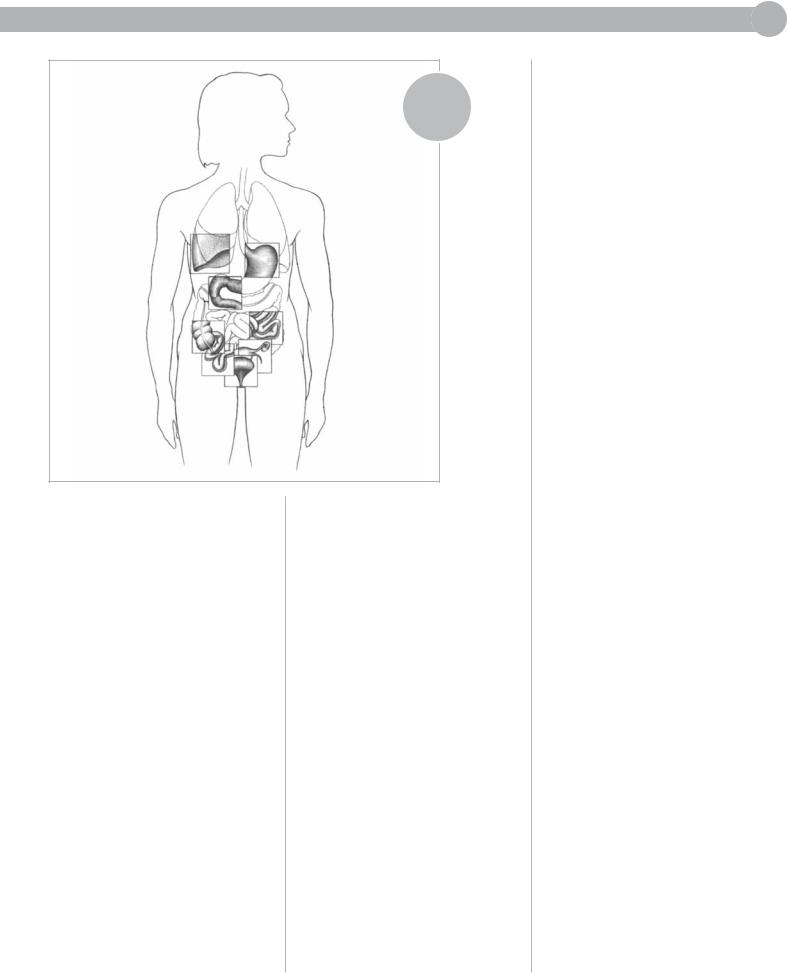
Tensile strength affects the tissue's ability to withstand injury but is not related to the length of time it takes the tissue to heal. As collagen accumulates during the reparative phase, strength increases rapidly, but it is many months before a plateau is reached.2 Until this time, the wound requires extrinsic support from the method used to bring it together—usually sutures. While skin and fascia (the layer of firm connective tissue covering muscle) are the strongest tissues in the body, they regain tensile strength slowly during the healing process. The stomach and small intestine, on the other hand, are composed of much weaker tissue but heal rapidly. Variations in tissue strength may also be found within the same organ. Within the colon, for example, the sigmoid region is approximately twice as strong as the cecum—but both sections heal at the same rate. Factors that affect tissue strength include the size, age, and weight
of the patient, the thickness of tissue, the presence of edema, and duration (the degree to which the tissue has hardened in response to pressure or injury).
PATIENT FACTORS THAT AFFECT WOUND HEALING
The goal of wound management is to provide interventions that
efficiently progress wounds through the biologic sequence of repair or regeneration. The patient's overall health status will affect the speed of the healing process. The following are factors that should be considered by the surgical team prior to and during the procedure.2-4
AGE—With aging, both skin and muscle tissue lose their tone and elasticity. Metabolism also slows, and circulation may be impaired. But aging alone is not a major factor in chronic wound healing. Aging and chronic disease states often go together, and both delay repair processes due to delayed cellular response to the stimulus of injury, delayed collagen deposition, and decreased tensile strength in the remodeled tissue. All of these factors lengthen healing time.
WEIGHT—Obese patients
of any age have excess fat at the wound site that may prevent securing a good closure. In addition, fat does not have a rich blood supply, making it the most vulnerable of all tissues to trauma and infection.
NUTRITIONAL STATUS—
Overall malnutrition associated with chronic disease or cancer,
CHAPTER 1 3
FIGURE 1
RELATIVE
TISSUE
STRENGTH
Lower |
|
|
|
||||||||||
respiratory |
|
|
|
||||||||||
tract (Weak) |
|
|
|
|
Stomach |
||||||||
|
|
|
|||||||||||
|
|
|
|
|
|
|
|
|
|
|
|
|
|
Duodenum |
|
|
(Weak) |
||||||||||
|
|
|
|||||||||||
(Strong) |
|
|
|
|
|
|
Small |
||||||
|
|
|
|
|
|||||||||
|
|
|
|
|
|
|
|
|
|
|
|
|
|
Cecum |
|
|
intestine |
||||||||||
|
|
||||||||||||
(Weak) |
|
|
|
|
|
|
|
|
|
(Weak) |
|||
|
|
|
|
|
|||||||||
Ileum |
|
|
Female |
||||||||||
(Weak) |
|
|
|
|
|
|
|
|
|
reproductive |
|||
|
|
|
|
|
|
|
|||||||
|
|
|
|
|
|
|
|
|
|
|
|
|
organs |
|
|
|
|
|
|
|
|
|
|
|
|
|
(Weak) |
|
|
|
|
|
|
|
|
|
|
|
|
|
Bladder |
|
|
|
|
|
|
|
|
|
|
|
|
|
|
|
|
|
|
|
|
|
|
|
|
|
|
|
(Weak) |
or specific deficiencies in carbohydrates, proteins, zinc, and vitamins A, B, and C can impair the healing process. Adequate nutrition is essential to support cellular activity and collagen synthesis at the wound site.
DEHYDRATION—If the patient's system has been depleted of fluids, the resulting electrolyte imbalance can affect cardiac function, kidney function, cellular metabolism, oxygenation of the blood, and hormonal function. These effects will not only impact upon the patient's overall health status and recovery from surgery but may also impair the healing process.
INADEQUATE BLOOD SUPPLY TO THE WOUND SITE—Oxygen is necessary for
cell survival and, therefore, healing. Skin healing takes place most rapidly in the face and neck, which receive the greatest blood supply, and most slowly in the extremities. The presence of any condition that compromises the supply of blood to the wound, such as poor circulation to the limbs in a diabetic patient or arteriosclerosis with vascular compromise, will slow and can even arrest the healing process.
IMMUNE RESPONSES—
Because the immune response protects the patient from infection, immunodeficiencies may seriously compromise the outcome of a surgical procedure. Patients infected with HIV, as well as those who have recently undergone chemotherapy or who have taken prolonged high
dosages of catabolic steroids, have debilitated immune Some patients have allergies to specific suturing materials, alloys, or latex. These, on the other hand, will cause a height ened immune response in the form of an allergic reaction. This may also interfere with healing process. Therefore, the surgeon should always check beforehand on a patient's allergies.
CHRONIC DISEASE—
A patient whose system has already been stressed by illness, especially endocrine disorders, diabetes, localized infection, or
injuries will heal more slowly will be more vulnerable to post surgical wound complications. All of these conditions merit concern, and the surgeon must consider their effects upon the tissues at the wound site, as well as their potential impact upon the patient's overall recovery from the procedure. Malignancies, in addition, may alter the cellular structure of tissue and influence the surgeon's choice of methods and closure materials.
RADIATION THERAPY—
Radiation therapy to the site prior to or shortly after surgery can produce impairment of healing and
to substantial wound complica tions. Surgical procedures for malignancies must be planned to minimize the potential for these problems.

4 WOUND HEALING
PRINCIPLES factors that affect the healing
can be controlled by the team in the operating room,
the obstetrical team in labor and or by the emergency team
trauma center. Their first is to maintain a sterile aseptic technique to prevent
. Organisms found
a patient's own body most cause postoperative but microorganisms
by medical personnel also a threat. Whatever the source,
presence of infection will deter
. In addition to concerns sterility, the following must
taken into consideration when and carrying out an procedure.3
THE LENGTH AND
DIRECTION OF THE INCISION—A properly planned incision is sufficiently long to afford sufficient optimum exposure. When deciding upon the direction of the incision, the surgeon must bear the following in mind:
direction in which wounds naturally heal is from side-to- side, not end-to-end.
arrangement of tissue fibers in the area to be dissected will vary with tissue type.
best cosmetic results may be achieved when incisions are made parallel to the direction of the tissue fibers. Results may vary depending upon the tissue
layer involved.
DISSECTION TECHNIQUE—
When incising tissue, a clean incision should be made through the skin with one stroke of evenly applied pressure on the scalpel. Sharp dissection should be used to cut through remaining tissues. The surgeon must preserve the integrity of as many of the underlying nerves, blood vessels, and muscles as possible.
TISSUE HANDLING—
Keeping tissue trauma to a minimum promotes faster healing. Throughout the operative procedure, the surgeon must handle all tissues very gently and as little as possible. Retractors should be placed with care to avoid excessive pressure, since tension can cause serious complications: impaired blood and lymph flow, altering of the local physiological state of the wound, and predisposition to microbial colonization.
HEMOSTASIS—Various mechanical, thermal, and chemical methods are available to decrease the flow of blood and fluid into the wound site. Hemostasis allows the surgeon to work in as clear a field as possible with greater accuracy. Without adequate control, bleeding from transected or penetrated vessels or diffused oozing on large denuded surfaces may interfere with the surgeon's view of underlying structures.
Achieving complete hemostasis before wound closure also will prevent formation of postoperative hematomas. Collections of
blood (hematomas) or fluid (seromas) in the incision can prevent the direct apposition of tissue needed for complete union of wound edges. Furthermore, these collections provide an ideal culture medium for microbial growth and can lead to serious infection.
When clamping or ligating a vessel or tissue, care must be taken to avoid excessive tissue damage. Mass ligation that involves large areas of tissue may produce necrosis, or tissue death, and prolong healing time.
MAINTAINING MOISTURE IN TISSUES—During long procedures, the surgeon may periodically irrigate the wound with warm physiologic (normal) saline solution, or cover exposed surfaces with saline-moistened sponges or laparotomy tapes to prevent tissues from drying out.
REMOVAL OF NECROTIC TISSUE AND FOREIGN MATERIALS—Adequate debridement of all devitalized tissue and removal of inflicted foreign materials are essential
to healing, especially in traumatic wounds. The presence of fragments of dirt, metal, glass, etc., increases the probability
of infection.
CHOICE OF CLOSURE MATERIALS—The surgeon must evaluate each case individually, and choose closure material which will maximize the opportunity for healing and minimize the likelihood of infection. The proper closure
material will allow the surgeon to approximate tissue with as little trauma as possible, and with enough precision to eliminate dead space. The surgeon's personal preference will play a large role in the choice of closure material; but the location of the wound, the arrangement of tissue fibers, and patient factors influence his or her decision as well.
CELLULAR RESPONSE TO CLOSURE MATERIALS—
Whenever foreign materials such as sutures are implanted in tissue, the tissue reacts. This reaction will range from minimal to moderate, depending upon the type of material implanted. The reaction will be more marked if complicated by infection, allergy, or trauma.
Initially, the tissue will deflect the passage of the surgeon's needle and suture. Once the sutures have been implanted, edema of the skin and subcutaneous tissues will ensue. This can cause significant patient discomfort during recovery, as well as scarring secondary to ischemic necrosis. The surgeon must take these factors into consideration when placing tension upon the closure material.
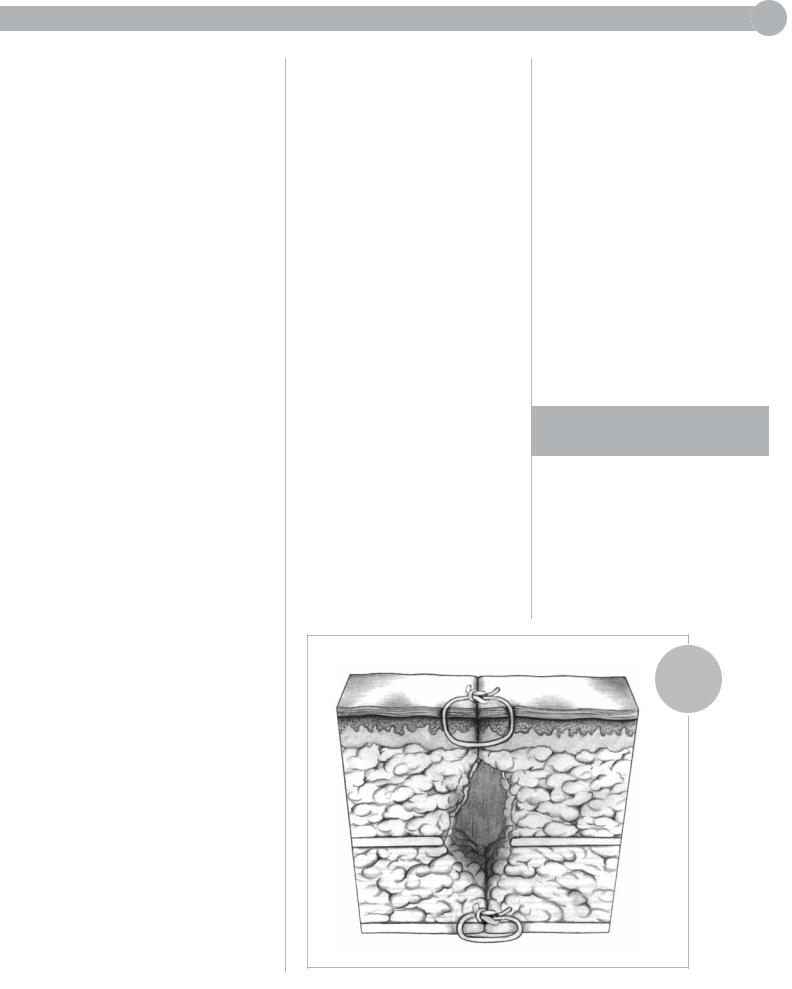
ELIMINATION OF DEAD SPACE IN THE WOUND—
Dead space in a wound results from separation of portions of the wound beneath the skin edges that have not been
closely approximated, or from air or fluid trapped between layers of tissue. This is especially true in the fatty layer which tends to lack
CHAPTER 1 5
blood supply. Serum or blood may collect, providing an ideal medium for the growth
of microorganisms that cause infection. The surgeon may elect to insert a drain or apply a pressure dressing to help eliminate dead space in the wound postoperatively.
CLOSING TENSION—
While enough tension must be applied to approximate tissue and eliminate dead space, the sutures must be loose enough to prevent exaggerated patient discomfort, ischemia, and tissue necrosis during healing.
POSTOPERATIVE DISTRACTION FORCES—
The patient's postoperative activity can place undue stress upon a healing incision. Abdominal fascia will be placed under excessive tension after surgery if the patient strains to cough, vomit, void, or defecate.
Tendons and the extremities also be subjected to excessive tension during healing. The surgeon must be certain that the approximated wound is adequately immobilized to prevent suture disruption
for a sufficient period of time after surgery.
IMMOBILIZATION—
Adequate |
of the |
approximated |
not |
necessarily of |
anatomic |
part, is |
surgery |
for efficient |
minimal |
scar formation. |
|
CLASSIFICATION OF
The Centers for |
|
and Prevention |
an |
adaptation of the |
College |
of Surgeons' |
|
schema, divides |
|
into 4 classes: |
|
clean- |
|
contaminated |
dirty |
FIGURE 2
DEAD SPACE
IN A WOUND