

BASIC_SURGICAL_TECHNIQUES budapest
.pdfinclusion of the neighboring tissues. This intervention (requiring the harmonized movements of the operator and the assistant) consists of three phases: soaking, clamping and ligation. First, the asistant applys only a pressure with the sponge and soaks up the blood (so, he does not cause a temporary vasoconstriction). The operator grasps the bleeding vessel with a Péan. The tip of the Péan should always faces the person who will do the ligation. The scrub nurse gives the thread while she is keeping the two ends of it stretched. The thread used for ligation should be as thin as possible. After applying the first basic knot, the assistant releases the Péan but the surgeon stretches the thread further. After the 2nd knot, the operator cuts the thread as follows: the scissors are slid down to the knot and rotated a quarter turn. The least possible amount of the thread should stay in the wound (foreign body!). It is not advisable to use a ligation directly beneath the skin because it disturbs the healing process of the wound.
Suturing
Transverse, transfixing,”figure of 8” stitch (sutura circumvoluta). In cases of large-caliber vessels or diffuse bleeding, non-absorbable (e.g. silk, polyethylene or wire) and absorbable (e.g. catgut, polyglycolic acid (Dexon) or polyglactic (Vicryl) suture materials can be used. A double stitch (suture twice) is applied under the bleeding tissue to form an “8”shaped loop and the knot is then tied.
Preventive hemostasis (planned hemostasis)
It happens with ligatures. In the operating field, the vessel should be clamped with two Péans, the part of the vessel located between them is cut, and the two ends of the vessels should be tied separately. Deschamp needle and the Payr probe can be used for the same purpose.
Clips
Clips are metal or plastic. Applied with the help of disposable or non-disposable devices.
Bone wax
This is a sterile mixture of beeswax, almond oil and salicylic acid. It adheres readily to the bloody bone surfaces, thereby achieving local hemostasis of the bone. For example it is used to stop bleeding after cutting the sternum.
Expedients
Suction, drainage (Hemovac, Jackson-Pratt, etc.) to remove body fluids and air. This facilitates the emptying of dead spaces, improves tissue regeneration, and blocks the development of edema and hematoma.
Other devices or mechanical methods for handling bleeding
-Rubber bands for digits
-Esmarch bandage
-Penrose drain
-Vessel loops
-Pneumatic tourniquets
-Pressure dressings, packing (compression), tamponades, and sand bag
5.3.2. Thermal methods
61
Low temperature – hypothermia
Hypothermia (a hypothermia blanket, ice, cold solutions for stomach bleeding) Cryosurgery: -20 to -180 °C cryogenic heat. Its mechanism:
-dehydration and denaturation of fatty tissue
-decreases the cellular metabolism/O2 demand
-leads to vasoconstriction.
Heat (high temperature)
It based on protein denaturation.
Electrosurgery
-In Paquelin (Claude André Paquelin (1836-1905), French surgeon) electrocauterization (which stops bleeding by “burning” the bleeding vessels), the tissue is not part of the circuit. In diathermy, the patient is in the circuit. Electrical current incises/excises or destroys the tissue. The area is automatically sterilized and burned.
-Essence: hemostasis + an aseptic technique
-Parts of the electrosurgical unit: generator, cable and neutral (indifferent) electrode, parts which are connecting to the wires: knife, needle, loop, blade. The effect depends on the current intensity and wave-form used. Coagulation is produced by interrupted (damped) pulses of current (50– 100/s) and a square wave-form. Cutting is produced by continuous (undamped) current and a sinus wave-form.
-The contemporary generators are working in an alternative manner [e.g. the surgeon regulates the cutting or coagulating functions.With the same electrode he can coagulate (at higher voltages) and cut (at lower voltages)]. The diathermy is not suitable for skin incision because it leads to burnning injury of the skin. We use it only for deeper tissues.
Monoplar diathermy
Only one (the active) electrode is connected to the cutting/coagulating device. The electric current is passing through the patient between this active electrode and the indifferent (neutral) electrode which is located out of the surgical territory and touching a large skin surface. This elecrode is placed at the time of positionning the patient on operating table.
Bipolar diathermy
In bipolar diathermy, two electrodes are combined in the instrument (e.g. forceps), and the current passes between the tips and not through the patient.
Local effectrosurgery
Electrocoagulation: a needle or disc touches the tissue directly, and burns the tissue (a grayish discharge). The tissues are expelled after 5-15 days. Use: bleeding coagulation. Electrofulguration: lighting or spark: he needle does not touch the tissue directly (it is 1–2 mm away). Use: “Spray” function – control of diffuse bleeding.
Electrodesiccation: the needle is inserted into the tissues. Use: to destroy warts and polyps. Electrosection: with a knife, blade or electrode. Use: excision or incision.
Laser surgery
62
Laser surgery is based on the emission of radiation by light amplification through a tube at a microscopic level. Use: coagulation and vaporization (carbon or steam) in delicate and fine tissues (eyes: retina detachment repair, brain, spinal cord, or gastrointestinal tract). The operator must wear safety goggles. Suction of the steam (CO2) is necessary.
5.3.3. Hemostasis with chemical and biological methods
Characteristics: Easy handling, quick absorption, non-toxic, and local effects without systemic consequences. Expected consequences: vasoconstriction, coagulation and a hygroscopic effect. Aethoxysclerol (polydocanol): This is not used for active coagulation. Main indications:
small superficial skin varices (injection into the veins) and esophagus varix sclerotization (given to the proximity of the varix).
Absorbable gelatin: Gelfoam, Lyostypt or Spongostan: powder or compressed-pad form. Made from purified gelatin solution. Adsorption capacity: 45 times more than its own weight. Absorption takes place in 20-40 days.
Absorbable collagen: Collastat®: This is in the form of a hemostatic sponge, applied dry to the oozing or bleeding site. Its use is contraindicated when there is an infection or in areas where blood has pooled.
Microfibrillar collagen: Avitene®: This is a powder-like, absorbable material from a bovine source; it is applied dry. It stimulates the adhesion of platelets and the deposition of fibrin. It functions as a hemostatic agent only when applied directly to source of bleeding. It is applied to oozing surfaces, including bone and areas of bleeding difficult to reach.
Oxidized cellulose: Oxycel®, Surgicel®: made of cellulose, able to adsorb a large amount of blood, with blood make an artificial thrombus. They are absorbed in 7–30 days.
Oxytocin: This is a hormone produced by pitutary gland, but is also prepared synthetically. Use: e.g. bleeding from uterus.
Epinephrine: This hormone is secreted by the adrenal gland, is also prepared synthetically. It is a vasoconstrictor. It is rapidly dispersed and has a short duration of action.
Thrombin: This enzyme is extracted from the bovine blood. It combines rapidly with fibrinogen to form a clot. It is available in liquid (spray) and powder forms. It must not be allowed to enter large vessels. It is for topical use only and is never injected.
Novel hemostatic agents: Indications: External bleeding where the conventional pressure dressings fail. It is not used in those places were you can apply a tourniquet.
1.HemCon: It is available as a chitosan-based product, made from shrimp shell polysaccharide + vinegar. This is a firm 7x7 cm dressing that is sterile and individually packaged. It adheres to a bleeding wound, and exerts vasoconstrictive properties.
2.QuikClot: This granular zeolite absorbs fluid, acts as a selective sponge for water, dehydrates blood, has handling properties similar to those of sand, and can generate significant heat during the adsorption process.
63
6. OPERATION (ACUTE, ELECTIVE, PREPARATION OF THE PATIENT,
SURGICAL EXPOSURES)
6.1. Preparations for an operation
”Salus aegroti suprema lex esto” = ”The well-being of the patient is the most important law." Aim: to perform the right operation, for the right reasons, on a right patient, and at a right time.
-From the financial and hygienic standpoints, the patient’s preoperative hospital stay should be as short as possible (Hospitalisation, iatrogenia, contamination)
-If it is possible, the patient should be admitted a day before operation or even at same day.
Careful examination of each patient individually is an important factor. The standpoint of ”Surgery is the aim!” should be neglected. We can think of increased surgical morbidity (and accompanying cardiacvascular, hepatic, and renal diseases) as the age of the patient is increasing.
6.2. Surgical indications, contraindications and risks
Indications
Proper evaluation of the surgical disease and risks:
-Vital indications: These are involved in the case of life-saving procedures. The patient can be treated only with an operation (100% mortality without operation). Example: rupture of an abdominal aorta aneurysm
-Absolute indications: These are involved in urgent procedures. The disease can be treated exclusively with an operation. The time can be chosen between narrow limits. Example: mechanical ileus.
-Relative indications: These are factors in elective procedures, e.g. programmed operations. The disease can be treated with or without surgery. The time of surgery can be choosen. Example: hernia.
Contraindications
In the cases of vital and absolute indications: only in moribund patients.
In the case of relative indication: decompensated accompanying diseases, does the surgery improve the survival?
Surgical risks
Surgical risks = risks of surgery itself + anesthesiological risks. The preoperative examinations must answer the questions of both surgeon and anesthesiologist, allowing them to give their agreed opinion in writing.
1.Low-risk surgery: Minor operations belong in this group (e.g. inguinal hernia repair), where the expected blood loss is less than 200 ml.
2.Medium-risk surgery: Surgical interventions of medium severity can be classified here (the expected blood loss is less than 1000 ml), e.g. colon resection.
3.High-risk surgery: Extended abdominal and thoracic operations (e.g. liver and lung resections) fall into this category. The blood loss exceeds 1000 ml. The patient needs postoperative intensive care and treatment. The rates of postoperative morbidity and mortality are high.
64

The extent of the operation is a determinator:
Operations on body surfaces are running with the smallest risks. The risks of the operation increase if there is an opening of a body cavity. Operations done over the hollow organs are running with higher riks. The opening of many hollow organs is running with more risks. Operations in which 2 body cavities are opened at the same time are runnig with the highest risks.
Factors which increase the surgical risks:
•Acute surgery
•Duration > 2 hours
•> 65 years old
•Pregnancy
•Malignant diseases
•Malnutrition
•Alcohol consumption
•Smoking
•Acute disturbancies
•hypovolaemia
•dehydration
•shock
•Acute inflammations
•respiratory
•urinary
•gastrointestinal
•sepsis
•Trombosis
•Acute organ insufficiencies:
•Heart
•Lung
•Kidney
•Liver
Acute endocrine disorder
Organ alterations:
• |
Cardiorespiratory |
|
• |
Hypertention |
|
• |
Nervous system alterations |
|
• |
Diabetes mellitus |
|
• |
Chronic Uraemia |
|
• |
Cirrhosis |
|
• |
Susceptibility for infection |
|
• |
Immunosuppresion |
|
• |
Thromboembolic predisposition |
|
|
|
|
|
|
|
• |
Chronic disorders: |
|
|
• |
Hypovolaemia |
|
• |
Anaemia |
• |
Chronic inflammations |
|
|
• |
Respiratory(bronchitis) |
|
• |
Urinary |
|
• |
Gastrointestinal (ulcer) |
• |
Trombosis |
|
• |
Allergia |
|
|
|
|
• |
Organ insufficiencies |
|
|
O |
Heart |
|
O |
Lung |
|
O |
Kidney |
|
O |
Liver |
• |
Endocrine disorders |
|
• |
Immunological disorders |
|
Pregnancy as a surgical risk factor |
||
• |
Hemophilia |
|
- acute or chronic systemic risk factors next to the pregnancy + surgical diseases |
||
- decreased maternal physiological reservoirs (respiration, circulation, metabolism) |
||
65
-altered anatomical relations
-atypical symptoms
-possibilty for foetal diseases
Menstration
In the past, there was a tendency to avoid surgery on a menstrating woman due to increased psychic unstability, possible increase in bleeding tendency, and increased hygienic demands seen during this period. Nowdays, the menstration is not considered as an obstacle to do the operation.
Overfeeding as a factor increasing the surgical risk
-respiratory disturbancy(usually restrictive): deteriorating the gas exchange, increased respiratory function
-decresed cardiac reservoirs
-difficulty with intubation (regurgitation)
-disturbancies with wound healing
-thromboembolism
Immunological factors
Immunosuppresion (transplanted patient), use of cytostatics (tumorous patient), AIDS and so on increased possibilty for infection, frequent wound healing disturbancies.
Oncological patients’ own problems
-chemotherapeutic agents
-radiotherapy (local inflammation)
-decreased function of the immune system
-paraneoplastic syndromes e.g. deep venous thrombosis
Increased age as a factor influencing the surgical risk-elderly patient
-elderly patient: age > 65 years old (the biological age is important and not the calender age)
-to assess the expected benefits, risks, and the patient’s survival
-to estimate the interactions between the drugs, used by patient for a long period of time and the necessary drugs which are given during the perioperative period
-cardiopulmonary deficiency is the cause of the death in most cases
Estimation of the surgical risks
It means to examine the followings:
-cardiovascular state,
-respiratory system,
-metabolic state,
-renal function,
-liver function,
-endocrine balance,
-homeostasis,
-immune system.
Examinations
-physical examination
-laboratory examination
-radiological examinations (US, CT, MRI, isotop, DSA, and so on)
-instrumental examination (endoscopy, biopsy, cytological examinations)
66
Heart and circulation:
-pulse, blood pressure
-ECG
-Echocardiography
-coronarography
-isotop
Increased cadiac risks:
-aortic stenosis, mitral stenosis
-dysrhythmia
-AMI: within 3 weeks mortality: 25%
-AMI: within 6 weeks mortality: 5%
-DM silent ischemia 25%
Lung, breathing:
-Chest X-ray
-Respiratory function
-Blood gas analysis (preparation for the surgery: respiratory physiotherapy and inspiratory treatment)
Laboratory examinations:
-blood count
-blood group
-bleeding and clotting times
-liver function tests
-renal function tests
-examination of the metabolic processes
-fluid and electrolyte balance
-plasma protein level
Diet regulations
If it is possible, you should prevent the deterioration of the nutritional status of the patient during the processes of the preoperative examination and preparation. Sometimes, the nutritional therapy is a part of preoperative preparation. To have a safe general anesthesia, the patient should avoid eating (fasting 6 hours prior to surgery).
Preoperative nutritional therapy
First, you should consider the natural oral feeding. If it is not possible, then the nasogastric, duodenal or jejunal tubes are the most appropriate ways of feeding. The parentral feeding is done through the peripheral or central venous catheters. The burned, tumorous, polytraumatized, and septic patients need the highst amount of energy.
Slag deprivation
-Diet: liquids for 2-3 days or a low-residue diet
-Enema: In the case of major abdominal surgeries (or those operations which involve the intestinal system), there is a need to make the intestinal tract empty.
-to make the stomach empty: In the case of pyloric stenosis, the nasogastric tube can remove the gastric contents and also lead to the gastric lavage(antibiotic).
Urinary catheter
67
It is needed in the case of long-lasted operations which are running with loss of a large amount of fluids.
Thrombosis prophylaxis
-Drugs:
-Heparin derivatives: Na-heparin, Ca-heparin, low molecular weight heparins
-Platelet aggregation inhibitors (e.g. Aspirin and Colfarit)
-Coumarin derivatives (e.g. Syncumar)
-Physical:
-early mobilization
-compression (elastic bandages)
-bed-side bicycle
-keeping the lower extremities at a high level
Psychic preparartion
That is natural for the patient to fear of the operation and its unwanted consequences. The surgeon should deals with the patient’s psychic state. He/she should carefully evaluate the indications and contraindications and choose the best possible intevention.
Legal aspects of the operations
-informing the patient (tumorous patients!)
-patient’s written consent (this should also include those decisions which may be made by the surgeon intraoperatively).
-in the case of children, the parents or the legal representative should give the informed written consent.
6.3 Surgical approaches
6.3.1. Laparotomy on the anterior abdominal wall
The direction of the incision can be: verical, transverse, or oblique. Vertical incisions:
-upper, lower, middle, or total median laparotomy
-paramedian laparotomy
-vertical transrectal laparotomy
-pararectal laparotomy
Transverse incisions:
-horizontal transrectal laparotomy
-Pfannensteil incision
Oblique incisions:
-McBurney-incision
-inguinal transmuscular laparotomy
-paracostal laparotomy (Kocher incision)
-subcostal laparotomy
Vertical incisions
Upper median laparotomy
The incision is made from xyphoid process to the umbilicus. Advantages: insures a quick and wide exposure, quickly and easily can be elongated and closed. Disadvantages: the fibrous tissue of the white line (linea alba) is cut and the the sutures are upon tensions at both sides
68
which can be a cause for a later postoperative hernia (Figure 67. A).
Lower median laparotomy
The incision is made from umbilicus to symphysis pubis. The advantages and disadvantages are the same as those for an upper median laparotomy. About 2/3 of sterile wound disruption happens folowing such a this incision (Figure 67. B).
Middle median laparotomy
An 8-10 cm long incision. Half of it is located above and the other half below the umbilicus. At the umbilcus the incision is curved towards the left side. Advantage: from a small incision we can inspect both the upper and the lower part of the abdominal cavity.
Total median laparotomy
The incision is made from xyphoid process to the syphysis pubis. It gives an excellent exposure but injures the statistic of the abdominal wall significantly. The patient is predisposed to the postoperative wound disruption. It also makes the postoperative coughing difficult, increases the danger of pneumonia, and can cause constipation. This incision is generally used in the case of extended abdominal operations (Figure 67. A+B).
Paramedian laparotomy
It is generally used only above the umbilicus. About 2 cm right (an parallel) to the midline cut the skin, subcutaneous tissue and the anterior leaflet of the rectus sheath. The rectus muscle is retracted. Following this, the posterior rectus sheath is also cut. The later scar will be strong and the possibilty for development of a henia is rare (Figure 67. C).
Transrectal laparotomy
About 2-3 cm right to the midline cut the skin, subcutaneous tissue, and the anterior leaflet of the rectus sheath. Then, separate the fibers of the rectus muscle from each other bluntly to be able to cut the posterior leaflet of the rectus sheath (together with transversalis fascia and the parietal peritoneum)(Figure 67. D).
Pararectal laparotomy
A vertical incision is parallel to the rectus muscle. Due to the denervation of the muscles the abdominal wall becomes significanly weakened. It is danger for development of a huge hernia. It is not advisable (Figure 67. E).
Lateral transmuscular laparotomy
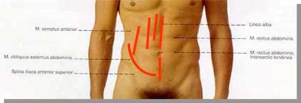
The incision is made starting from a point located 2-3 cm lateral to the external edge of the rectus muscle. The longest incision of such starts at the lower edge of the 10 th rib and runs till the level of the anterior sup. ilac spine. The” pararectal” and ”an incision made alongside the semilunar line of Spiegel” are not the ideal incisions because they weaken the abdominal wall significantly. They are not advisable (Figure 67. F).
69
D C A
E
F
B
Figure 67. Vertical laparotomies
A.Upper median laparotomy, B. Lower median laparotomy, A+B Total median laparotomy, C. Paramedian laparotomy, D. Transrectal laparotomy, E. Pararectal laparotomy, F. lateral transmuscular laparotomy
Transverse and oblique laparotomies
They cause less injury to the nerves of the abdominal wall muscles. In this way, the possibilties for postoperative sterile wound disruption and later hernia are less.
Upper transverse laparotomy
The incision is made at the area beween the xyphoid proc. and the umbilicus, starting from one external edge of the rectus muscle and ending at its other edge (It is at the border line beween the middle and lower 2/3) (Figure 68. A). This incision can be made larger by elongating it at both of its lateral sides (even up to the middle axillary lines). This incision rarely injures the abdominal wall muscles. The innervations of these muscles are not injured and the wound heals with development of a strong scar. In upper abdominal surgeries we can combine the upper median incision with a transverse incision. Such this incision is called: the Mercedes-Benz incision.
Lower transverse laparotomy
This a mild concave incision a few centimeter below the umbilicus (Figure 68. B). One or both rectus muscles are cut. Sometimes, we only cut the rectus sheaths and do not incise the muscle itself. The incision can be elongated laterally.
Paracostal laparotomy
A curved incision started from xyphoid proc. to the lateral side of the abdominal wall. It is located 2-3 cm below and parallel with the costal margins (Figure 68. C). The possibilty for development of a postoperative hernia is high. On the left side, this incision is helpful in performing a splenectomy. On right side it can be used to perform an open cholecystectomy. Nowdays, with application of the laparoscopic cholecystectomy we can avoid the postoperative pain and complications of such this incision!
70