

Kaplan USMLE - Step 2 CK Lecture Notes 2017- Surgery
.pdfUSMLE Step 2 CK λ Surgery
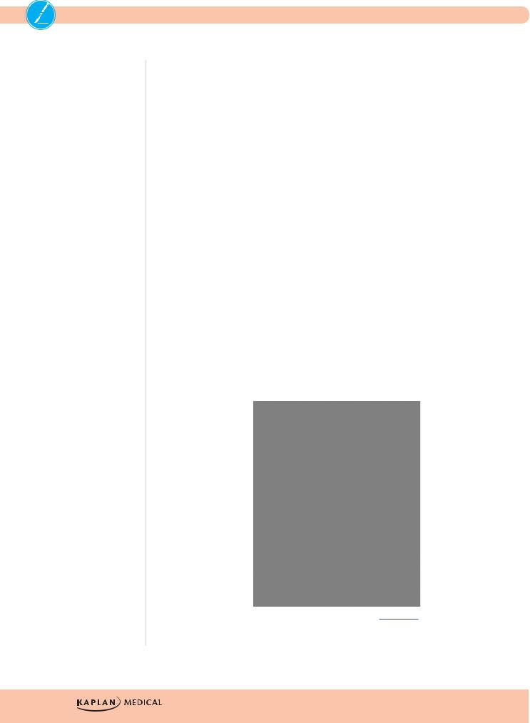
Copyright 2007 Bates, M.D. - Custom Medical Stock Photo.
All rights reserved.
Figure I-2-5. Intertrochanteric Fracture of the Hip Noted on X-ray
Femoral shaft fracture is often treated with intramedullary rod fixation.
•If bilateral and comminuted, it may produce enough internal blood loss to lead to shock (external fixation may help while the patient is stabilized).
•If open, it is an orthopedic emergency, requiring OR irrigation and closure within 6 hours.
•If multiple, fat embolism syndrome may develop, in which severe respiratory distress occurs secondary to marrow fat entering the blood stream and embolizing to the pulmonary vasculature.
•Treatment is supportive care.
Knee injury typically produces swelling of the knee; knee pain without swelling is unlikely to be a serious knee injury. Collateral ligament injury is usually sustained when the force of impact is the side of the knee, a common sports injury. Medial blows disrupt the lateral ligament and vice versa.
•The knee will be swollen and there is localized pain by direct palpation on the affected side.
•With the knee flexed 30°, passive abduction or adduction will produce pain on the torn ligaments and allow further displacement than the normal leg.
24

Chapter 2 λ Orthopedics
•Abduction demonstrates the medial injuries (valgus stress test), whereas adduction diagnoses the lateral injuries (varus stress test). Isolated injuries are treated with a hinged cast.
•When several ligaments are torn, surgical repair is preferred.
Anterior cruciate ligament injury is more common than posterior injury.
•There is severe knee swelling and pain.
•With the knee flexed 90°, the leg can be pulled anteriorly, like a drawer being opened (anterior drawer test).
•A similar finding can be elicited with the knee flexed at 20° by grasping the thigh with one hand, and pulling the leg with the other (Lachman test).
Posterior cruciate ligament injury produces the opposite findings. MRI is diagnostic. Sedentary patients may be treated with immobilization and rehabilitation, whereas athletes require arthroscopic reconstruction.
Meniscal tear is difficult to diagnose clinically and on x-rays, but is beautifully demonstrated on MRI.
•Protracted pain and swelling after a knee injury
•Possible “catching and locking” which limits knee motion, and a “click” when the knee is forcefully extended
•Repair is done, trying to save as much meniscus as possible
•Complete meniscectomy leads to the late development of degenerative arthritis
Injuries to the medial meniscus, medial collateral, and anterior cruciate often occur simultaneously.
Tibial stress fracture is seen in young men subjected to forced marches. There is tenderness to palpation over a very specific point on the bone, but x-rays are initially normal. Treat with a cast, and repeat the x-rays in 2 weeks. Non–weight bearing with crutches is another option.
Leg fracture involving the tibia and fibula is often seen when a pedestrian is hit by a car. Physical exam shows angulation; x-rays are diagnostic. Casting takes care of the ones that are easily reduced; intramedullary nailing is needed for the ones that cannot be aligned. The lower leg (along with the forearm) is one of the most common locations for development of the compartment syndrome. Increasing pain after a long leg cast has been applied always requires immediate removal of the cast and appropriate assessment.
Rupture of the Achilles tendon is seen in out-of-shape middle-age men who subject themselves to severe strain (tennis, for instance). As they plant the foot and change direction, a loud popping noise is heard (like a rifle shot), and they fall clutching the ankle. Limited plantarflexion is still possible; but pain, swelling, and limping bring them to medical attention. Palpation of the tendon reveals a gap. Casting in equinus position allows healing in several months; surgery achieves a quicker cure.
Fracture of the ankle occurs when falling on an inverted or everted foot. In either case, both malleoli break. AP, lateral, and mortise x-rays are diagnostic. Open reduction and internal fixation is needed if the fragments are displaced.
25
USMLE Step 2 CK λ Surgery
Orthopedic Emergencies
Compartment syndrome occurs most frequently in the forearm or lower leg.
•Precipitating events include prolonged ischemia followed by reperfusion, crushing injuries, or other types of trauma.
•In the lower leg, by far the most common cause is a fracture with closed reduction.
•The patient has pain and limited use of the extremity; the compartment feels very tight and tender to palpation.
•The most reliable physical finding is excruciating pain with passive extension.
•Pulses may be normal.
•Emergency fasciotomy is required for treatment.
Pain under a cast is always handled by removing the cast and examining the limb.
Open fracture in which a broken bone protrudes from the wound requires irrigation in the OR and suitable reduction within 6 hours from the time of the injury. It is called compound or open fracture.
Posterior dislocation of the hip occurs when the femur is driven backward, such as in a headon car collision where the knees hit the dashboard. The patient has hip pain and lies in the stretcher with the leg shortened, adducted, and internally rotated (in a broken hip the leg is also shortened, but it is externally rotated). Because of the tenuous blood supply of the femoral head, emergency reduction is needed to avoid avascular necrosis.
Gas gangrene occurs with deep, penetrating, dirty wounds. In about 3 days the patient is extremely sick, looking toxic and moribund. The affected site is tender, swollen, discolored, and has gas crepitation. Treatment includes IV penicillin, extensive emergency surgical debridement, and possibly hyperbaric oxygen.
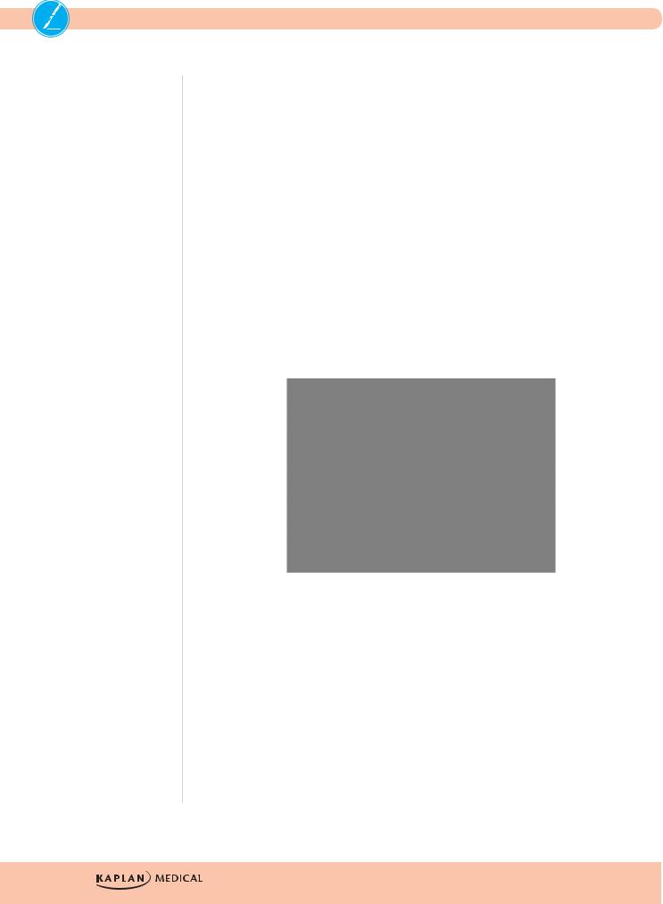
phil.cdc.gov.
Figure I-2-6. Gangrene of the Toes
26

Chapter 2 λ Orthopedics
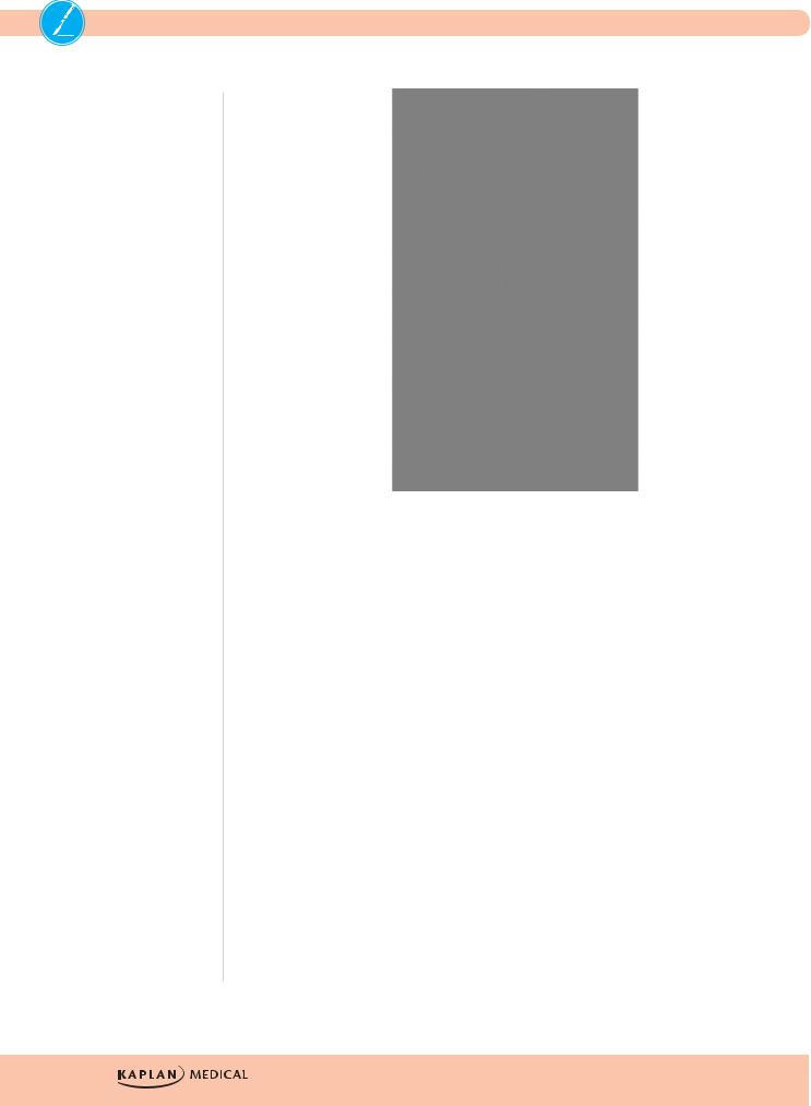
Reproduced with permission from SRS-X, the SRS Educational Resource, the Scottish Radiological Society, www.radiology.co.uk.
Figure I-2-7. Gas Gangrene due to Clostridium Perfringens Infection
Associated neurovascular injuries
The radial nerve can be injured in oblique fractures of the middle to distal thirds of the humerus. If a patient comes in unable to dorsiflex (extend) the wrist, and regains function when the fracture is reduced and the arm is placed on a hanging cast or coaptation sling, no surgical exploration is needed. However, if nerve paralysis develops or remains after reduction, the nerve is entrapped and surgery has to be done.
Popliteal artery injury can occur in posterior dislocations of the knee. Following reduction of the dislocation, the popliteal artery must be evaluated with U/S, because even if distal pulses which were absent return following reduction of the dislocation, there may be an intimal flap or local dissection that may need either further evaluation with CT angiogram or surgical exploration. If pulses remain absent or an obvious injury is identified on U/S, surgical exploration is indicated. Delayed restoration of flow may require a prophylactic fasciotomy.
Injury patterns—the second hidden fracture
The direction of force that produces an obvious injury may produce another one that is less obvious and needs to be sought.
27
USMLE Step 2 CK λ Surgery
•Falls from a height landing on the feet may have obvious foot or leg fractures, but fractures of the lumbar or thoracic spine may be less obvious and must be assessed.
•Head-on automobile collisions may produce obvious injuries in the face, head, and torso, but if the knees hit the dashboard, the femoral heads may be driven backward into the pelvis or out of the acetabulum and thus cause a fracture or dislocation.
The presence of facial fractures or closed head injuries mandates evaluation of the cervical spine initially with CT scan and further with MRI if pain or neurological symptoms persist.
Common Hand Problems
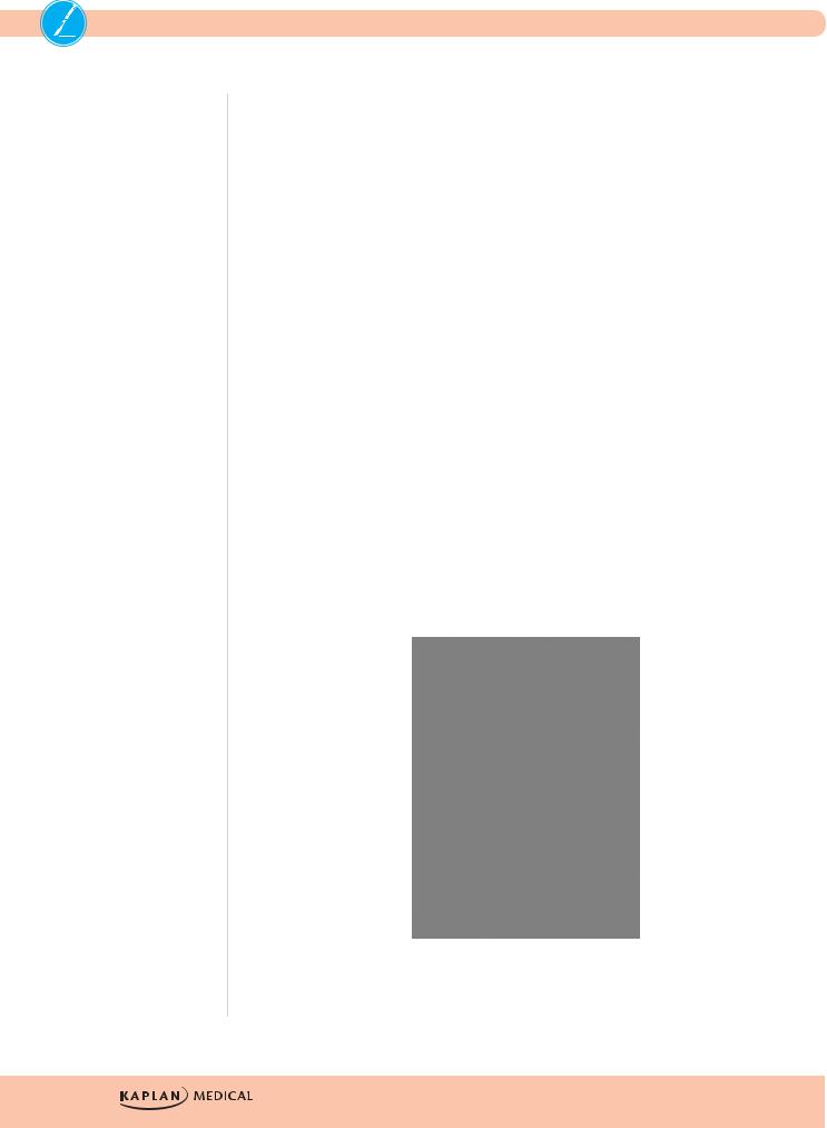
Carpal tunnel syndrome occurs following performance of repetitive hand work such as typing and presents with numbness and tingling in both hands in the distribution of the median nerve (radial 3½ fingers). The symptoms can be reproduced by hanging the hand limply for a few minutes, or by tapping, percussing or pressing the median nerve over the carpal tunnel (Tinel’s sign). The diagnosis is clinical, but the American Academy of Orthopaedic Surgery recommends that wrist x-rays (including carpal tunnel view) be done to rule out other pathology. Initial treatment is splinting and anti-inflammatory agents. If these conservative measures fail, surgery is indicated following electromyography and nerve conduction velocity.
Figure I-2-8. Thenar Atrophy is a Feature of Carpal Tunnel Syndrome
Trigger finger is more common in women and presents with acute finger flexion and the inability to extend it unless pulled with the other hand, which results in a painful “snap.” Steroid injection is the first line of therapy; surgery is the treatment of last resort.
De Quervain tenosynovitis is seen in young mothers who, as they carry their baby, force their hand into wrist flexion and thumb extension to hold the baby’s head. They complain of pain along the radial side of the wrist and the first dorsal compartment. On physical exam the pain can be reproduced by asking her to hold the thumb inside her closed fist, then forcing the wrist into ulnar deviation. Splint and anti-inflammatory agents can help, but steroid injection is most effective. Surgery is rarely needed.
28

Chapter 2 λ Orthopedics
Dupuytren contracture occurs in older men of Norwegian ancestry and in alcoholics. There is contracture of the palm of the hand, and palmar fascial nodules can be felt. Surgery may be needed when the hand can no longer be placed flat on a table.
A felon is an abscess in the pulp of a fingertip, caused by a neglected penetrating injury. Patients complain of throbbing pain, and have all the classic findings of an abscess, including fever. Because the pulp is a closed space with multiple fascial trabecula, pressure can build up and lead to tissue necrosis; thus surgical drainage is urgently indicated.
Gamekeeper thumb is an injury of the ulnar collateral ligament sustained by forced hyperextension of the thumb (historically suffered by gamekeepers when they killed rabbits by dislocating their necks with a violent blow with the extended thumb—nowadays seen as a skiing injury when the thumb gets stuck in the snow or the ski strap during a fall). On physical exam there is collateral laxity at the thumb-metacarpophalangeal joint, and if untreated it can be dysfunctional and painful, and lead to arthritis. Casting is usually effective.
Jersey finger is an injury to the flexor tendon sustained when the flexed finger is forcefully extended (as in someone unsuccessfully grabbing a running person by the jersey). When making a fist, the distal phalanx of the injured finger does not flex with the others.
Mallet finger is the opposite: the extended finger is forcefully flexed (a common volleyball injury), and the extensor tendon is ruptured. The tip of the affected finger remains flexed when the hand is extended, resembling a mallet. For both of these injuries, splinting is usually the first line of treatment.
Traumatically amputated digits are surgically reattached whenever possible. The amputated digit should be cleaned with sterile saline, wrapped in a saline-moistened gauze, placed in a sealed plastic bag, and the bag placed on a bed of ice. The digit should not be placed in antiseptic solutions or alcohol, should not be put on dry ice, and should not be allowed to freeze. With the use of electric nerve stimulation to preserve muscular function, entire amputated extremities can be reattached.
Back Pain
Lumbar disk herniation occurs most commonly at L4–L5 or L5–S1. Peak age incidence is the fourth decade of life.
•Patients often describe several months of vague aching pain (the “discogenic pain” produced by pressure on the anterior spinal ligament) before they have the sudden onset of the “neurogenic pain” precipitated by a forced movement.
•The latter is extremely severe, “like an electrical shock that shoots down the leg” (exiting on the side of the big toe in L4–L5, or the side of the little toe in L5–S1), and it
is exacerbated by coughing, sneezing, or defecating (if the pain is not exacerbated by those activities, the problem is not a herniated disk). Patients cannot ambulate, and they hold the affected leg flexed.
•Straight leg-raising test reproduces excruciating pain and MRI confirms the diagnosis.
•Treatment for most patients is bed rest, physical therapy, and pain control, enhanced by a regional nerve block; surgical intervention is needed if neurologic deficits are progressing; emergency intervention is needed in the presence of the cauda equine syndrome (distended bladder, flaccid rectal sphincter, or perineal saddle anesthesia).
29
USMLE Step 2 CK λ Surgery
Copyright 2007 Bates, M.D. - Custom Medical Stock Photo.
Figure I-2-9. Spine MRI Showing Lumbar
Disc Herniation of L4-L5 Interspace
Ankylosing spondylitis is seen in men in the third and fourth decades of life who complain of chronic back pain and morning stiffness. The pain is worse at rest, and improves with activity. Symptoms are progressive, and x-rays reveal a “bamboo spine.” Anti-inflammatory agents and physical therapy are effective. Many of these patients have the HLA B-27 antigen, which is also associated with uveitis and inflammatory bowel disease.
Metastatic malignancy should be suspected in the elderly who have progressive back pain that is worse at night and unrelieved by rest or positional changes. Weight loss is often an additional finding. The most common pathology is lytic breast cancer metastases in women and blastic prostate metastases in men. Most lesions are identifiable on x-ray, but MRI is a more sensitive diagnostic tool.
Leg Ulcers
Diabetic ulcer is typically indolent and located at pressure points (heel and metatarsal head). It starts because of the neuropathy, and does not heal because of the microvascular disease. It can sometimes heal with good blood glucose control and wound care, but often becomes chronic and sometimes leads to amputation due to osteomyelitis.
30

Chapter 2 λ Orthopedics
Copyright 2007 Biomedical Communications - Custom Medical Stock Photo.
All rights reserved.
Figure I-2-10. Gross Appearance of a Large Diabetic Foot Ulcer
Ulcer from arterial insufficiency is usually as far away from the heart as it can be, i.e., at the tip of the toes. It looks dirty, with a pale base devoid of granulation tissue. The patient has other manifestations of arteriosclerotic occlusive disease (absent pulses, trophic changes, claudication, or rest pain). Workup begins with Doppler studies looking for a pressure gradient, though in the presence of microvascular disease this may not be present (and these lesions are less amenable to surgical therapy). Further evaluation with CT angiogram may be necessary, and ultimately, formal angiography leading to angioplasty, stenting, or surgical revascularization.
Venous stasis ulcer develops in chronically edematous, indurated, and hyperpigmented skin above the medial malleolus. The ulcer is painless, with granulating bed. The patient has varicose veins, and suffers from frequent bouts of cellulitis. Duplex scan is useful in the workup. Treatment revolves around physical support to keep the veins empty: support stockings, Ace bandages, and Unna boot. Surgery may be required (vein stripping, grafting of the ulcer, injection sclerotherapy); endovascular ablation with laser or radiofrequency may also be used.
wikipedia.org.
Figure I-2-11. Venous Stasis Ulcers
31
USMLE Step 2 CK λ Surgery
Marjolin’s ulcer is a squamous cell carcinoma of the skin that has developed in a chronic leg ulcer. The classic setting is one of many years of healing and breaking down, such as seen in untreated third-degree burns that underwent spontaneous healing, or in chronic draining sinuses secondary to osteomyelitis. A dirty-looking, deeper ulcer develops at the site, with heaped up tissue growth around the edges. Biopsy is diagnostic. Treatment is wide local excision and skin grafting if necessary.
Foot Pain
Plantar fasciitis is a very common but poorly understood problem affecting older, overweight patients who complain of disabling, sharp heel pain every time their foot strikes the ground.
•The pain is worse in the mornings.
•X-rays show a bony spur matching the location of the pain, and physical exam shows exquisite tenderness to palpation over the spur, although the bony spur is not likely the cause of the problem as many asymptomatic people have similar spurs.
•Spontaneous resolution occurs over several months, during which time symptomatic treatment is offered.
Morton’s neuroma is an inflammation of the common digital nerve at the third interspace, between the third and fourth toes. The neuroma is palpable and exquisitely tender to palpation. The cause is typically the use of pointed, high heel shoes (or pointed cowboy boots) that force the toes to be bunched together. Management includes analgesics and more sensible shoes, but surgical excision can be performed if conservative management fails.
Gout produces the typical swelling, redness, and exquisite pain of sudden onset at the first metatarsal-phalangeal joint in a middle-age obese man with high serum uric acid. Uric acid crystals are identified in fluid from the joint. Treatment for the acute attack is indomethacin and colchicine; treatment for chronic control is allopurinol and probenicid.
Copyright 2007 NMSB - Custom Medical Stock Photo.
Figure I-2-12. Gross Appearance of Acute Gout
32
Chapter 2 λ Orthopedics
TUMORS
Children and Young Adults
Primary malignant bone tumors are diseases of young people. They present with persistent low-grade pain for several months.
•Osteogenic sarcoma is the most common primary malignant bone tumor.
––It is seen in ages 10–25, usually around the knee (lower femur or upper tibia).
––A typical “sunburst” pattern is often described on x-rays.
•Ewing sarcoma is the second most common.
––It affects younger children (ages 5–15) and it grows in the diaphyses of long bones.
––A typical “onion skinning”–type pattern is often seen on x-rays.
Adults
Most malignant bone tumors in adults are metastatic, from the breast in women (lytic lesions) and from the prostate in men (blastic lesions). Localized pain is an early finding. X-rays can be diagnostic, CT scans give more information, and MRI is even more sensitive. Lytic lesions commonly present as pathologic fractures.
•Multiple myeloma is seen in old men and presents with fatigue, anemia, and localized pain at specific places on several bones. X-rays are diagnostic, showing multiple, punched-out lytic lesions.
––They also have Bence-Jones protein in the urine and abnormal immunoglobulins in the blood, best demonstrated by serum proptein electrophoresis (SPEP).
––Treatment is chemotherapy; thalidomide can be used in the event that chemotherapy fails.
•Soft tissue sarcoma has relentless growth of soft tissue mass over several months. It is firm and typically fixed to surrounding structures.
––It can metastasize hematogenously to the lungs but does not invade the lymphatic system.
––MRI delineates the extent of the mass and invasion of local structures.
––Incisional biopsy to obtain tissue is diagnostic.
––Treatment includes wide local excision, radiation, and chemotherapy.
Copyright 2007 - Custom Medical Stock Photo.
All rights reserved.
Figure I-2-13. Shoulder X-ray Showing
Punched-out Lesions of Multiple Myeloma
33