

7 Microbial Genetics: Replication and Expression of Genetic Information, 191
Molecular Basis of Heredity, 192 Structure of DNA, 194
Nucleotides—Building Blocks of the Genetic Code, 194
Unity of the Microbial World
Chapter Outline
Microorganisms 3
What is a Microorganism?
Newsbreak: French Revolution Leads to the Metric
System Organizational Structure of Microorganisms
Cells of Living Organisms Historical Perspective: Discovery of Microorganisms
Unicellular and Multicellular Organisms
Prokaryotic and Eukaryotic Cells
Acellular Nonliving Viruses Highlight: Practical Significance of
Organizational Differences Among Microorganisms Importance of Microorganisms to Humankind 14 Highlight: A Practical View of Microbiology Microorganisms and Disease Newsbreak: Origins of Vaccination Newsbreak: Source of Cholera in London Beneficial Uses of Microorganisms Newsbreak: Biotechnology to Cure Global Warming
Preview to Chapter ?
In this chapter we will:
Study unifying characteristics of microorganisms.
Define the scope of the science of microbiology.
Describe the attributes of microorganisms.
Distinguish microorganisms from plants and animal
Examine the structural organization of microorganis
Identify major characteristics of viruses, prokaryotic eubacteria and archaebacteria, and eukaryotic fungi, algae, and protozoa.
Gain insight into the relevance of microorganisms tc human health and well-being.
Learn the following key terms and names:
algae microscope
archaebacteria multicellular
bacteria nucleus
cell organelles
cellular metabolism pathogens
DNA (deoxyribonucleic plasma membrane
acid) prokaryotic cells
eubacteria protozoa
eukaryotic cells tissues ['tɪʃuː]
fungi unicellular
microbiology viruses ['vīrəs] ['vaɪ(ə)rəs] microorganism
MICROORGANISMS
Many of us equate the terms microorganisms and germs. This is not surprising since we continuously battle microorganisms to maintain our health. Unseen microorganisms exert a powerful influence on our lives. By learning about microorganisms you will be able to understand why we sometimes suffer infections and disease. You also will be able to see how modern medical practices attempt to control and to eliminate disease-causing microorganisms and to treat infectious diseases when they do occur. At the same time you may be surprised that, while the general layman's view is that all microorganisms are harmful to human health, most microorganisms do not cause disease. Our skin, hair, tongue, intestines, and other body parts are literally swarming with bacteria that do us no harm. In fact, they are advantageous, or even necessary to us. Life on earth depends on microorganisms that have an enormous capacity to degrade and to recycle materials.
What is a Microorganism?
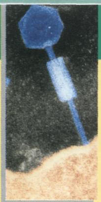
As you begin your study of microbiology—the science that deals with microorganisms — you may ask, exactly what is a microorganism and how do microorganisms differ from other organisms? As implied by the word microorganism (from the Greek word micro, meaning small), microorganisms are very small life forms—so small that individual microorganisms usually cannot be seen without magnification (FIG. 1-1). These microorganisms include the viruses, bacteria, fungi, algae, and protozoa. They are the organisms that microbiologists study (FIG. 1-2).
To describe size, scientists use the metric system measure of length that is based on a meter (a meter is approximately a yard). Fractions of a meter are described by using prefixes such as milli (one thousandth), micro (one millionth), and nano (one billionth). A millimeter (mm) is a thousandth of a meter

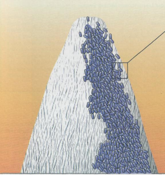
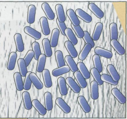
FIG. 1-1 Microorganisms are extremely small, so they are invisible to the naked eye. Most bacteria are 0.5 to 2 µm in length. A million bacteria can fit on the tip of a pin. Individual bacteria can only be seen by using a microscope to magnify the image.
4 С H A P T E R 1 UNITY OF THE MICROBIAL WORLD
French Revolution Leads to the Metric System
The metric system was introduced during the French Revolution, in part because of concerns with differences in the measuring systems used in various parts of France but equally to demonstrate that a new governmental regime was in charge. The fundamental unit of measuring length is the metric system, the meter (m), was defined as one ten-millionth the distance from the earth's north pole to the equator. The official measurement was made in 1799 geometrically between Dunkirk and Barcelona. A platinum meter was deposited in the Archives of the Republic as the official standard mea-
sure of length. Although French proponents of the reform in weights and measures sought a system based on some natural universal unit to be used throu ghoul the world, England rejected the metric system as impractical. Jefferson, although enthusiastic about such г change for the United States, rejected the French systerc and tried to develop his own system of measures. Thus while the rest of the world uses the metric system, the United States and Britain do not generally use this sys tern of measure, except for scientists—who, the work over, use the metric system.
or approximately 0.004 inches; a micrometer (|µm) is a millionth of a meter or approximately 0.000004 inches, and a nanometer (nm) is a billionth of a meter or approximately 0.000000004 inches.
The smallest object that can be seen with the naked eye is about a tenth of a millimeter (0.1 mm), which is equal to 100 micrometers (100 µm). Most microorganisms are smaller than this. Largest bacterial cells,
for example, are only 5 µm in length (0.005 mm), small bacterial cells are about 0.1 µm in length (µm less than a millionth of a meter). Therefore generally must magnify the images of most bac about 1,000 times just to be able to see them, viruses are smaller yet. The largest viruses almost 0.1 µm and the smallest viruses are 0.01 (less than a millionth of a meter). Thus the imag
MICROORGANISMS 5
viruses must be magnified 10,000 to 100,000 times to be seen.
Microorganisms are defined by their small size, since they generally are invisible to the naked eye and can only be seen with a microscope.
Organizational Structure of Microorganisms
Cells of Living Organisms
The cell is the fundamental unit of all living systems, including all living microorganisms. Cells have a boundary layer, called the plasma membrane, that separates the living cell from the external surroundings. The plasma membrane controls the flow of materials into and out of the cell. It permits the cell to maintain the highly organized state that is a major characteristic of living systems. If the plasma membrane is damaged and fails to regulate the flow of materials, the cell dies and life ceases. To protect this essential structure, many cells have a rigid cell wall surrounding the plasma membrane.
Cells are the fundamental functional units of living organisms.
All cells have a plasma membrane that separates the living system from the nonliving surroundings.
Cells carry out the essential functions of life, including processing of energy and materials for growth and replication of hereditary information for reproduction. Cells contain the molecule DNA (deoxyribonucleic acid), which is the universal substance of living cells that passes hereditary information to offspring cells. The DNA of a cell specifies the potential characteristics of that cell and is often called the "master molecule of life" because it directs the activities of the cell. In all living cells there is a flow of information from the DNA through molecules of RNA (ribonucleic acid) to direct the synthesis of proteins. The synthesis of proteins occurs at numerous ribosomes within the cell. Proteins are the action molecules of life, catalyzing chemical reactions within living cells. By specifying the proteins that a cell makes, the information contained in the DNA directs cellular metabolism, the process in which living cells utilize energy and transform materials into the structures necessary for growth and reproduction. All living cells must carry out metabolism to sustain life, one of the essential functions of which is to generate ATP (adenosine triphosphate) as a central currency of cellular energy. Thus cells uniformly have structures that regulate the flow of materials, the storage and expression of genetic information, and the ability to carry out metabolism (FIG. 1-3).
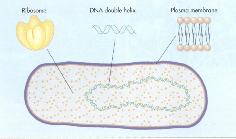
FIG. 1 -3 The cell is the fundamental unit of all living organisms. It is separated from its surroundings by a plasma membrane that regulates the flow of materials into and out of the cell. The organism's hereditary information is contained within the cell in molecules of DNA. Proteins that catalyze the metabolism of the living organism are synthesized at ri-bosomes within the cell.
6 CHAPTER 1 UNITY OF THE MICROBIAL WORLD
Discovery of Microorganisms

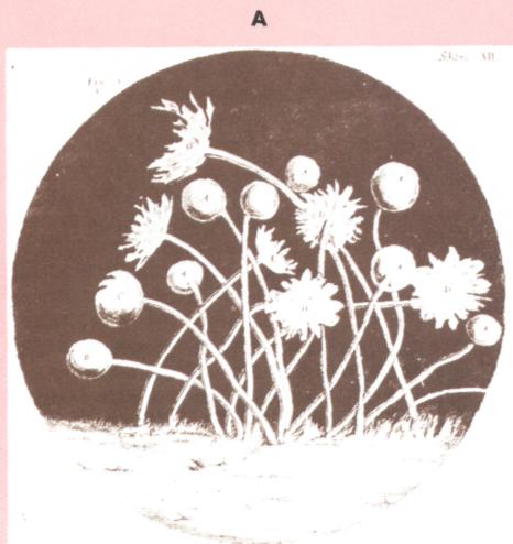
The first microscopes were simple ground glass lenses that magnified images of previously unseen objects. By the late seventeenth century, microscopes permitted magnifications of several hundred times, making it possible to discover the microbial world. Among the first to observe the previously invisible microbial world was Robert Hooke, an English scientist. His detailed drawings of fungi, made in 1667, reflect hours of tedious observations (FIG. A).
Antonie van Leeuwenhoek, an amateur scientist and official winetaster of Delft, Holland, made the first recorded observations of bacteria in 1670 (FIG. B). Van Leeuwenhoek's interest in microscopes was probably related to the use of magnifying glasses by drapers to examine fabrics. His hobby was microscopy and he made over 100 microscopes, each consisting of a simple glass lens (FIG. C). These microscopes were little more than magnifying glasses, each capable of magnifying an image about 300 times, so that bacteria could barely be seen as fuzzy images. Leeuwenhoek must have had great patience and persistence to squint through the lens of his handheld microscope at dimly lighted specimens
Robert Hooke made microscopic observations and provided the earliest descriptions of many fungi. Various species of fungi can clearly be identified in his drawings made from 1635 to 1703 and recorded in his book, Micrographia.
Antonie van Leeuwenhoek (1632-1723), here seen holding one of his microscopes, opened the door to the hidden world of microorganisms when he described bacteria. Although he was only an amateur scientist, Leeuwenhoek's keen interest in optics and his diligence allowed him to make this important discovery.
8 CHAPTERl UNITY OF THE MICROBIAL WORLD
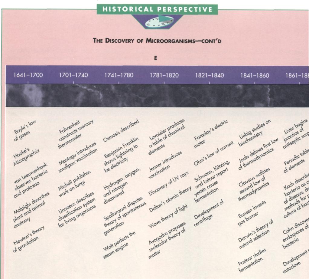
Time line of some scientific discoveries important to the development of microbiology. The study of microorganisms, which began in the late 1600s with the observations of Leeuwenhoek, developed in the mid-nineteenth century into a true science through the studies of Robert Koch and Louis Pasteur. Major developments have been made in the twentieth century, including the discovery of antibiotics and the molecular basis of heredity. Today microbiology is a flourishing field of science.

Cells have genetic information in the form of DNA that directs the form and function of the cell and passes hereditary information from one generation to the next.
Cells carry out metabolism that transforms energy and materials for growth and reproduction.
Unicellular and Multicellular Organisms
Living organisms are composed of one or more cells. The human body, for example, is composed of trillions of cells. Many microorganisms are unicellular, meaning that the entire organism is composed of a single cell. Most bacteria, for example, are unicellular. Each bacterial cell comprises the entire organism and is capable of independently carrying out metabolism for growth and reproduction to form progeny (offspring). Some microorganisms are multicellular, meaning that they are composed of many cells. Most multicellular microorganisms—such as the fungi that can often be seen as fuzzy filaments growing on bread—can be viewed as aggregates of cells with each individual cell having the properties of the entire organism. Each individual cell retains the capacity of reproducing to form the entire organism.
Microorganisms do not exhibit the advanced structural organization of plants and animals that form specialized groups of cells called tissues. Microorganisms lack tissues. The cells of a plant or animal tissue function together as integrated units. In animals, for example, we find epithelial tissue covering the external body surface, connective tissue binding together and supporting other tissues, muscle tissue moving parts of the animal's body, and nerve tis-
sue transmitting messages from one part of the ani mal to another. The cells of these differentiated tis sues usually lose the ability to generate the entire or ganism.
Microorganisms are more simply organized than plants and animals.
Microorganisms lack differentiated tissues.
Prokaryotic and Eukaryotic Cells
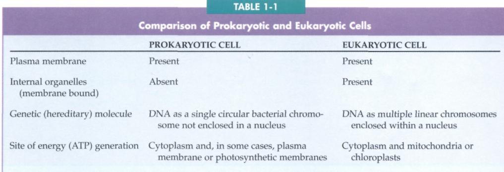
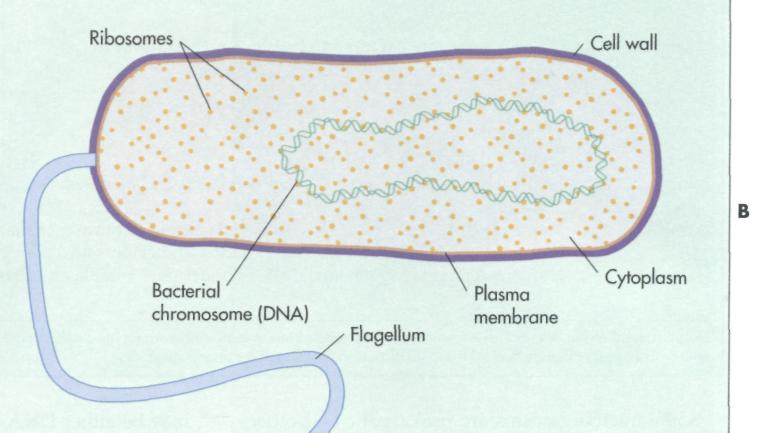
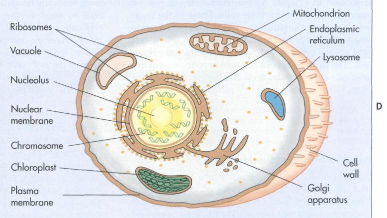
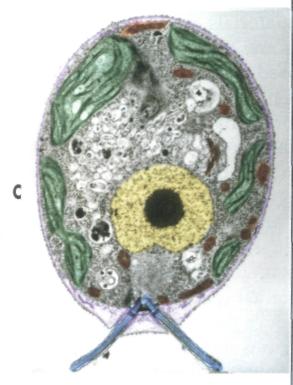
Among all living organisms there are two fundamen tally different types of cells: prokaryotic cells (fron the Greek word meaning before a nucleus) and eukaryotic cells (from the Greek word meaning having a nucleus) (FIG. 1-4). Both prokaryotic cells and eu karyotic cells carry out all the essential life functions exchange of materials with the environment, energprocessing, and reproduction. However, the struc tures of these types of cells, discussed in detail ir Chapter 5, are quite different (Table 1-1).
A prokaryotic cell has a much simpler interna structure than a eukaryotic cell. Eukaryotic cells have numerous membrane-bound compartments, callec organelles, that perform specialized functions. Pro karyotic cells do not contain membrane-bound or ganelles. Of prime importance is the fact that the DNA (the substance that encodes the hereditary information of the cell) is not separated within a specialized organelle from the rest of the cell contents ir a prokaryotic cell. The DNA of a eukaryotic cell, ir contrast, is contained within an organelle called the nucleus.
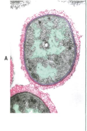
A, Colorized micrograph of a prokaryotic cell of the bacterium Pseudomonas aeruginosa (32,400X).
B, Drawing of a prokaryotic cell.
C, Colorized micrograph of a eukaryotic cell of the green alga Chlamy-domonas reinhardtii (6,750x).
D, Drawing of an algal eukaryotic cell.