

Se “lugansk state medical university”
Chair of internal medicine with basis of pulmonology
Subject №15: «peptic ulcer disease»
Amount of education hours: 2
CONSPECT OF THE LECTURE
History ofepigastric pain present in 80-90 % of patient, but is nonspecific.
Burning epigastric pain exacerbated by fasting and improved with meals is a symptom complex associated with peptic ulcer disease (PUD).
Anulcer is defined as disruption of the mucosal integrity of the stomach and/or duodenum leading to a local defect or excavation due to active inflammation. Ulcers occur within the stomach and/or duodenum and are often chronic in nature.
Acid peptic disorders are very common in the United States, with 4 million individuals (new cases and recurrences) affected per year. Lifetime prevalence of PUD in the United States is ~12% in men and 10% in women. Moreover, an estimated 15,000 deaths per year occur as a consequence of complicated PUD. The financial impact of these common disorders has been substantial, with an estimated burden on direct and indirect health care costs of ~$10 billion per year in the United States.
Why does the ulcer appear ? Let remember the physiology of gastric secretion.
Physiology of Gastric Secretion
Hydrochloric acid and pepsinogen are the two principal gastric secretory products capable of inducing mucosal injury. Acid secretion should be viewed as occurring under basal and stimulated conditions. Basal acid production occurs in a circadian pattern, with highest levels occurring during the night and lowest levels during the morning hours. Cholinergic input via the vagus nerve and histaminergic input from local gastric sources are the principal contributors to basal acid secretion. Stimulated gastric acid secretion occurs primarily in three phases based on the site where the signal originates (cephalic, gastric, and intestinal).
- Sight, smell, and taste of food are the components of the cephalic phase, which stimulates gastric secretion via the vagus nerve.
- The gastric phase is activated once food enters the stomach. This component of secretion is driven by nutrients (amino acids and amines) that directly stimulate the G cell to release gastrin, which in turn activates the parietal cell via direct and indirect mechanisms. Distention of the stomach wall also leads to gastrin release and acid production.
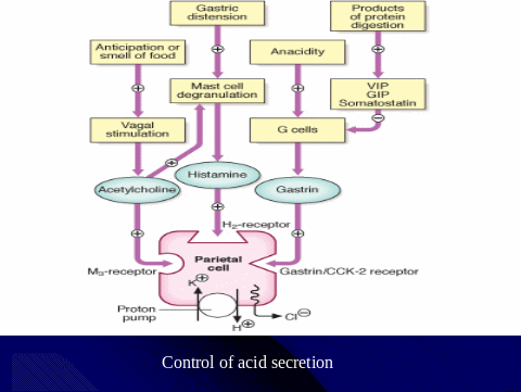
- The last phase of gastric acid secretion is initiated as foodenters the intestine and is mediated by luminal distention and nutrient assimilation. A series of pathways that inhibit gastric acid production are also set into motion during these phases. The gastrointestinal hormone somatostatin is released from endocrine cells found in the gastric mucosa (D cells) in response to HCl. Somatostatin can inhibit acid production by both direct (parietal cell) and indirect mechanisms [decreased histamine release from enterochromaffin-like (ECL) cells and gastrin release from G cells. Additional neural (central and peripheral) and hormonal (secretin, cholecystokinin) factors play a role in counterbalancing acid secretion. Under physiologic circumstances, these phases are occurring simultaneously.
The gastric epithelium is under a constant assault by a series of endogenous noxious factors including HCl, pepsinogen/pepsin, and bile salts. In addition, a steady flow of exogenous substances such as medications, alcohol, and bacteria encounter the gastric mucosa. A highly intricate biologic system is in place to provide defense from mucosal injury and to repair any injury that may occur.
The mucosal defense system can be envisioned as a three-level barrier, composed of preepithelial, epithelial, and subepithelial elements. The first line of defense is a mucus-bicarbonate layer, which serves as a physicochemical barrier to multiple molecules including hydrogen ions. Mucus is secreted in a regulated fashion by gastroduodenal surface epithelial cells. It consists primarily of water (95%) and a mixture of lipids and glycoproteins. Mucin is the constituent glycoprotein that, in combination with phospholipids (also secreted by gastric mucous cells), forms a hydrophobic surface with fatty acids that extend into the lumen from the cell membrane. The mucous gel functions as a nonstirred water layer impeding diffusion of ions and molecules such as pepsin. Bicarbonate, secreted by surface epithelial cells of the gastroduodenal mucosa into the mucous gel, forms a pH gradient ranging from 1 to 2 at the gastric luminal surface and reaching 6 to 7 along the epithelial cell surface. Bicarbonate secretion is stimulated by calcium, prostaglandins, cholinergic input, and luminal acidification.
Surface epithelial cells provide the next line of defense through several factors, including mucus production, epithelial cell ionic transporters that maintain intracellular pH and bicarbonate production, and intracellular tight junctions. If the preepithelial barrier were breached, gastric epithelial cells bordering a site of injury can migrate to restore a damaged region (restitution). This process occurs independent of cell division and requires uninterrupted blood flow and an alkaline pH in the surrounding environment. Several growth factors including epidermal growth factor (EGF), transforming growth factor (TGF) α, and basic fibroblast growth factor (FGF) modulate the process of restitution. Larger defects that are not effectively repaired by restitution require cell proliferation. Epithelial cell regeneration is regulated by prostaglandins and growth factors such as EGF and TGF-α. In tandem with epithelial cell renewal, formation of new vessels (angiogenesis) within the injured microvascular bed occurs. Both FGF and vascular endothelial growth factor (VEGF) are important in regulating angiogenesis in the gastric mucosa.
An elaborate microvascular system within the gastric submucosal layer is the key component of the subepithelial defense/repair system. A rich submucosal circulatory bed provides HCO3-, which neutralizes the acid generated by parietal cell secretion of HCl. Moreover, this microcirculatory bed provides an adequate supply of micronutrients and oxygen while removing toxic metabolic by-products.
Prostaglandins play a central role in gastric epithelial defense/repair (Fig. 274-4). The gastric mucosa contains abundant levels of prostaglandins. These metabolites of arachidonic acid regulate the release of mucosal bicarbonate and mucus, inhibit parietal cell secretion, and are important in maintaining mucosal blood flow and epithelial cell restitution.
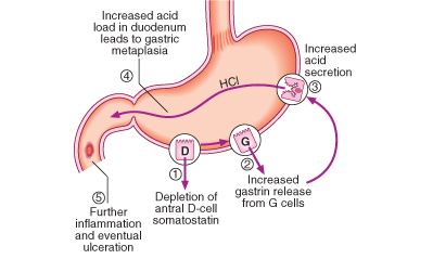
P. Seguence of events in the pathophysiology of duodenal ulceration
PATHOPHYSIOLOGIC BASIS OF PEPTIC ULCER DISEASE
Multiple factors play a role in the pathogenesis of PUD. The two predominant causes are H. pylori infection and NSAID ingestion. PUD not related to H. pylori or NSAIDs may be increasing. Independent of the inciting or injurious agent, peptic ulcers develop as a result of an imbalance between mucosal protection/repair and aggressive factors. Gastric acid plays an essential role in mucosal injury.
PUD encompasses both gastric and duodenal ulcers.
Ulcers are defined as a break in the mucosal surface >5 mm in size, with depth to the submucosa. Duodenal ulcers (DUs) and gastric ulcers (GUs); share many common features in terms of pathogenesis, diagnosis, and treatment, but several factors distinguish them from one another.
Epidemiology
DUODENAL ULCERS
DUs are estimated to occur in 6 to 15% of the western population. The incidence of DUs declined steadily from 1960 to 1980 and has remained stable since then. The death rates, need for surgery, and physician visits have decreased by >50% over the past 30 years. The reason for the reduction in the frequency of DUs is likely related to the decreasing frequency of Helicobacter pylori. Before the discovery of H. pylori, the natural history of DUs was typified by frequent recurrences after initial therapy. Eradication of H. pylori has greatly reduced these recurrence rates.
GASTRIC ULCERS
GUs tend to occur later in life than duodenal lesions, with a peak incidence reported in the sixth decade. More than half of GUs occur in males and are less common than DUs, perhaps due to the higher likelihood of GUs being silent and presenting only after a complication develops. Autopsy studies suggest a similar incidence of DUs and GUs.
MISCELLANEOUS PATHOGENETIC FACTORS IN ACID PEPTIC DISEASE
Cigarette smoking has been implicated in the pathogenesis of PUD. Not only have smokers been found to have ulcers more frequently than do nonsmokers, but smoking appears to decrease healing rates, impair response to therapy, and increase ulcer-related complications such as perforation. The mechanism responsible for increased ulcer diathesis in smokers is unknown. Theories have included altered gastric emptying, decreased proximal duodenal bicarbonate production, increased risk for H. pylori infection, and cigarette-induced generation of noxious mucosal free radicals. Acid secretion is not abnormal in smokers. Despite these interesting theories, a unifying mechanism for cigarette-induced peptic ulcer diathesis has not been established.
Genetic predisposition has also been considered to play a role in ulcer development. First-degree relatives of DU patients are three times as likely to develop an ulcer; however, the potential role of H. pylori infection in contacts is a major consideration. Increased frequency of blood group O and of the nonsecretor status have also been implicated as genetic risk factors for peptic diathesis. However, H. pylori preferentially binds to group O antigens. Therefore, the role of genetic predisposition in common PUD has not been established.
Psychological stress has been thought to contribute to PUD, but studies examining the role of psychological factors in its pathogenesis have generated conflicting results. Although PUD is associated with certain personality traits (neuroticism), these same traits are also present in individuals with nonulcer dyspepsia (NUD) and other functional and organic disorders. Although more work in this area is needed, no typical PUD personality has been found.
Diet has also been thought to play a role in peptic diseases. Certain foods can cause dyspepsia, but no convincing studies indicate an association between ulcer formation and a specific diet. This is also true for beverages containing alcohol and caffeine. Specific chronic disorders have been associated with PUD. Those with a strong association are (1) systemic mastocytosis, (2) chronic pulmonary disease, (3) chronic renal failure, (4) cirrhosis, (5) nephrolithiasis, and (6) α1-antitrypsin deficiency. Those with a possible association are (1) hyper- parathyroidism, (2) coronary artery disease, (3) polycythemia vera, and (4) chronic pancreatitis.
Multiple factors play a role in the pathogenesis of PUD. The two predominant causes are H. pylori infection and NSAID ingestion. PUD not related to H. pylori or NSAIDs may be increasing. Independent of the inciting or injurious agent, peptic ulcers develop as a result of an imbalance between mucosal protection/repair and aggressive factors. Gastric acid plays an essential role in mucosal injury.
Pathophysiology
It is now clear that H. pylori and NSAID-induced injury account for the majority of DUs. Gastric acid contributes to mucosal injury but does not play a primary role.
DUODENAL ULCERS
Many acid secretory abnormalities have been described in DU patients. Of these, average basal and nocturnal gastric acid secretion appear to be increased in DU patients as compared to control; however, the level of overlap between DU patients and control subjects is substantial. The reason for this altered secretory process is unclear, but H. pylori infection may contribute to this finding. Accelerated gastric emptying of liquids has been noted in some DU patients but is not consistently observed; its role in DU formation, if any, is unclear. Bicarbonate secretion is significantly decreased in the duodenal bulb of patients with an active DU as compared to control subjects. H. pylori infection may also play a role in this process.
GASTRIC ULCERS
As in DUs, the majority of GUs can be attributed to either H. pylori or NSAID-induced mucosal damage. GUs that occur in the prepyloric area or those in the body associated with a DU or a duodenal scar are similar in pathogenesis to DUs. Gastric acid output (basal and stimulated) tends to be normal or decreased in GU patients. When GUs develop in the presence of minimal acid levels, impairment of mucosal defense factors may be present.
H. PYLORI AND ACID PEPTIC DISORDERS
Gastric infection with the bacterium H. pylori accounts for the majority of PUD. This organism also plays a role in the development of gastric mucosal-associated lymphoid tissue (MALT) lymphoma and gastric adenocarcinoma. Although the entire genome of H. pylori has been sequenced, it is still not clear how this organism, which is in the stomach, causes ulceration in the duodenum, or whether its eradication will lead to a decrease in gastric cancer.
The Bacterium initially named Campylobacter pyloridis, is a gram-negative microaerophilic rod found most commonly in the deeper portions of the mucous gel coating the gastric mucosa or between the mucous layer and the gastric epithelium. It may attach to gastric epithelium but under normal circumstances does not appear to invade cells. It is strategically designed to live within the aggressive environment of the stomach. It is S-shaped (~0.5 × 3 µm in size) and contains multiple sheathed flagella. Initially, H. pylori resides in the antrum but, over time, migrates toward the more proximal segments of the stomach.
The bacterium, initially named Campylobacter pyloridis, is a gram-negative microaerophilic rod found most commonly in the deeper portions of the mucous gel coating the gastric mucosa or between the mucous layer and the gastric epithelium. It may attach to gastric epithelium but under normal circumstances does not appear to invade cells. It is strategically designed to live within the aggressive environment of the stomach. It is S-shaped (~0.5 × 3 µm in size) and contains multiple sheathed flagella. Initially, H. pylori resides in the antrum but, over time, migrates toward the more proximal segments of the stomach.
Two factors that predispose to higher colonization rates include poor socioeconomic status and less education
Transmission of H. pylori occurs from person to person, following an oral-oral or fecal-oral route. The risk of H. pylori infection is declining in developing countries. The rate of infection in the United States has fallen by >50% when compared to 30 years ago.
Pathophysiology
H. pylori infection is virtually always associated with a chronic active gastritis, but only 10 to 15% of infected individuals develop frank peptic ulceration. The basis for this difference is unknown. Initial studies suggested that >90% of all DUs were associated with H. pylori, but H. pylori is present in only 30 to 60% of individuals with GUs and 70% of patients with DUs. The pathophysiology of ulcers not associated with H. pylori or NSAID ingestion [or the rare Zollinger-Ellison syndrome (ZES)] is unclear.
The particular end result of H. pylori infection (gastritis, PUD, gastric MALT lymphoma, gastric cancer) is determined by a complex interplay between bacterial and host factors
About 20,000 patients die each year from serious gastrointestinal complications from NSAIDs. Unfortunately, dyspeptic symptoms do not correlate with NSAID-induced pathology. Over 80% of patients with serious NSAID-related complications did not have preceding dyspepsia. In view of the lack of warning signs, it is important to identify patients who are at increased risk for morbidity and mortality related to NSAID usage. Even 75 mg/d of aspirin may lead to serious gastrointestinal ulceration, thus no dose of NSAID is completely safe.