198 CHAPTER 7 MICROBIAL GENETICS: REPLICATION AND EXPRESSION OF GENETIC INFORMATION
in
DNA are single-ring structures called pyrimidines
and the
other two (A and G) are double-ring structures called purines.
The
charge interactions between purines and pyrimidines allow them to
form weak hydrogen bonds (FIG. 7-7). Chemically the most stable
hydrogen bonding occurs when guanine forms three hydrogen bonds with
cytosine and when adenine forms two hydrogen bonds with
thymine. The proper alignment to form these hydrogen bonds occurs
only when the sugar-phosphate backbones of the two DNA chains run in
opposing directions and are twisted together to form the double
helix.
DNA,
which stores and transmits cellular hereditary
The
hydrogen bonding of A to T and C to G is called base
pairing. It
is this complementarity that establishes the basis for the double
helical arrangement of DNA and for the accurate replication of
the DNA macromolecule. This is essential for passage of hereditary
information from one generation to the next. It also means that in
the double helical DNA molecule, the amount of adenine is always the
same as the amount of thymine, and the amount of guanine is
always the same as the amount of cytosine (A = T and G = C).
FIG.
7-7 Hydrogen
bonding occurs between nucleotide base pairs. Adenine forms two
hydrogen bonds with thymine. Guanine forms three hydrogen bonds with
cytosine.
Base
pairing occurs between complementary nucleotides—adenine
pairs with thymine and guanine pairs with cytosine.
REPLICATION
OF DNA
When
a cell divides, its hereditary information is passed to the next
generation. Replication of the hereditary information involves
synthesizing new DNA molecules that have the same nucleotide
sequences as those of the parental organism. The transfer
of hereditary information is possible because DNA has a unique
chemical structure in which the two chains of the DNA double helix
are complementary
in
nucleotide sequence. Wherever a G is found in one chain, a C is
found in the other, and wherever a T is present in one chain, its
complementary chain will have an A. A nucleotide sequence of ATCG in
one chain has a corresponding sequence of TAGC in the other chain.
The nucleotide sequence in one chain specifies the sequence in the
other. The information in DNA is, thus, accurately replicated so
that an exact copy is passed from one generation to the next.
The
order of nucleotides in each chain of a double helical DNA molecule
specifies the order of nucleotides in the new complementary
chains.
Semiconservative
DNA Replication
The
process by which a double helical DNA molecule is copied to form a
duplicate DNA macromolecule is
called
semiconservative
replication.
It is so
named because during replication each of the chains of nucleotides
in the DNA being replicated remains intact, The two chains of
nucleotides in the double-stranded DNA molecule are conserved—and
a new, complementary chain is assembled for each one. Each of
the conserved parental DNA chains serves as the template that
specifies the sequence of nucleotides in the newly synthesized
strands.
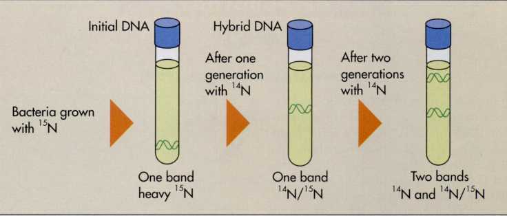
Semiconservative
replication was demonstrated experimentally by Matthew Meselson and
Franklin Stahl at the California Institute of Technology in 1958
(FIG. 7-8). They grew a culture of Escherichia
coli in
a medium in which the sole source of nitrogen was the heavy isotope
15N. The heavy nitrogen was incorporated into the
nucleotides of DNA during bacterial reproduction, so that the DNA of
these bacteria became heavier than usual. They then transferred
these bacteria to a medium containing the normal lighter isotope
14N. At various time intervals they collected cells and
analyzed the DNA to determine if it was "heavy" (15N
label), light (14N label), or intermediate (mixture of
15N and 14N label). For these analyses they
used an ultracentrifuge—an instrument that spins its contents at
high speed—which caused materials tot separate out according to
their different densities.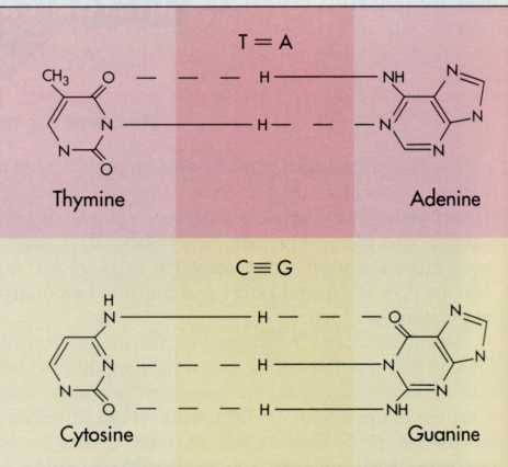
Information, is a double helical molecule.
FIG.
7-8 The semiconservative nature of DNA replication was demonstrated
by labelling DNA in one generation by the incorporation of heavy
nitrogen (15N) and following the fate of this tagged DNA
from one generation to the next, using density gradient
ultracentrifugation. The location of the bands obtained by
ultracentrifugation, that is, the distance that the DNA moves, which
is a function of the molecular weight of the DNA, permitted the
tracking of the fate of the heavy DNA when the cells were grown in
the presence of normal light nitrogen (14N). The banding
pattern obtained in these experiments, which is illustrated in the
figure, proved that DNA replication occurs by a semiconservative
method.
Denser
molecules move farther than lighter molecules in cesium
chloride density gradient centrifugation, so DNA containing 15N
moves a greater distance than DNA containing only 14N.
The movement is such that bands of DNA can be distinguished
corresponding to light, heavy, and intermediate DNA.
Initially
Meselson and Stahl detected only one band. This band corresponded to
heavy DNA in which both chains of the DNA contained the 15N
label. After sufficient time for one complete round of DNA
replication, again only one band of DNA was detected, but now the
band was at an intermediate level between all-light isotope and
all-heavy isotope DNA. This intermediate band was exactly what was
predicted by the hypothesis that DNA replication is
semiconservative. Each DNA double helix had one chain from the
parental DNA that contained the heavy 15N isotope and one
newly synthesized chain that contained only the light 14N
isotope. Also as predicted, after sufficient time for a second
round of DNA replication, Meselson and Stahl observed two bands of
DNA, one intermediate and the other light. This occurred because
when the intermediate DNA containing one light and one heavy chain
replicated, it contributed one heavy chain to form another
intermediate DNA macromolecule and one light chain to form a
new all-light DNA macromolecule. This experiment confirmed that
DNA replication is semiconservative as suggested by the
Watson-Crick model of the DNA double helix.
DNA
replication is semiconservative, producing two "holf-old,
half-new" DNA macromolecules every time the DNA is duplicated.
Steps
in DNA
Replication
Unwinding
the DNA Double Helix—Replication Forks
The
first step in semiconservative DNA replication is to pull apart a
portion of the DNA helix. This enables each of the chains to act as
a template (pattern) to direct the synthesis of a new
complementary chain of nucleotides. This can occur because hydrogen
bonds are relatively weak. Thus the two chains can separate without
breaking apart the covalently linked nucleotides of the chains,
which would destroy the information encoded within them. This
establishes the basis for one chain serving as a template for the
synthesis of a new chain of DNA with a sequence of nucleotides
that is exactly complementary.
The
chains do not entirely separate before DNA replication. Rather, a
localized region of the DNA unwinds because the two parental
DNA chains are pulled apart by specific enzymes. This creates a
region of two single strands and provides space for individual
nucleotides to align opposite their complementary bases for the
synthesis of new chains. This region of localized DNA synthesis is
called a replication
fork
(FIG.
7-9, p. 201). At the replication fork, enzymes link nucleotides
to form a new DNA strand that is complementary to the original
template DNA.
The
DNA double helix unwinds to form a replication fork where DNA
synthesis occurs.
In
eukaryotic cells, multiple replication forks form at different
locations. Simultaneous synthesis of different portions of the
DNA is thus made possible. In a bacterial cell, DNA replication is
initiated at only
REPLICATION
OF DNA 199