

Introduction
Genome sequencing
Genome anatomy and physiology
Genomic variation
Medicine: Mendelian and chromosomal disorders
Medicine: common diseases and traits
Medicine: cancer
Human history
New frontiers
References
Acknowledgements
Author information
Supplementary information
Comments
The view from 2000
Our knowledge of the contents of the human genome in 2000 was surprisingly limited. The estimated count of protein-coding genes fluctuated wildly. Protein-coding information was thought to far outweigh regulatory information, with the latter consisting largely of a few promoters and enhancers per gene. The role of non-coding RNAs was largely confined to a few classical cellular processes. And, the transposable elements were largely regarded as genomic parasites.
A decade later, we know that all of these statements are false. The genome is far more complex than imagined, but ultimately more comprehensible because the new insights help us to imagine how the genome could evolve and function.
Protein-coding genes
Since the early 1970s, the total number of genes (the vast majority assumed to be protein-coding) had been variously estimated at anywhere from ~35,000 to well over 100,000, based on genetic load arguments, hybridization experiments, the average size of genes, the number of CpG islands and shotgun sequencing of expressed sequence tags. The HGP paper suggested a total of 30,000–40,000 protein-coding genes, but the estimate involved considerable guesswork owing to the imperfections of the draft sequence and the inherent difficulty of gene identification.
Today, the human genome is known to contain only ~21,000 distinct protein-coding genes21. Generating a reliable gene catalogue required eliminating the many open reading frames (ORFs) that occur at random in transcripts, while retaining those that encode bona fide proteins. The key insight was to identify those ORFs with the evolutionary signatures of bona fide protein-coding genes (such as amino-acid-preserving substitutions and reading-frame-preserving deletions) and prove that most ORFs without such conservation are not newly arising protein-coding genes. Recent RNA-Seq projects have confirmed the gene catalogue, while illuminating alternative splicing, which seems to occur at >90% of protein-coding genes and results in many more proteins than genes.
The proteome is now known to be similar across placental mammals, with about two-thirds of protein-coding genes having 1:1 orthologues across species and most of the rest belonging to gene families that undergo regular duplication and divergence—the invention of fundamentally new proteins is rare.
Conserved non-coding elements
The most surprising discovery about the human genome was that the majority of the functional sequence does not encode proteins. These features had been missed by decades of molecular biology, because scientists had no clue where to look.
Comparison of the human and mouse genomes showed a substantial excess of conserved sequence, relative to the neutral rate in ancestral repeat elements4. The excess implied that at least 6% of the human genome was under purifying selection over the past 100 million years and thus biologically functional. Protein-coding sequences, which comprise only ~1.5% of the genome, are thus dwarfed by functional conserved non-coding elements (CNEs). Subsequent comparison with the rat and dog genomes confirmed these findings5, 6.
Although the initial analysis provided a bulk estimate of the amount of conserved sequence, it could only pinpoint the most highly conserved elements. Among them are nearly 500 ultraconserved elements (200 bases or more perfectly conserved across human, mouse and rat), most of which neither overlap protein-coding exons nor show evidence of being transcribed22. On the basis of statistical measures of constraint, tens of thousands of additional highly conserved non-coding elements (HCNEs) were identified5, 23. In many cases the evolutionary origins of these HCNEs could be traced back to the common ancestor of human and fish. HCNEs preferentially reside in the gene deserts that often flank genes with key functions in embryonic development5, 22, 23. Large-scale screens of these sequences in transgenic mice revealed that they are highly enriched in tissue-specific transcriptional enhancers active during embryonic development24, revealing a stunning complexity of the gene regulatory architecture active in early development24 (Fig. 1).
Figure 1: Evolutionary conservation maps.
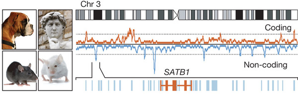
Comparison among the human, mouse, rat and dog genomes helps identify functional elements in the genome. The figure shows the density of protein-coding sequences (red) and the most highly conserved non-coding sequences (blue) along chromosome 3. Highly conserved non-coding sequences are enriched in gene-poor regions, each of which contained a gene involved in early development (such as SATB1, shown). Images courtesy of iStock Photo.
Full size image (62 KB)
Download PowerPoint slide (440K)
Figures/tables index
Next figure
Sequencing additional genomes has gradually increased our power to pinpoint the less stringently conserved CNEs. Recent comparison with 29 mammalian genomes has identified millions of additional conserved elements, comprising about two-thirds of the total conserved sequence.
Evolutionary analyses of CNEs have also enabled the discovery of distinct types of functional elements, including regulatory motifs present in the promoters and untranslated regions of co-regulated genes, insulators that constrain domains of gene expression, and families of conserved secondary structures in RNAs. Nonetheless, the function of most CNEs remains to be discovered.
Nature of evolutionary innovation
Although some CNEs show deep conservation across vertebrate evolution, most evolve and turn over at a faster pace than protein-coding sequences. At least 20% of the CNEs conserved among placental mammals are absent in marsupial mammals, compared to only ~1% for protein-coding sequences8. These elements arose in the period between our common ancestor with marsupial mammals (~180 million years (Myr) ago) and our common ancestor with placental mammals (~90 Myr ago), or else were ancestral elements lost in the marsupial lineage. The proportion of CNEs having detectable conservation with birds is much lower (~30% detectable, ~310 Myr ago) and with fish is near zero (~450 Myr ago). The more rapid change of CNEs provides experimental support for the notion25 that evolution of species depends more on innovation in regulatory sequences than changes in proteins.
Extrapolation from the marsupial–placental comparison indicates that at least ~20% of functional non-coding elements in human should be absent in mouse. Interestingly, ChIP-Seq studies report even greater differences in the localization of transcription factor binding sites between mammals. However, physical binding may not imply biological function: the evolutionary and biochemical data still need to be reconciled.
Transposons as drivers of evolutionary innovation
The HGP paper included a detailed analysis of transposon-derived sequences, but largely viewed transposons as a burden on the genome.
Comparative genomics, however, began to change this picture. The first hint was a handful of families of CNEs that had clearly been derived from transposons26. Comparison of placental and marsupial genomes then revealed that at least 15% of the CNEs that arose during the period from 180 Myr ago to 90 Myr ago were derived from transposon sequences8; the true total is likely to be considerably larger, because the flanking transposon-derived sequences will have degenerated in many cases. In retrospect, the advantage seems obvious. First, most transposons contain sequences that interact with the host transcriptional machinery, and therefore provide a useful substrate for evolution of novel regulatory elements. Second, a regulatory control that evolved at one locus could give rise to coordinated regulation across the genome by being picked up by a transposon, scattered around the genome and retained in advantageous locations. Over evolutionary timescales, transposons may earn their keep.
Small non-coding RNAs
The HGP paper analysed all known classes of functional human non-protein-coding RNAs (ncRNAs), which consisted largely of those supporting protein translation (ribosomal, transfer and small nucleolar RNAs) and transcript splicing (small nuclear RNAs).
In late 2000, vertebrates were found to harbour an important new type of ncRNA first discovered in C. elegans. Called microRNAs (miRNAs), these products bind target mRNAs and decrease their stability27. Today, the human genome is known to encode ~100 evolutionarily conserved families of miRNAs. Genomic analysis proved critical in identifying the target mRNAs: evolutionarily conserved 7-base sequences in 3′ untranslated regions complementary to bases 2–8 of conserved miRNAs28. A typical conserved miRNA has ~200 target mRNAs with conserved binding sites. A few dozen miRNAs have been shown to have key regulatory roles, such as in cancer and development. Many of the others may help to fine-tune gene expression, although some may be too subtle to have detectable phenotypes in laboratory experiments. Recently, a new class of small RNAs, called PIWI-interacting RNAs, has been discovered that functions through a similar molecular machinery—they act to silence transposons in the germline.
Ubiquitous transcription
In 2000, transcription was thought to be largely confined to regions containing protein-coding genes. Only a handful of non-classical large functional ncRNAs was known, such as telomerase RNA, 7SL signal recognition RNA, Xist and H19, and these were regarded as quirky exceptions. Pioneering studies of the human sequence soon began to provide hints that additional large RNA molecules might exist. Hybridization of RNA to microarrays of genomic sequence suggested that more than 10% of the genome was represented in mature transcripts, with most lying outside protein-coding exons29, and random cDNA sequencing turned up many transcripts that could not be linked to protein-coding genes30 (Fig. 2). With increasingly sensitive assays, it was concluded by 2007 that virtually every nucleotide in the euchromatic genome was likely to be represented in primary (unspliced) transcripts in at least some cell type at some time31. Many of these transcripts, however, have extremely low expression levels and show little evolutionary conservation; these may represent ‘transcriptional noise’ (that is, reproducible, tissue-specific transcription from loci with randomly occurring weak regulatory signals). Exactly how much of the ubiquitous transcription is biologically functional remains controversial.
Figure 2: Chromatin state maps.

The genomic sites of chromatin modifications or protein binding can be mapped, using chromatin immunoprecipitation (ChIP) and massively parallel sequencing. The figure highlights chromatin marks associated with the active promoters (green) and actively transcribed regions (blue), in a region on chromosome 22. The four features shown correspond to two active protein-coding (dark grey), one inactive protein-coding (light grey) and one long intergenic non-coding RNA (maroon). Image courtesy of B. Wong (ClearScience).
Full size image (33 KB)
Download PowerPoint slide (412K)
Previous figure
Figures/tables index
Next figure
Large intergenic non-coding RNAs
Epigenomic maps facilitated the discovery of a large class of thousands of genes encoding evolutionarily conserved (and thus clearly functional) transcripts, now called large intergenic non-coding RNAs (lincRNAs)32. The genes were pinpointed because they carry the distinctive chromatin patterns of actively transcribed genes but lack any apparent protein-coding capacity32. On the basis of their expression patterns, they have diverse roles in processes such as cell-cycle regulation, immune responses, brain processes and gametogenesis. A substantial fraction binds chromatin-modifying proteins and may modulate gene expression, for example, in the HOX complex33 and in the p53-response pathway. Although their mechanism of action remains to be elucidated, lincRNAs may act analogously to telomerase RNA by serving as ‘flexible scaffolds’34 that bring together protein complexes to elicit a specific function.
lincRNAs are not the end of the ncRNA story. RNA-Seq studies have begun to define catalogues of antisense RNAs that overlap protein-coding genes18. Unlike lincRNAs, these transcripts show little evolutionary conservation (beyond the coding region)18 and may function by base-pairing with the overlapping transcript or simply by causing chromatin changes through the act of transcription.
Epigenomic maps
Recognizing the distinctive functional domains in the genome of a cell is a key challenge, both for genome scientists and for the cell itself. With thousands of genome-wide epigenomic maps, it is now clear that functionally active domains are associated with specific patterns of epigenomic marks12, 13, 31, 35 (Fig. 2). For example, active promoters show DNase hypersensitivity, histone acetylation and histone 3 lysine 4 trimethylation; transcribed regions are marked by histone 3 lysine 4 trimethylation; and enhancers show binding of the p300 acetyltransferase. Other features are seen at exons, insulators and imprinting control regions. The binding sites of transcription factors can also be read out, given an antibody with adequate specificity.
Moreover, it is possible to study dynamic behaviour and developmental potential by comparing epigenomic maps from related cellular states. For example, bivalent chromatin domains (both histone 3 lysine 4 trimethylation and histone 3 lysine 27 trimethylation) mark genes that are poised to play key parts in subsequent lineage decisions36. Epigenomic maps can also reveal genes that serve as obstacles to cellular reprogramming, and DNA methylation maps are helping identify aberrant functions in cancer37. Ultimately, hundreds of thousands of epigenomic marks will be layered atop the genome sequence to provide an exquisite description of genomic physiology in a cell type.
Three-dimensional structure of the genome
Whereas general features of chromosomal packaging had been worked out through classical techniques such as X-ray diffraction, little was known about in vivo physical contacts between genomic loci more than a few kilobases apart.
The (one-dimensional) genome sequence enabled technologies for mapping the genome in three dimensions. Chromosome confirmation capture (3C) could test whether two loci are nearby in the nucleus, based on proximity-based ligation followed by locus-specific polymerase chain reaction38. It revealed, for example, that β-globin’s locus control region forms an ‘active hub’ involving physical contact between genomic elements separated by 100 kb or more.
New approaches, such as a method called Hi-C, extend 3C to examine all physical contacts in an unbiased genome-wide fashion39. It has revealed that the genome is organized into two compartments, corresponding to open and closed chromatin, and, at megabase scale, exhibits folding properties consistent with an elegant structure called a fractal globule.
The road ahead
The ultimate goal is to understand all of the functional elements encoded in the human genome. Over the next decade, there are two key challenges. The first will be to create comprehensive catalogues across a wide range of cell types and conditions of (1) all protein-coding and non-coding transcripts; (2) all long-range genomic interactions; (3) all epigenomic modifications; and (4) all interactions among proteins, RNA and DNA. Some efforts, such as the ENCODE and Epigenomics Roadmap projects, are already underway31. Among other things, these catalogues should help researchers to infer the biological functions of elements; for example, by correlating the chromatin states of enhancers with the transcriptional activity of nearby genes across cell types and conditions. These goals should be feasible with massively parallel sequencing and assay miniaturization, although they will require powerful ways to purify specific cell types in vivo, and the fourth goal will require a concerted effort to generate specific affinity reagents that recognize the thousands of proteins that interact with nucleic acids.
The second and harder challenge is to learn the underlying grammar of regulatory interactions; that is, how genomic elements such as promoters and enhancers act as ‘processors’ that integrate diverse signals. Large-scale observational data will not be enough. We will need to engage in large-scale design, using synthetic biology to create, test and iteratively refine regulatory elements. Only when we can write regulatory elements de novo will we truly understand how they work.
Genomic variation