MINISTRY OF EDUCATION AND SCIENCE OF UKRAINE
NATIONAL AVIATION UNIVERSITY
Institute of ecological safety
DEPARTMENT OF BIOTECHNOLOGY
Home work
Features of nucleic acids replication
Student: Litvin Irina
Group IES 204
Leader: Vasylchenko O.A
Kyiv 2013
Table of content
Inroduction.
Basics.
Replication Forks.
DNA Polymerase.
Okazaki Fragments.
Steps of DNA replication.
RNA Self Replication.
Conclusion.
References
1. Introduction
Deoxyribonucleic acid (DNA) is the carrier of genetic information for all living creatures. An organisms`s genome, made of DNA, encodes the genetic blueprint for building that organism. Though the process of replication , the entire genome is copied and passed on to each new cell made in the body. Replication is also the mechanism of heredity, which is the passing of genetic information from the parents to their children.
The duplex structure of DNA, discovered by Watson and Crick in 1953, is a double helix. Each of the two strands of the double helix is comprised of the nucleic acid bases adenine (A), guanine (G), cytosine (C) and thymine (T) attached to a backbone of sugar and phosphate molecules. Each purine base (A or G) on one strand will pair up with a pyrimidine base (C or T) on the other strand in a specific manner – A:T and G:C. Because the bases match up in this complementary fashion, if the two strands are separated each can act as a template for the copying, or replication, of the other.
Errors in the replication of DNA are normally fixed inside the cell. Those mistakes that go unrepaired are responsible for mutations, which can lead to altered proteins or gene regulatory sequences leading to cancer or other genetic defects. These same mutations are the mechanism driving evolution by continually providing new material upon which selective pressure can act.
2.Basics
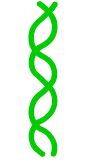
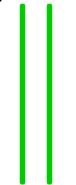
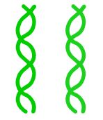
Before the mechanisms was understood, it was postulated that DNA replication could occur in two different ways. Conservative replication suggests that the helix is copied as an entire unit leaving the parent molecule intact. (Fig.1) The Watson and Crick template model supports a semi-conservative copying of DNA. In semi-conservative replication the parental helix would break apart. Each strand would then be copied individually making each daughter helix a hybrid of one parental and one new strand (Fig.2).
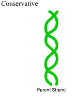
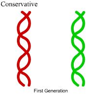

Fig.1 Conservative replication
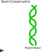
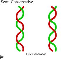
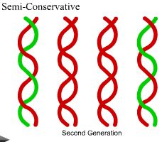
Fig.2 Semi-conservative replication
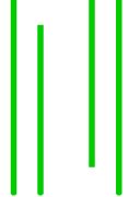
3. Replication Forks
The replication fork is a structure that forms within the nucleus during DNA replication. It is created by helicases, which break the hydrogen bonds holding the two DNA strands together. The resulting structure has two branching "prongs", each one made up of a single strand of DNA. These two strands serve as the template for the leading and lagging strands, which will be created as DNA polymerase matches complementary nucleotides to the templates. The templates may be properly referred to as the leading strand template and the lagging strand templates.
In DNA`s semi-conservative replication the original helix is split into two strands forming a replication fork. In the division of prokaryotic-circular DNA the theta model is used. From only one initiation site the replication forks move in opposing directions until they meet, severing the parental strands from one another.
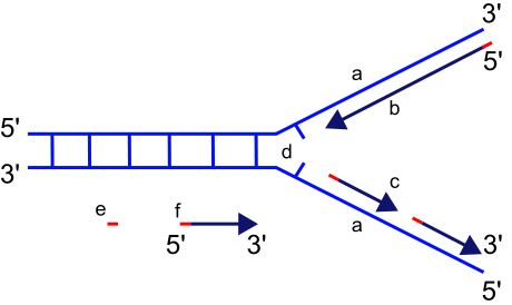
Scheme of the replication fork
a: template, b: leading strand, c: lagging strand, d: replication fork, e: primer,
f: Okazaki fragments.
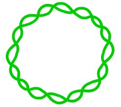
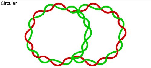
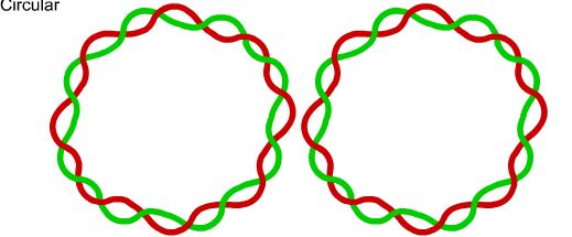
Circular
In the division of prokaryotic-circular DNA the theta (θ) model is used. From only one initiation site the replication forks move in opposing directions until they meet, severing the parental strands from one another.
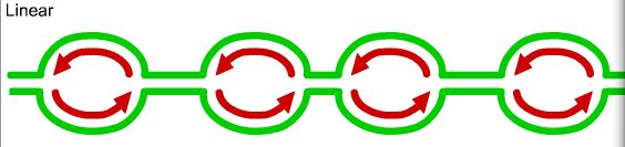
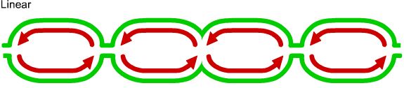
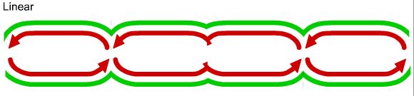
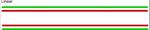
Linear
In linear-eukaryotic DNA several initiation sites are used. These sires form replication bubbles, which grow and join as the fork moves.