

Penetration of Zona Pellucida
Once inside the corona radiata, the sperm binds to the species-specific ZP3 receptor on the egg’s glycoprotein coat. This triggers the acrosomal reaction, or the release of enzymes stored in the sperm’s acrosome (e.g. acrosin). These enzymes help the sperm “drill through” the zona pellucida.
Once the sperm has penetrated the outer layers it fuses with the plasma membrane of the egg and releases its contents inside. The head and the tail of the sperm degrade, so that all mitochondria in the embryo (and all mitochondrial DNA) come from the mother. The touch of spermatozoon to an ovum surface causes reciprocal changes in a boundary layer of ovoplasma.
The third phase of fertilization begins.
Entry of a sperm into the egg causes changes that prevent polyspermy (fertilization of an egg by more than one sperm). These changes are known as the cortical reaction. Cortical granules move to a surface of a plasmatic membrane and their contents secretes in the space environmental ovum.
Fusion of Pronuclei
In ovoplasma the spermatozoon head turns on 180 degrees, approximated and turns in male pronucleus. The ovum nucleus turns in female pronucleus. The male and female pronuclei fuse and make a synkaryon. The fertilized egg is called zygote (“together”).
Cleavage
The zygote undergoes a number of ordinary mitotic divisions that increase the number of cells in the zygote but not its overall size. Each cycle of division takes about 24 hours. The individual cells are known as blastomeres. At the 32-cell stage the embryo is known as a morula (L. “mulberry”), a solid ball consisting of an inner cell mass and an outer cell mass. The inner cell mass will eventually become the embryo and fetus, while the outer cell mass will eventually become part of the placenta.
Blastocyst Formation Compaction
The cells on the outside of the morula form tight intercellular junctions and express ion channels to create an impermeable barrier.
Cavitation
A fluid-filled cavity forms inside the morula. This cavity is known as the blastocyst cavity or blastocoele, and the morula is now called a blastula or blastocyst. The inner cell mass is now known as the embryoblast and the outer cell mass becomes the trophoblast.
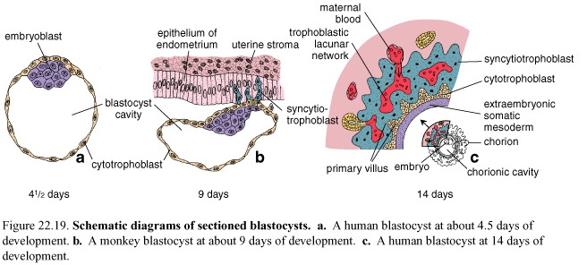
Figure 8. Schematic diagrams of sectioned blastocysts. a. A human blastocyst at about 4, 5 days of development. b. A monkey blastocyst at about 9 days of development. c. A human blastocyst at 14 days of development
Human cleavage is called:
-complete
-asynchronous
-nearly equal
Implantation
Hatching
The blastula sheds its zona pellucida. This is required for implantation to occur. One function of the zona pellucida is to prevent premature implantation.
Attachment and Invasion
The embryo attaches to and invades into the maternal endometrium. The trophoblast differentiates into the cytotrophoblast and the syncytiotrophoblast. The embryo typically implants in the posterior superior wall of the uterus. The response of the maternal endometrial cells to the invading embryo is called the decidual reaction.