

Nucleus
The nucleus is a membrane-limited compartment that contains the genome (genetic information) in eukaryotic cells.
The nucleus of nondividing cell (interphase cell) consists of the following components:
-chromatin, organized as euchromatin and heterochromatin
-nucleolus (or nucleoli)
-membranous nuclear envelope
-nucleoplasm
Chromatin
Chromatin is a complex of DNA and proteins. It is responsible for the characteristic basophilia of the nucleus. The densely staining material is highly condensed chromatin called heterochromatin and the lightly staining material is a dispersed form called euchromatin.
The proteins of chromatin include 5 basic proteins called histones and others called nonhistone proteins.
Euchromatin is indicative of active chromatin, i.e. chromatin that is stretched out so that the genetic information in the DNA can be read and transcribed.
The smallest units of chromatin structure are macromolecular complexes of DNA and histones called nucleosomes.
In dividing somatic (mitotic) cells, chromatin is condensed and organized into discrete bodies called chromosomes. Each chromosome is formed by two chromatids that are joined together at a point called the centromere.
With the exception of the mature gametes, the egg and sperm, human cells contain 46 chromosomes organized as 23 homologous pairs. Twenty-two of the pairs have identical chromosomes and are called autosomes. There is one pair of sex chromosomes, designated X and Y. The chromosomal number, 46, is found in most of somatic cells of the body and is called the diploid (2n) number. The mature sex cell, egg and sperm, as a result of meiosis have only 23 chromosomes, the haploid (1n) number as well as the haploid (1n) amount of DNA.
Karyotypes are chromosome pairs sorted according to their morphology. In females, only X chromosome (either of the two) is used by each cell, the inactive X is often visible as a clump of heterochromatin, termed sex chromatin, or the Barr body can be used to identify the sex of a fetus.
Nucleolus
 Figure
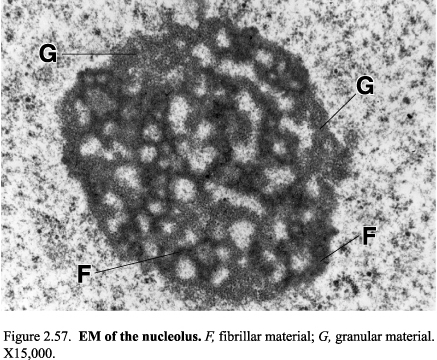
5. EM of the nucleolus; F, fibrillar component; G, granular component
Figure
5. EM of the nucleolus; F, fibrillar component; G, granular component
The nucleolus is the site of ribosomal RNA synthesis and initial ribosomal assembly. The nucleolus is a nonmembranous, intranuclear structure formed by filamentous and granular material.
The nucleolus consists of two pars: pars granulosa and pars fibrosa
The pars fibrosa is the most densely staining compartment of the nucleolus. The network formed by the pars granulosa is called the nucleonema.
Nucleoplasm
 Nucleoplasm
is the material enclosed by the nuclear envelope exclusive of the
chromatin and the nucleolus.
Nucleoplasm
is the material enclosed by the nuclear envelope exclusive of the
chromatin and the nucleolus.
Nuclear envelope
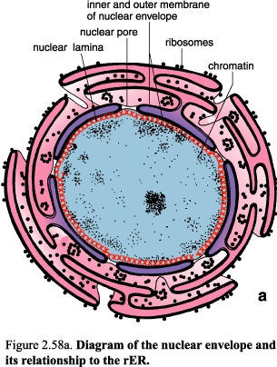
Figure 6. Diagram of the nuclear envelope and its relationship to the rER
The nuclear envelope formed by two unit membranes with a perinuclear cisternal space between them serves as a membranous boundary between the nucleoplasm and the cytoplasm of the interphase cell. The nuclear envelope has an array of perforation called nuclear pores.