

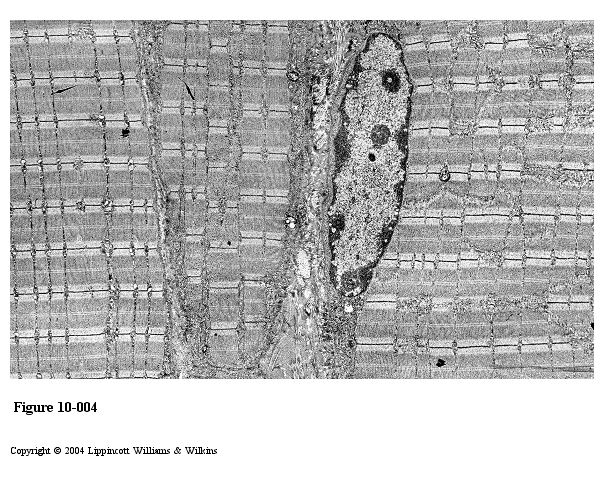
Cardiac muscle
It consists of long fibers that appear to branch and anastomose with neighboring fibers. Unlike skeletal muscle fibers, the fibers of cardiac muscle are formed by individual, mononucleated Cardiac muscle cells that are joined to one another in linear array.
Cardiac muscle fibers exhibit densely staining cross-bands, called intercalated discs that cross the fibers in a linear fashion.
The central location of the nucleus in cardiac muscle cells helps to distinguish them from multinucleated skeletal muscle fibers, whose nuclei lie immediately under the sarcolemma.
More about cardiac muscle you are going to study in cardiovascular system.
Smooth muscle
Smooth muscle is the simplest appearing muscle tissue.
Smooth muscle is the intrinsic muscle of the alimentary canal, blood vessels, genitourinary tract, respiratory tract and other hollow and tubular organs.
Smooth muscle generally occurs as bundles or sheets of elongate fusiform cells. The cells also called fibers. The cytoplasm of the smooth muscle contains actin and myosin. The nuclei of smooth muscle cells are located in the center of the cell and often have a corkscrew appearance in the longitudinal section. This is due to contraction of the cell during fixation.
The cytoplasmic organelles are concentrated at each end of the nucleus. The rest of the cytoplasm is seen to be filled with actin filaments interspersed with intermediate filaments. Cytoplasmic densities are seen among the filaments and in relation to the plasma membrane.
The cytoplasmic densities or dense bodies contain the protein α-actinic.
Smooth muscle cells have no T system.
Contraction and its control
Smooth muscle is specialized for slow, prolonged contraction in a wave-like manner, producing peristaltic movements as in the gastrointestinal tract and the male genital tract, or it may contract throughout producing extrusive movements as in the urinary bladder, the gall bladder and the uterus. Smooth muscle has spontaneous contractile activity.
Figure 29. A suggest model for smooth muscle cell contraction

Figure 30. Skeletal muscle, electron micrograph
Nervous tissue
Nervous tissue consists of two principal types of cells: nerve cells (neurons) and supporting cells.
The neuron or nerve cell is the functional unit of the nervous system. All neurons have a cell body and processes, the axon and dendrites.
The cell body of a neuron contains the nucleus and those organelles that maintain the nerve cell.
Most neurons have only one axon, usually the longest process extending from the cell; some axons may be more than 1 meter long. A neuron usually has many dendrites, processes that are shorter and usually thicker that an axon.
Neurons are classified on the basis of the number of processes extending from the cell body. Thus:
Multipolar neurons have only one axon and two or more dendrites
Bipolar neurons have one axon and one dendrite
Unipolar (actually pseudounipolar) neurons have one process the axon that divides close to the cell body into two long processes.
Figure 31. Diagram of a motor neuron
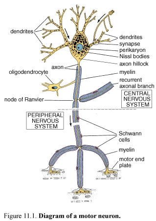
Cell body
The cell body of the neuron has the characteristics of a protein-secreting cell. The cell body or perikaryon is the usually large dilated region of the cell that contains a large euchromatic nucleus.
Nerve cell perikarya contain inclusions, Nissl bodies that stain intensely with basic dyes. Each Nissl body corresponds to a stack of rough endoplasmic reticulum (rER).
The perikaryon also contains numerous mitochondria, a large perinuclear Golgi apparatus, lysosomes, microtubules, neurofilaments, vesicles and inclusions.
Nissl bodies, free ribosomes and the Golgi complex extend into the dendrites but not into the axon. Neurons do not divide. They must last for a lifetime.