

Neutrophils
Figure16. EM of a human neutrophil
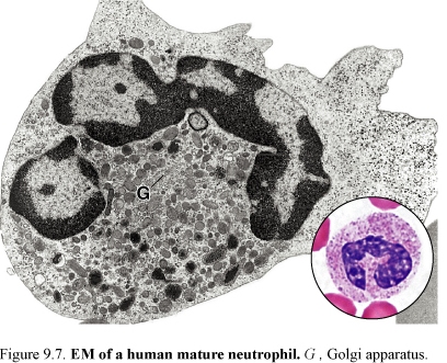
Neutrophils measure 10-12 μm in diameter. They constitute 60-70% of circulating leukocytes. Neutrophils are so called because of its ability of staining with acidic and basic dyes. The mature neutrophil has three to five lobes of nuclear material joined by thinner nuclear strands. The cytoplasm of the neutrophil contains two kinds of granules: specific granules and azurophilic granules.
Specific granules are small and most numerous. They contain bacteriostatic and bacteriocidal agents, such as lysozyme, as well as alkaline phosphatases.
Azurophilic granules are larger and less numerous. They contain peroxidase and acid phosphatases and lysosomal enzymes.
In the female, few neutrophils can be seen containing the inactive X chromosome (Bar body) appearing as a drumstick-shaped nuclear appendage.
Main functions are:
Neutrophils constitute a defense against invasion by microorganisms, especially bacteria. They are active phagocytes of small particles and have sometimes been called microphages to distinguish them from macrophages, which are larger cells.
Bacteria first adhere to the neutrophil surface and then are surrounded and engulfed by pseudopodia.
When a bacterium is phagocytized, the specific granules fuse with the membrane of phagosome and their bacteriocidal content is emptied into the phagosome.
Azurophilic granules fuse with the phagosome-specific granule complex somewhat later; the hydrolytic enzymes of the lysosomal azurophilic granules digest the microorganism.
Eosinophils
Figure17. EM of a human eosinophil; Cr, crystalline body; M, mitochondria
E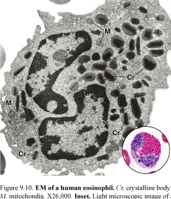 osinophils
are less numerous than neutrophils, consisting only 2-5% of
leukocytes in normal blood.
osinophils
are less numerous than neutrophils, consisting only 2-5% of
leukocytes in normal blood.
The cell has a diameter of 12-14 μm and contains a characteristic bilobed nucleus.
Eosinophilic specific granules have a crystalline core an arginine-rich protein called the major basic protein that probably accounts for the eosinophilia of these granules. The less dense material surrounding the internum is known as the externum or matrix. The granules contain histaminase, peroxidase, arylsulfatase and other hydrolytic enzymes. An increase in the absolute number of eosinophils in blood (eosinophilia) is associated with allergic reactions and helmintic (parasitic) infections.
Main functions are:
-eosinophils play an important role in the control of local responses in allergic reactions
-their specific granules contain many enzymes that degrade chemical mediators released by mast cells in association with hypersensitivity reactions
-eosinophils are thought to play an important role in the defense against certain types of parasites
Basophils
Basophils are the least numerous of the leukocytes, consisting less than 1% of the total leukocytes.
Basophils are about 10- 12 μm in diameter.
Figure18. EM of a human basophil; B, basophilic granules, M, myelin figures
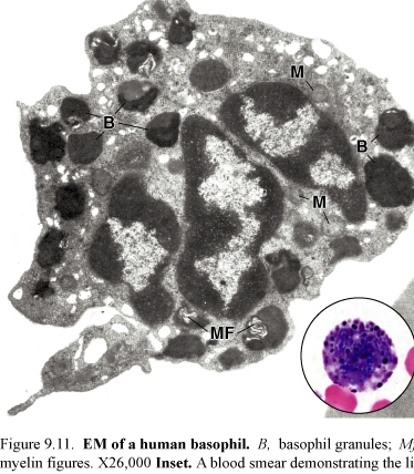
The nucleus is divided into irregular lobes, but the division usually obscured by the overlying specific granules.
Specific granules in basophils are fewer and more irregular in size and shape than the granules of the other granulocytes.
Basophilic specific granules contain heparin and histamine and are capable of generating leukotriens. They cause slow contraction of smooth muscles.
Histamine and slow-reacting substance of anaphylaxis are vasoactive agents that bring on the dilation of small blood vessels.
Main functions are:
-basophils may supplement the function of mast cells in immediate hypersensitivity reactions by migrating into connective tissues
-basophils have also a very weak phagocytic activity