

Functions of a blood
1. Convey nutrients and oxygen directly or indirectly to cells.
2. Carry wastes and carbon dioxide away from the cells.
3. Carry hormones and other regulatory agents to and from the cells and tissues of the body.
4. Have a major homeostatic role based on its thermoregulatory and buffering capacity
5. Transport humoral agents and cells that protect the body from infections, foreign proteins and transformed cells, i.e. cancer cells.
The formed elements of blood include.
1. Red blood cells, also called erythrocytes.
2. White blood cells, also called leucocytes
3. Platelets.
Plasma
Plasma is the liquid intercellular material or matrix. The relative volume of cells and plasma is about 45% and 55%, respectively.
This value is called a hematocrit. Plasma consists of 91-92% of water, 7-8% of proteins (fibrinogens, globulins and albumin) and 1-2% of other solutes.
Red blood cells
Figure14. SEM of the erythrocytes
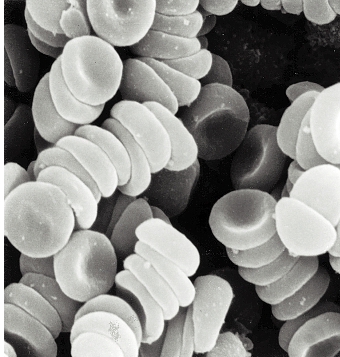 Erythrocytes,
which are anucleate, are packed with the 02
carrying protein hemoglobin (Hb). Under normal conditions, these
cells never leave the circulatory system. The normal number of the
erythrocytes for the women is 3,7-4,9×10¹²/L,
and for the men is 3,9-5,5×10¹²/L.
Erythrocytes,
which are anucleate, are packed with the 02
carrying protein hemoglobin (Hb). Under normal conditions, these
cells never leave the circulatory system. The normal number of the
erythrocytes for the women is 3,7-4,9×10¹²/L,
and for the men is 3,9-5,5×10¹²/L.
M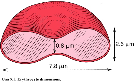 ost
mammalian erythrocytes are biconcave disks without nuclei. There are
some other shapes of the erythrocytes, such as spherical, flat, plane
and branched. This phenomenon is called poikylocytosis.
There are 2 types of it. It is called physiological
poikylocytosis when
80% of red blood cells have shape as biconcave disk and 20% other
shape. And it is called pathological
poikylocytosis
when more than 20% of erythrocytes have other shape.
ost
mammalian erythrocytes are biconcave disks without nuclei. There are
some other shapes of the erythrocytes, such as spherical, flat, plane
and branched. This phenomenon is called poikylocytosis.
There are 2 types of it. It is called physiological
poikylocytosis when
80% of red blood cells have shape as biconcave disk and 20% other
shape. And it is called pathological
poikylocytosis
when more than 20% of erythrocytes have other shape.
Figure15. Erythrocyte dimensions
Human erythrocytes are 7-8μm (micrometer) in diameter, 2, 6 μm thick at the rim and 0, 8 μm thick in the center.
A decreased number of erythrocytes in the blood are usually associated with anemia. An increased number of erythrocytes are called erythrocytosis or polycythemia.
Erythrocytes with diameters greater than 9 μm are called macrocytes, and those with diameters less than 6 μm are called microcytes. The presence of a high percentage of erythrocytes with great variations in size is called anisocytosis.
Erythrocytes are surrounded by a plasmalemma. It consists of about 40% lipid, 50% protein and 10% carbohydrate. Erythrocytes contain in their interiors about 33% of hemoglobin, the 02 carrying protein that accounts for their acidophilia.
Hb consists of four polypeptide chains complexed to iron-containing heme groups. Hb is designated as HbA, HbA2 and HbF. Adult Hb is about 96% HbA, 2% HbA2 and 2 % HbF. HbF is the principal form of Hb in the fetus.
Main functions are:
-the transport of oxygen from the lungs to the tissues of the body
-the transport of carbon dioxide from the tissues of the body to the lungs
White blood cells
Leukocytes are classified into two groups: granulocytes and agranulocytes.
All leucocytes are spherical cells. The increased number of leucocytes is called leucocytosis; the decreased number of leucocytes is called leucopenia.
According to the presence or absence of specific granules leucocytes are classified into:
-granular leucocytes (granulocytes)
-agranular leucocytes (agranulocytes)
Granulocytes have two types of granules: specific and azurophilic granules.
Granulocytes have nuclei with two or more lobes and include the neutrophils, eosinophils and basophils.
All granulocytes have a life span of a few days and dying by apoptosis (programmed cell death) in the connective tissue.
Agranulocytes do not have specific granules, but they do contain various numbers of azurophilic granules (lysosomes). The nucleus is round or indented. This group includes the lymphocytes and monocytes.