

Classification of epithelium
Epithelium is classified by cell arrangement and cell shape not by function.
Classification of epithelium is based on the combination of the number of the surface cells. Thus, epithelium is described as:
-simple, when it is one cell layer thick
-stratified, when it is two or more cell layers
The individual cells that compose an epithelium are described as:
-squamous, where the width of the cell is greater than its height
-cuboidal, where the width, depth and height are approximately the same
-columnar, where the height of the cell exceeds the width
1. Simple
squamous epithelium
is composed of one row of flat cells; all of them are resting on the
basement membrane. 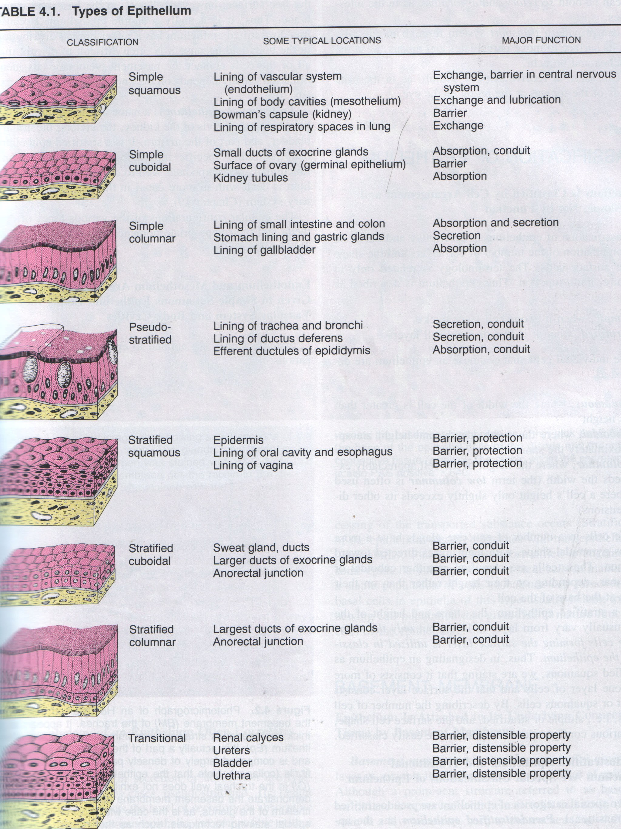
Simple squamous epithelium that covers the external surfaces of the digestive organs, lungs and heart is called mesothelium.
Simple squamous epithelium that lines the lumina of the heart chambers, blood vessels and lymphatic vessels is called endothelium.
The major functions are exchange, lubrication (mesothelium) and barrier.
2. Simple cuboidal epithelium is composed of one row of cuboidal cells; all of them are resting on the basement membrane.
Simple cuboidal epithelium lines small excretory ducts of exocrine glands. In the proximal convoluted tubules of the kidney, the apical surfaces of the simple cuboidal epithelium are lined with a brush border consisting of microvilli.
The major functions are absorption and barrier.
3. Simple columnar epithelium is composed of one row of columnar cells; all of them are resting on the basement membrane. Simple columnar epithelium lines small intestine and colon, stomach, gastric glands and gallbladder.
The major functions are absorption and secretion.
4. Pseudostratified epithelium has appearance of being stratified. Some of the cells do not reach the free surface; however, all of them are resting on the basement membrane. Thus, it is actually a simple epithelium. It lines the trachea and bronchi; ductus deferens and efferent ductules of epididymis.
The major functions are secretion absorption and conduit.
5. Stratified squamous epithelium contains multiple cell layers; only first layer is resting on the basement membrane. The basal cells are cuboidal to columnar; these cells give rise to cells that migrate toward the surface and become squamous.
There are two types of stratified squamous epithelium: nonkeratinized and keratinized.
Nonkeratinized epithelium exhibits live surface cells and covers moist cavities, such as the mouth, pharynx, esophagus, vagina and anal canal.
The major functions are barrier and protection.
Keratinized epithelium is found on exposed surfaces of the body, such as the skin. The surface layers contain nonliving, keratinized cells that are filled with the protein keratin.
The major functions are barrier and protection.
S tratified cuboidal and stratified columnar epithelium has a limited distribution in the body. Both types of epithelia line the larger excretory ducts of the pancreas, salivary glands and sweat glands. In these ducts, the epithelium exhibits two or more layers of cells.
The major functions are barrier and conduit.
Transitional epithelium lines the minor and major renal calyces, pelvis, ureter and bladder of the urinary system.
This type of epithelium changes shape and can resemble either stratified squamous or stratified cuboidal epithelia, depending on whether it is stretched or contracted. When transitional epithelium is contracted, the surface cells appear dome-shaped, when it is stretched, the epithelium appears squamous.
The major functions are barrier and distensible property.
Glands
Glands are composed of epithelial cells specialized to synthesize and secrete a specific product.
Typically, glands are classified into two major groups reflecting how their products are distributed:
-exocrine glands secrete their products into a surface through ducts or tubes. The ducts also composed of epithelial cells
-endocrine glands don’t have a duct system. They secrete their products into the connective tissue from which they enter the bloodstream in order to reach their target cells. The products of endocrine glands are called hormones.
Exocrine glands are classified as both unicellular and multicellular.
Unicellular glands are the simplest in structure. In this case the secretory component consists of single cells distributed among other cells that are not secretory. A typical example is the goblet cell.
Multicellular glands are composed of more than one cell and exhibit varying degrees of complexity. The end pieces of the gland contain the secretory cells. The portion of the gland connecting the secretory cells to the surface serves as a duct. The ducts of the multicellular exocrine glands may be non-branching or branching. So, accordingly we have two types of glands:
Simple glands have non-branching ducts.
Compound glands have branching ducts.
If the secretory potion is shaped like a tube, the gland is tubular.
If it is shaped like a flask, the gland is alveolar or acinar.
If the tube ends in a sac-like dilation the gland is tubuloalveolar.
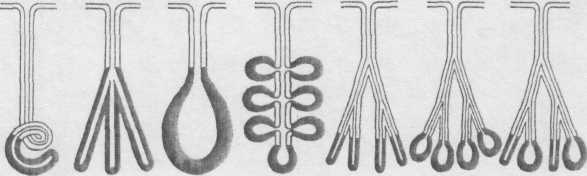 Tubular
secretory portions may be straight, coiled or branched. Alveolar
secretory potions may be single or branched.
Thus,
exocrine gland may be described as
Tubular
secretory portions may be straight, coiled or branched. Alveolar
secretory potions may be single or branched.
Thus,
exocrine gland may be described as
Simple coiled tubular
Simple branched tubular
Simple acinar
Simple branched acinar
Compound tubular
Compound acinar
Compound tubuloacinar
Classification according nature of secretion
Secretory units may be mucous, serous or mixed.
Mucous glands secrete mucinogens, large proteins. Examples of mucous glands include goblet cells and the minor salivary glands of the tongue and palate.
Serous glands such as pancreas secrete an enzyme-rich watery fluid.
Mixed glands contain acini that produce mucous secretions as well as acini that produce serous secretions.
According to the mode of secretion:
In other words, according to how the secretory cells release their secretion.
A) Merocrine glands;
In this type, the membranes of the secretory vesicles fuse with the cell membrane releasing their contents of secretion by exocytosis, without loss of any of the glandular cytoplasm. Nearly, all exocrine glands are of this type.
B) Holocrine glands;
In this type, the whole of the cell, together with its accumulated secretion, is shed and liberated out of the secretory unit. A good example of this type is the sebaceous gland of the skin.
C) Apocrine glands:
The secretory granules are liberated with some of the cytoplasm of the apical part of the secretor cell. Examples of these glands are the mammary glands and the special types of sweat glands.
Blood
Blood is a fluid connective tissue that circulates through the cardiovascular system.
Blood consists of formed elements, cells and their derivatives and a protein-rich matrix called plasma.