

Human_Histology
.pdf208 Textbook of Human Histology
Fig. 17.4: Relationship of podocytes to a glomerular capillary.
Note that the entire surface of the capillary is covered by processes of podocytes, the bare areas being shown only for sake of clarity
(Schematic representation)
Fig. 17.5: Relationship of a glomerular capillary to podocytes, basement membrane and mesangium (Schematic representation)
flow through the glomerulus. Other functions attributed to mesangial cells include phagocytosis, and maintenance of glomerular basement membrane. The mesangium becomes prominent in a disease called glomerulonephritis.
Glomerular Capsule
The glomerular capsule is a double-layered cup, the two layers of which are separated by the urinary space. The outer layer is lined by squamous cells. With the light microscope the inner wall also appears to be lined by squamous cells, but the EM shows that these cells, called podocytes, have a highly specialized structure. The urinary space becomes continuous with the lumen of the renal tubule at the urinary pole of the renal corpuscle. This pole lies diametrically opposite the vascular pole.
Podocytes
The podocytes are so called because they possess foot-like processes. Each podocyte has a few primary processes that give the cell a star-shaped appearance (Fig. 17.4). These processes are wrapped around glomerular capillaries and interdigitate with those of neighboring podocytes. Each primary process terminates in numerous secondary processes also called pedicels (or end feet) that rest on the basal lamina. The cell body of the podocyte comes in contact with the basal lamina only through the pedicels.
Glomerular Basement Membrane
As compared to typical membranes the glomerular basement membrane is very thick (about 300 nm). It is made up of three layers. There is a central electron dense layer (lamina densa), and inner and outer electron lucent layers (lamina rara interna and externa). The lamina densa contains a network of collagen (type IV) fibrils, and thus acts as a physical barrier. The electron lucent layers contain
the glycosaminoglycan heparan sulfate. This bears the negative charges referred to above. The glomerular basement membrane is, therefore, both a physical barrier and an electrical barrier to the passage of large molecules.
As shown in Figure 17.5, the glomerular basement membrane (and overlying podocyte cytoplasm) does not go all round a glomerular capillary. The gap is filled in by mesangium. A thin membrane continuous with the lamina rara interna may separate endothelial cytoplasm from mesangium. In the interval between capillaries the basement membrane is in contact with mesangium.
The Renal Tubule
The renal tubule is divisible into several parts that are shown in Figure 17.2 and Plate 17.3. Starting from the glomerular capsule (in proximodistal sequence). They are:
(a) the proximal convoluted tubule; (b) the loop of Henle consisting of a descending limb, a loop, and an ascending limb; and (c) the distal convoluted tubule, which ends by joining a collecting tubule. Along its entire length the renal tubule is lined by a single layer of epithelial cells that are supported on a basal lamina.
Proximal Convoluted Tubule
The junction of the proximal convoluted tubule with the glomerular capsule is narrow and is referred to as the neck. The proximal convoluted tubule is made up of an initial part having many convolutions (lying in the cortex), and of a terminal straight part that descends into the medulla to become continuous with the descending limb of the loop of Henle.
The neck is lined by simple squamous epithelium continuous with that of the glomerular capsule (Some texts refer to the neck as part of the glomerulus).

Chapter 17 The Urinary System 209
PLATE 17.3: %$ '
%$
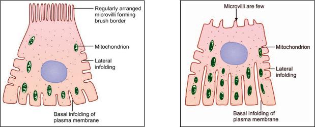
The proximal convoluted tubules are 40–60 μm in diameter. They have a relatively small lumen. They are lined by cuboidal (or columnar) cells having a prominent brush border. The nuclei are central and euchromatic. The cytoplasm stains pink (with hematoxylin and eosin). The basal part of the cell shows a vertical striation.
With the EM the lining cells of the proximal convoluted tubules show microvilli on their luminal surfaces. The striae, seen with the light microscope near the base of each cell, are shown by EM to be produced by infoldings of the basal plasma membrane, and by numerous mitochondria that lie longitudinally in the cytoplasm intervening between the folds (Fig. 17.6). The presence of microvilli, and of the basal infoldings greatly increases the surface area available for transport. Adjacent cells show some
A high power view of a part of the renal medulla shows # # ! < versely
They are lined by a cuboidal epithelium, the cells of which# ! !
( # ' ! ! = >$% # ! similar in appearance to those of blood capillaries
( # # ' ! !
#$# # # ' ,
Key
. #
. 3
. 3 #
0 " ' <
1 " ' <
3 3 !
lateral interdigitations. Numerous enzymes associated with ionic transport are present in the cytoplasm.
Loop of Henle
The descending limb, the loop itself, and part of the ascending limb of the loop of Henle are narrow and thin walled. They constitute the thin segment of the loop. The upper part of the ascending limb has a larger diameter and thicker wall and is called the thick segment.
The thin segment of the loop of Henle is about 15–30 μm in diameter. It is lined by low cuboidal or squamous cells. The thick segment of the loop is lined by cuboidal cells.
The loop of Henle is also called the ansa nephroni. With the EM the flat cells lining the thin segment of the loop of Henle show very few organelles indicating that the
210 Textbook of Human Histology
Fig. 17.6: Some features of the ultrastructure of a cell lining a proximal convoluted tubule (Schematic representation)
Fig. 17.7: Some features of ultrastructure of a cell lining a distal convoluted tubule (Schematic representation)
cells play only a passive role in ionic movements across them. In some areas the lining epithelium may show short microvilli, and some basal and lateral infoldings.
The length of the thin segment of the loop of Henle is variable. The loops of nephrons having glomeruli lying deep in the cortex (juxtamedullary glomeruli) pass deep into the medulla. Those associated with glomeruli lying in the middle of the cortical thickness extend into the medulla to a lesser degree, so that part of the loop of Henle lies in the cortex. Some loops (associated with glomeruli placed in the superficial part of the cortex) may lie entirely within the cortex.
Distal Convoluted Tubule
The distal convoluted tubule has a straight part continuous with the ascending limb of the loop of Henle, and a convoluted part lying in the cortex. At the junction between the two parts, the distal tubule lies very close to the renal corpuscle of the nephron to which it belongs.
The distal convoluted tubules are 20–50 μm in diameter. They can be distinguished (in sections) from the proxi-
mal tubules as (Fig. 17.7).They have a much larger lumen
The cuboidal cells lining them do not have a brush border
They stain less intensely pink (with eosin).
The structure of the (ascending) thick segment of the loop of Henle is similar to that of distal convoluted tubules. The cells of the distal convoluted tubules resemble those of the proximal convoluted tubules with the following differences.
They have only a few small microvilli.
The basal infoldings of plasma membrane are very prominent and reach almost to the luminal surface of the cell. This feature is characteristic of cells involved in the active transport of ions. Enzymes concerned with active transport of ions are present in the cells.
At the junction of the straight and convoluted parts of the distal convoluted tubules, the cells show specializations that are described below in connection with the juxtaglomerular apparatus.
Note: Students may be confused by somewhat different terminology used in some books. The straight part of the proximal convoluted tubule is sometimes described as part of the loop of Henle, and is termed the descending thick segment, in distinction to the (ascending) thick segment. Some workers regard the thin segment alone to be the loop of Henle. They include the descending and ascending thick segments with the proximal and distal convoluted tubules respectively.
Collecting Tubule
The terminal part of the distal convoluted tubule is again straight. This part is called the junctional tubule or connecting tubule, and ends by joining a collecting duct.
The smallest collecting tubules are 40–50 μm in diameter, and the largest as much as 200 μm. They are lined by a simple cuboidal, or columnar, epithelium. Collecting tubules can be easily distinguished from convoluted tubules as follows.Collecting tubules have larger lumina. In transverse
sections their profiles are circular in contrast to the irregular shapes of convoluted tubules.
The lining cells have clear, lightly staining cytoplasm, and the cell outlines are usually distinct. They do not have a brush border.

The walls of collecting tubules (in the proximal part of the collecting system) are lined by two types of cells. The majority of cells (called clear cells) have very few organelles, a few microvilli and some basal infoldings. The lining epithelium also contains some dark cells (or intercalated cells). These have microvilli, but basal infoldings are not seen. They contain numerous mitochondria.
The cells of the collecting ducts do not have microvilli, or lateral infoldings of plasma membrane. Very few organelles are present in the cytoplasm.
"
($)
In the renal corpuscle water and various small molecules to the urinary space of the glomerular capsule. Theoretically constituted by (a) the capillary endothelium, (b) by the cells (podocytes) forming the glomerular (or visceral) layer of the glomerular capsule, and (c) by a glomerular basement membrane that intervenes between the two layers of cells named above, and represents the fused basal laminae of follows.
Firstly, the endothelial cells show numerous fenestrae or pores that are larger than pores in many other situations. The fenestrae are not closed by membrane. As a endothelial cells do not form an effective barrier.
Between the areas of attachment of individual pedicels there are gaps in which the basal lamina is not covered by podocyte cytoplasm. Filtration takes place through the basal lamina at these gaps that are, therefore, calledor (Fig. 17.8). These slits are
. From what has been said through podocyte cytoplasm.
Chapter 17 The Urinary System 211
Contd...
in the basement membrane and in podocyte processes. (Loss of this charge, in some diseases, leads to excessive leakage of protein through the barrier).
Defects in the glomerular basement membrane are responsible for the nephrotic syndrome in which large amounts of protein are lost through urine. The regular arrangement of
podocyte processes is also disorganized in this condition.
Juxtaglomerular Apparatus
Juxtaglomerular apparatus is a specialised organ situated near the glomerulus of each nephron (juxta = near). The juxtaglomerular apparatus is formed by three different structures.
Juxtaglomerular cellsMacula densaLacis cells
Juxtaglomerular Cells
A part of the distal convoluted tubule (at the junction of its straight and convoluted parts) lies close to the vascular pole of the renal corpuscle, between the afferent and efferent arterioles. In this region the muscle cells in the wall of the afferent arteriole are modified. They are large and rounded (epithelioid) and have spherical nuclei. Their cytoplasm contains granules that can be stained with special methods. These are juxtaglomerular cells. They are innervated by unmyelinated adrenergic nerve fibers. Juxtamedullary cells are regarded, by some, as highly modified myoepithelial cells as they contain contractile filaments in the cytoplasm.
The granules of the juxtaglomerular cells are seen by EM to be membrane bound secretory granules. They contain an enzyme called renin.
The juxtaglomerular cells also probably act as baroreceptors reacting to a fall in blood pressure by release of renin. Secretion of renin is also stimulated by low sodium blood levels and by sympathetic stimulation.
Note: In addition to renin the kidney produces the hormone erythropoietin (which stimulates erythrocyte production). Some workers have claimed that erythropoietin is produced by juxtaglomerular cells, but the site of production of the hormone is uncertain.
Fig. 17.8: Filtration slits (Schematic representation)
It follows, therefore, that the only real barrier across which! " greatly enhanced by the presence of a high negative charge
Contd...
Macula Densa
The wall of the distal convoluted tubule is also modified at the site of contact with the arteriole. Here the cells lining it are densely packed together, and are columnar (rather than cuboidal as in the rest of the tubule). These cells form the macula densa. The cells of the macula densa lie in close contact with the juxtaglomerular cells.
212 Textbook of Human Histology
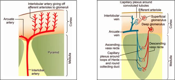
Fig. 17.9: Arrangement of arteries within the kidney (Schematic representation)
Lacis Cells
In addition to the renin producing cells, and the macula densa, the juxtaglomerular apparatus has a third component: these are lacis cells. These cells are so called as they bear processes that form a lace-like network. They are located in the interval between the macula densa and the afferent and efferent arterioles. The function of lacis cells is unknown.
Mode of Action of Juxtaglomerular Apparatus
The juxtaglomerular apparatus is a mechanism that controls the degree of resorption of ions by the renal tubule. It appears likely that cells of the macula densa monitor the ionic constitution of the fluid passing across them (within the tubule). The cells of the macula densa appear to influence the release of renin by the juxtaglomerular cells.
Renin acts on a substance called angiotensinogen present in blood and converts it into angiotensin I. Another enzyme (present mainly in the lungs) converts angiotensin I into angiotensin II. Angiotensin II increases blood pressure. It also stimulates the secretion of aldosterone by the adrenal cortex, thus influencing the reabsorption of sodium ions by the distal convoluted tubules, and that of water through the collecting ducts. In this way it helps to regulate plasma volume and blood pressure.
Renal Blood Vessels
At the hilum of the kidney each renal artery divides into a number of lobar arteries (one for each pyramid). Each
Fig. 17.10: Behavior of efferent arterioles of glomeruli in the# (Schematic representation)
lobar artery divides into two (or more) interlobar arteries that enter the tissue of the renal columns and run toward the surface of the kidney. Reaching the level of the bases of the pyramids, the interlobar arteries divide into arcuate arteries (Fig. 17.9). The arcuate arteries run at right angles to the parent interlobar arteries.
They lie parallel to the renal surface at the junction of the pyramid and the cortex. They give off a series of interlobular arteries that run through the cortex at right angles to the renal surface to end in a subcapsular plexus. Each interlobular artery gives off a series of arterioles that enter glomeruli as afferent arterioles. Blood from these arterioles circulates through glomerular capillaries that join to form efferentarteriolesthat emerge from glomeruli.
The behavior of efferent arterioles leaving the glomeruli differs in the case of glomeruli located more superficially in the cortex, and those lying near the pyramids. Efferent arterioles arising from the majority of glomeruli (superficial) divide into capillaries that surround the proximal and distal convoluted tubules. These capillaries drain into interlobular veins, and through them into arcuate veins and interlobar veins. Efferent arterioles arising from glomeruli nearer the medulla (juxtamedullary glomeruli) divide into 12 to 25 straight vessels that descend into the medulla. These are the descending vasa recta (Fig. 17.10). Side branches arising from the vasa recta join a capillary plexus that surrounds the descending and ascending limbs of the loop of Henle (and also the collecting tubules). The capillary plexus consists predominantly of vessels running longitudinally along the tubules. It is drained by ascending
Chapter 17 The Urinary System 213
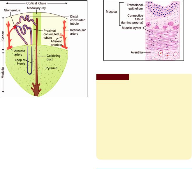
Fig. 17.11: The concept of cortical lobules
(Schematic representation)
vasa recta that run upwards parallel to the descending vasa recta to reach the cortex. Here they drain into interlobular or arcuate veins. The parallel orientation of the ascending and descending vasa recta and their close relationship to the ascending and descending limbs of the loops of Henle is of considerable physiological importance.
From this account of the renal blood vessels it will be clear that two sets of arterioles and capillaries intervene between the renal artery and vein. The first capillary system, present in glomeruli, is concerned exclusively with the removal of waste products from blood. It does not supply oxygen to renal tissues. Exchanges of gases (oxygen, carbon dioxide) between blood and renal tissue is entirely through the second capillary system (present around tubules).
It has been said that in most tissues the blood supply exists to provide service to the parenchyma (in the form of the supply of oxygen and nutrients, and the removal of carbon dioxide and other waste products). In the kidney, on the other hand, the parenchyma exists to provide service to blood (by removal of waste products in it).
It has also been held that interlobular arteries divide the renal cortex into small lobules. Each lobule is defined as the region of cortex lying between two adjacent interlobular arteries. A medullary ray, containing a collecting duct, runs vertically through the middle of the lobule (midway between the two arteries) (Fig. 17.11). Glomeruli lie in a zone adjacent to the arteries while other parts of the nephron lie nearer the center of the lobule.
Fig. 17.12: Layers of ureter (Schematic representation)
Clinical Correlation
Glomerulonephritis or Bright’s disease is the term used for diseases that primarily involve the renal glomeruli. It is convenient to classify glomerular diseases into 2 broad groups:
Primary glomerulonephritis in which the glomeruli are the predominant site of involvement.
Secondary glomerular diseases include certain systemic and hereditary diseases which secondarily affect the glomeruli.
Nephrotic syndrome is a constellation of features in different diseases having varying pathogenesis; it is characterized by
Hydronephrosis is the term used for dilatation of renal pelvis and
ofurine. Hydronephrosis develops if one or both the pelviudilatation and hypertrophy of the urinary bladder but no hydronephrosis. Hydroureter nearly always accompanies hydronephrosis. Hydronephrosis may be unilateral or bilateral.
THE URETERS
Ureters are muscular tubes that conduct urine from renal pelvis to the urinary bladder. The wall of the ureter has three layers:
An inner lining of mucous membraneA middle layer of smooth muscle
An outer fibrous coat: adventitia (Fig. 17.12 and Plate 17.4).
Mucous Membrane
The mucous membrane has a lining of transitional epithelium that is 4 to 5 cells thick and an underlying connective tissue, lamina propria.
The mucosa shows a number of longitudinal folds that give the lumen a star-shaped appearance in transverse section. The folds disappear when the ureter is distended.
214 Textbook of Human Histology
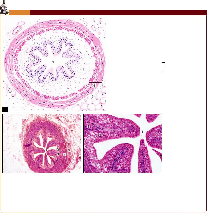
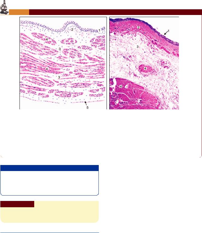
PLATE 17.4: *
Key
. "
0
1 " Mucosa2 B
9 3
|
|
|
|
|
|
|
|
C |
|
* &
# ? !
!
% # <
! # , ! , # in the gut
! @ %
Muscle Coat
The muscle coat is usually described as having an inner longitudinal layer and an outer circular layer of smooth muscle. A third layer of longitudinal fibers is present outside the circular coat in the middle and lower parts of the ureter. The layers are not distinctly marked off from each other. Some workers have reported that the musculature
of the ureter is really in the form of a meshwork formed by branching and anastomosing bundles of muscle fibers.
Adventitia
Adventitia is the outer fibrous coat consisting of loose connective tissue. It contains numerous blood vessels, nerves, lymphatics, and some fat cells.
Chapter 17 The Urinary System 215
PLATE 17.5: *
|
|
|
|
|
|
|
|
|
|
* +
The urinary bladder is easily recognized because the mucous !
The epithelium rests on lamina propria
! # layers between which there is a layer of circular or oblique , ! ! #
The outer surface is lined in parts by peritoneum (serosa) #
"
$ % # prevented by the oblique path followed by the terminal part of the ureter, through the bladder wall. When the musculature of the bladder contracts this part of the ureter is compressed. This mechanism constitutes a physiological sphincter.
Clinical Correlation
Ureterocele is cystic dilatation of the terminal part of the ureter which lies within the bladder wall. The cystic dilatation lies beneath the bladder mucosa and can be visualized by cystoscopy.
THE URINARY BLADDER
Urinary bladder is a muscular bag, where urine is stored temporarily and is discharged periodically via urethra during micturition.
Key
.
0 Lamina propria
1
2 (
9 ( !
The wall of the urinary bladder consists of three layers:An inner mucous membrane
A thick coat of smooth muscleAn outer serous layer (Plate 17.5).
Mucous Membrane
The mucous membrane is lined by transitional epithelium. There is no muscularis mucosae.
In the empty bladder the mucous membrane is thrown into numerous folds that disappear when the bladder is distended. Some mucous glands may be present in the mucosa specially near the internal urethral orifice.
When the bladder is distended (with urine) the lining epithelium becomes thinner. This results from the ability of the epithelial cells to change shape and shift over one another.

216Textbook of Human Histology
Note: The transitional epithelium lining the urinary bladder (and the rest of the urinary passages) is capable of withstanding osmotic changes caused by variations in concentrations of urine. It is also resistant to toxic substances present in urine.
Muscle Coat
The muscle layer is thick. The smooth muscle in it forms a meshwork. Internally and externally the fibers tend to be longitudinal. In between them there is a thicker layer of circular (or oblique) fibers. Contraction of this muscle coat is responsible for emptying of the bladder. That is why it is called the detrusor muscle. Just above the junction of the bladder with the urethra the circular fibers are thickened to form the sphincter vesicae.
Serous Layer
The superior surface of the bladder is covered by mesothelium of peritoneum, forming serous layer. The inferior part of the bladder is covered with adventitia which is made of fibroelastic connective tissue carrying blood vessels, nerves and lymphatics.
Clinical Correlation
Ectopia vesicae is a rare condition owing to congenital is associated with splitting of the overlying anterior abdominal wall. This results in exposed interior of the bladder. There may be prolapse of the posterior wall of the bladder through the defect in the anterior bladder and abdominal wall. The condition in males is often associated with epispadias in which the urethra opens on the dorsal aspect of penis.
Cystitis is the is rare since the normal bladder epithelium is quite resistant to infection. Cystitis may occur by spread of infection from upper from the urethra such as in instrumentation. Cystitis is caused!"# is more common in females because of the shortness of urethra which is liable to faecal contamination and due to mechanical trauma during sexual intercourse.
The Urethra
Urethra is a tube that carries urine from bladder to the exterior. In males, semen also passes through the urethra.
Although the male urethra is much longer than the female urethra, the structure of the two is the same. The wall of the urethra is composed of:
Mucous membraneSubmucosaMuscle layer.
In the case of the male, the prostatic urethra is surrounded by prostatic tissue; and the penile urethra by erectile tissue of the corpus spongiosum.
Mucous Membrane
The mucous membrane consists of a lining epithelium that rests on connective tissue. The epithelium varies in different parts of the urethra. Both in the male and female, the greater part of the urethra is lined by pseudostratified columnar epithelium. A short part adjoining the urinary bladder is lined by transitional epithelium, while the part near the external orifice is lined by stratified squamous epithelium.
The mucosa shows invaginations or recesses into which mucous glands open.
Submucosa
The submucosa consists of loose connective tissue.
Muscle Coat
The muscle coat consists of an inner longitudinal layer and an outer circular layer of smooth muscle. This coat is better defined in the female urethra. In the male urethra it is well defined only in the membranous and prostatic parts, the penile part being surrounded by occasional fibers only.
In addition to this smooth muscle the membranous part of the male urethra, and the corresponding part of the female urethra are surrounded by striated muscle that forms the external urethral sphincter.
Male Reproductive System
The male reproductive system consists of (Fig. 18.1)A pair of testis
Genital ducts: the epididymis, the ductus deferens, and ejaculatory ducts
Accessory sex glands: a pair of seminal vesicle, a single prostate, and a pair of bulbourethral glands
Male urethra andPenis.
TESTIS
General Structure of Testis
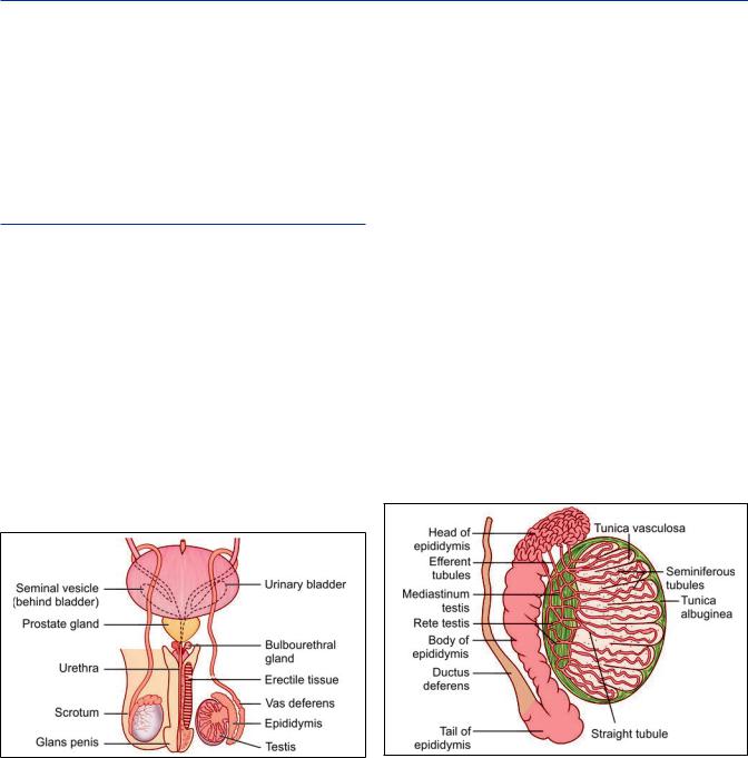
The adult testes are paired ovoid organs placed outside the bodyinscrotum.Thisisidealfornormalspermatogenesisas the temperature is 2 to 3°C less than the body temperature. The right and left testes produce the male gametes or spermatozoa. Each testis is about 4 cm long. The testis lies within a double layered serous sac called the tunica vaginalis. The outermost layer of the organ is formed by a dense fibrous membrane called the tunica albuginea (Fig. 18.2 and Plate 18.1).
The tunica albuginea consists of closely packed collagen fibers amongst which there are many elastic fibers. In the posterior part of the testis the connective tissue of the tunica albuginea expands into a thick mass that projects into the substance of the testis. This projection is called the
mediastinum testis. The visceral layer of tunica vaginalis covers the tunica albuginea, except in the region of the mediastinum testis.
Numerous septa pass from the mediastinum testis to the tunica albuginea, and divide the substance of the testis into a number of lobules. Each lobule is roughly conical, the apex of the cone being directed toward the mediastinum testis. Each lobule contains one or more highly convoluted seminiferous tubules.
Near the apex of the lobule the seminiferous tubules lose their convolutions and join one another to form about 20 to 30 larger, straight tubules (or tubuli recti) (Fig. 18.2). These straight tubules enter the fibrous tissue of the mediastinum testis and unite to form a network called the rete testis. At its upper end the rete testis gives off 12 to 20 efferent ductules. These ductules pass from the upper part of the testis into the epididymis. This duct is highly coiled on itself and forms the body and tail of the epididymis. The epididymis has a head, a body and a tail. The head of the epididymis is made up of highly convoluted continuations of the efferent ductules. At the lower end of the head of the epididymis these tubules join to form a single tube called the duct of the epididymis. At the lower end of the tail of the epididymis the duct becomes continuous with the ductus deferens.
Fig. 18.1: Male reproductive system (Schematic representation)
Fig. 18.2: Basic structure of the testis (Schematic representation)