

MusculoSkeletal Exam
.pdfChapter 1 Introduction
Table 1.1 Paradigms for osteoarthritis and rheumatoid arthritis.
Paradigm for osteoarthritis |
Paradigm for rheumatoid arthritis |
Male |
Female |
Laborer |
20–40 years old |
50+ years old |
Symmetrical small joint involvement |
Large joint involvement |
Associated swelling, fever, rash, morning stiffness |
Asymmetrical involvement |
Abating with use |
Pain in proportion to activity |
|
|
|
Paradigms may also be created for specific tissues (i.e., joints, tendons, muscles, etc.). The paradigm for a joint condition such as osteoarthritis would be welllocalized pain, swelling, stiffness on sedentary posturing, and pain increasing in proportion to use, whereas a paradigm for a mild tendon inflammation (tendinitis) may be painful stiffness after sedentary posturing that becomes alleviated with activity and gentle use. A paradigm for ligament injury would include a history of a specific traumatic event, together with the resultant loss of joint stability demonstrated on active and passive tensile loading of a joint.
The reader is encouraged to create his or her own paradigms for various conditions, paradigms that include the entire portrait of an injury or disease process with which a given patient or tissue may be compared. In this process, it will become obvious that it is not sufficient to limit one’s expertise to the localization of complaints to an anatomical region. It is also necessary to be able to discriminate between the involvement of specific structures that may lie in close proximity within that region (i.e., bursae and tendons overlying a joint).
It can be concluded therefore that an accurate physical examination is as critical to the process of diagnosis as is a complete and accurate history of a patient’s complaints. An accurate physical examination demands a thorough knowledge and familiarity with anatomy and function.
What are the Components of the Musculoskeletal System?
The musculoskeletal system is composed of bone, cartilage, ligaments, muscle, tendons, synovium, bursae,
Figure 1.3 (opposite) Conditioning is the adaptation of a biological system to the controlled application of increasing stress at a frequency, intensity, and duration within the system’s tolerance limit, with a resultant increase in the system’s tolerance limit.
and fascia. This system is derived embryologically from the mesenchyme and is composed of soft and hard connective tissues. These tissues have evolved to serve two basic functions: structural integrity and stable mobility. The tissues are composite materials made up of cells lying within the extracellular matrix they produce.
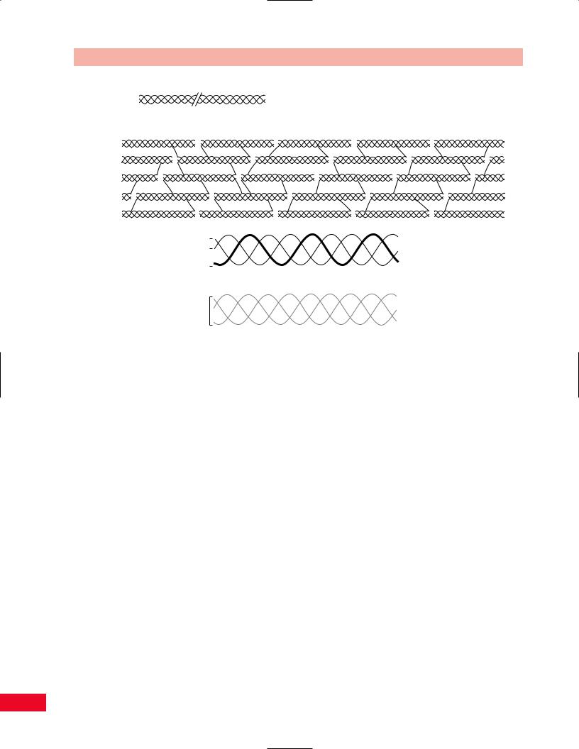
Collagen, a long linear protein (Figure 1.4A), is the most abundant of the extracellular materials found in connective tissues. The foundation of collagen is a repetitive sequence of amino acids that form polypeptide chains. Three such chains are then braided together to form a triple helical strand called tropocollagen. These strands join to make microfibrils; long linear structures specifically designed to resist tensile loading. The microfibrils are bonded together through chemical cross-linking to form collagen fibers. The degree of cross-linking determines the physical properties of a specific collagen fiber. The more cross-linking that exists, the stiffer the fiber will be. The degree of collagen cross-linking is in part genetically and in part metabolically determined. This explains why some people are much more flexible than others. Vitamin C is critical for the formation of cross-links. As such, scurvy, a clinical expression of vitamin deficiency, is characterized by “weak tissues.” Hypermobility of joints (i.e., ability to extend the thumbs to the forearms, ability to hyperextend at the knees and elbows, excessive subtalar pronation with flat, splayed feet) is a clinical manifestation of genetically determined collagen cross-linking (Figure 1.4B).
Different types of collagen exist for different categories of tissues. These types are defined by the specific composition of the polypeptide chains that form the strands of the collagen molecules. Type I collagen is found in connective tissue such as bone, tendons, and ligaments. Type II is found uniquely in articular hyaline cartilage. Other collagen types exist as well (Figure 1.4C).
If collagen represents the fiber in the composite structure of connective tissue, ground substance represents the “filler” between the fibers. The main components
5
Introduction Chapter 1
A
B
1 Chains 

C
2 Chain 

Type I collagen
All 1 Chains
Type II collagen
Figure 1.4 (A) Collagen is a linear protein made of alpha chains that wind into a triple helix. (B) Collagen fibrils are formed by the cross-linking of collagen monomer proteins. (C) The different types of collagen are determined by the number of alpha-1 and alpha-2 collagen monomers that join to form a triple-helix collagen molecule. For example, two alpha-1 chains and one alpha-2 chain that join to form a triple helix make type 1 collagen, which is found in bone, tendon, ligament, fascia, skin, arteries, and the uterus. Type 2 collagen, which is found in articular cartilage, contains three alpha-1 chains. There are at least 12 different collagen types.
of ground substance are aggregates of polyglycan macromolecules. An example of such a macromolecule is the proteoglycan hyaluronic acid, found in articular cartilage. Hyaluronic acid is a molecule of more than 1 million daltons. It is composed of a long central core from which are projected many protein side chains containing negatively charged sulfate radicals. It can best be visualized as a bristle brush from which many smaller bristle brushes are projected (Figure 1.5). These strongly negative sulfate radicals make the hyaluronic acid molecule highly hydrophilic (water attracting). This ability to attract and hold water allows the connective tissue ground substance to function as an excellent hydrostatic bearing surface that resists compression load.
Immobilization reduces the diffusion and migration of nutrients throughout the connective tissues. This in turn compromises cellular activity and upsets the normal homeostatic balance of collagen and ground substance turnover. The result is an atrophy of collagen fibers and a diminution of ground substance (Cantu and Grodin, 2001), with subsequent deterioration of the connective-tissue macrofunction (i.e., chondromalacia patellae).
Bone
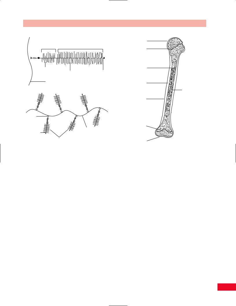
Bone provides the structure of the body. It is the hardest of all connective tissues. One-third of bone is comprised of collagen fibers and two-thirds mineral salts, primarily calcium hydroxyapatite. Bone is formed in response to stress. Although genetically determined, the size and shape of a bone are dependent on environmental factors for its full expression. This response of bone to its loading history has been termed Wolff’s law. There are two major types of bone: cortical and cancellous. All bones are covered by highly vascularized and innervated tissue called periosteum, except when they are within the synovial cavity of a joint (Figure 1.6).
Cortical bone is very dense, highly calcified, and uniquely constructed to resist compression loads. It can also resist tensile bending and torsional loads, but much more poorly. This is a direct function of cortical bone’s ultrastructure, which is a composite of flexible collagen fibers and rigid mineral crystals. Cortical bone is usually found within the diaphysis of long bones. It has a hollow central cavity which is termed the medullary canal or marrow cavity.
6
Keratan |
|
sulfate-rich |
Chrondroitin sulfate-rich |
region |
region |
Keratan |
|
|
sulfate |
Chrondroitin sulfate |
Protein core |
chain |
chain |
|
Hyaluronic acid (HA)
Proteoglycan "bristle brushes"
Link protein
Protein side |
Hyaluronic acid |
|
chain core |
||
central "core" |
||
|
Sulfate radicals
(chrondroitin and keratan sulfate)
Figure 1.5 The proteoglycan aggregate is formed on a backbone of hyaluronic acid and has the appearance of a bristle brush.
Chapter 1 Introduction
Articular cartilage
Line of epiphyseal cartilage
Trabeculae of
spongy bone
Compact or cortical bone
Marrow cavity
Periosteum
Line of epiphyseal cartilage
Articular cartilage
Figure 1.6 The structure of a typical long bone.
At the end of long bones and at the sites of tendon and ligament attachments, bones tend to expand and cortical bone gives way to a more porous structure, termed cancellous or trabecular bone. The trabeculae of cancellous bones lie in the direction of transmitted loads. They act as conduits of load from the articular surface to the underlying diaphyseal cortical bone. Overload of the trabeculae will, on a microscopic scale, duplicate overload of an entire bone (i.e., fracture). This overload, because of the innervation that exists within a bone, will give rise to pain (arthritic discomfort due to mechanical overload secondary to joint deformity or erosion of articular cartilage). The resultant healing of these microfractures leads to increased calcium deposition, hence subchondral sclerosis noted around articular joints on x-ray films, and hypertrophy of stressed sites such as the midshaft of the tibia secondary to stress fractures occurring from overuse in distance running.
Cartilage
Cartilage is a connective tissue made of cells (chondroblasts and chondrocytes) that produce an extracellular matrix of proteoglycans and collagen fibers with a
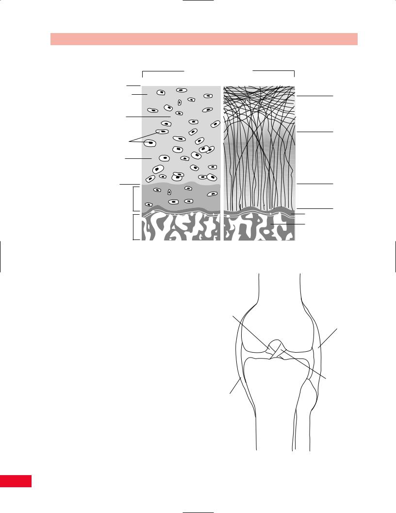
high water content. The tensile strength of cartilage is due to the collagen component. Its resistance to compression is due to the ability of proteoglycan to attract and hold water. Cartilage types include articular or hyaline cartilage (Figure 1.7); fibrocartilage, which exists at the attachment sites of ligaments, tendons, and bones; fibroelastic cartilage, found in menisci and intravertebral discs; and growth-plate cartilage, located in the physis of immature bones. With age, cartilage tends to decrease in water content and the number of cross-links among collagen molecules increases. The result is that cartilage tissue becomes more brittle, less supple, and less able to resist tensile, torsional, and compression loading. Hence, cartilage becomes more vulnerable to injury with age.
Articular cartilage lines the spaces in synovial joints. It is attached to the underlying bone by a complex interdigitation analogous to that of a jigsaw puzzle. Regeneration of this cartilage is slow and inconsistent in terms of restoration of articular integrity. It can be replaced by a less mechanically efficient fibrocartilage after injuries have occurred. There are no blood vessels within articular cartilage and nutrition is solely dependent on the loading and unloading of the joint, which allows water-soluble nutrients and waste products to
7
Introduction Chapter 1
Composition and Structure of Cartilage
Articular hyaline cartilage
Histology |
Orientation of collagen fibers |
|
Lamina splendens |
Zone I |
|
H2O in and out due to |
Tangential |
|
|
||
pressure of joint surfaces |
|
|
on one another |
Zone II |
|
Matrix |
||
Oblique |
||
|
||
Chondrocytes |
|
|
in lacunae |
|
|
Ground |
Zone III |
|
substance |
Vertical |
|
Tidemark |
|
|
Calcified cartilage |
Zone IV |
|
Vertical |
||
|
End plate |
|
Subchrondral bone |
Trabecular |
|
bone |
||
|
Figure 1.7 The composition and structure of articular hyaline cartilage. Water moves in and out of the cartilage due to the pressure of the joint surfaces on one another and attraction of the water by the ground substance. Note the orientation of the collagen fibers.
enter and leave the cartilaginous matrix through a |
|
porous surface layer. |
|
The fibroelastic cartilage of the intervertebral disc |
|
allows for very minimal movement between adjacent |
Posterior |
vertebrae while providing shock absorption. Due to |
cruciate |
the orientation of the fibers, they are more vulnerable |
ligament |
|
|
to flexion and rotational forces. Fibroelastic cartilage |
|
is also present in the menisci of the knee. Here it func- |
|
tions not only to absorb shock but also to increase the |
|
functional surface area of the joint, thereby providing |
|
additional stability. Due to its elastin content, fibro- |
|
elastic cartilage is resilient and able to return to its |
|
prior shape following deformation. |
|
Ligaments
Ligaments are the static stabilizers of joints. They con- |
Medial |
nect bones to bones (Figure 1.8). Ligaments and other |
collateral |
capsular structures of the joint are made of dense, |
ligament |
|
|
organized connective tissue. Ligaments contain collagen |
|
Figure 1.8 (right) The ligaments of the knee. Due to the inherent instability of the joint, ligaments are necessary to prevent motion in all planes. They act as the primary stabilizers of the joint and are assisted by the muscles and other connective tissues.
Lateral collateral ligament
Anterior cruciate ligament
8
Chapter 1 Introduction
STRESS, (load/cross-section area)
Breaking point
Anterior longitudinal ligament
Breaking point
Ligamentum flavum
STRAIN, (extension/original length)
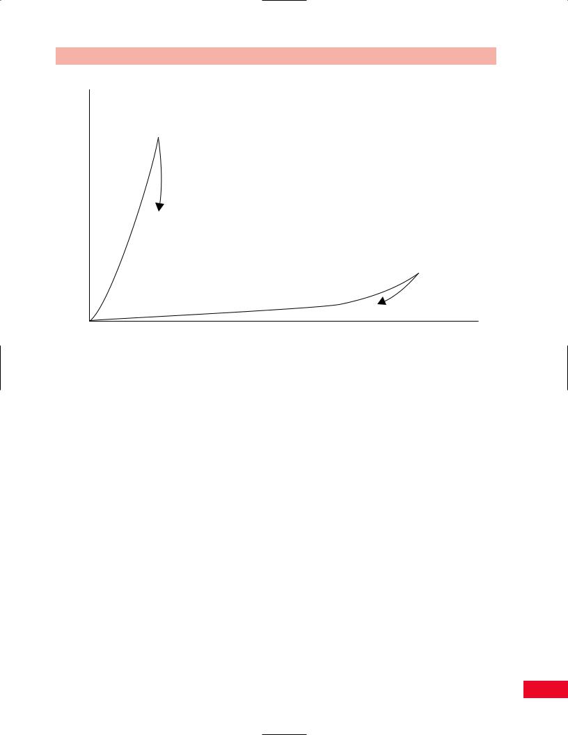
Figure 1.9 The mechanical response of stress and strain on the anterior longitudinal ligament and the ligamentum flavum. The anterior cruciate ligament, having more collagen than elastin, can handle a larger load, but will only stretch a short amount before breaking. The ligamentum flava, having more elastin than collagen, cannot tolerate a very large load, but can stretch a lot before breaking.
and a variable amount of elastin. The collagen provides tensile strength to the ligaments and elastin provides suppleness. The fibers of collagen are arranged more or less parallel to the forces that the ligament is intended to resist. Most ligaments and capsular tissues enter the bone as a progression from collagen fibers to fibrocartilage to calcified cartilage and then finally bone. Some ligaments (and tendons) attach to the periosteum first, which then attaches to the bone. The site of ligament failure is a function of the load it experiences. Ligaments resist slow loading better than rapid loading. Therefore, rapid loading may produce an intraligamental lesion, whereas a slower pattern of loading will create injuries at or near the bone–ligament interface.
Elastin is a protein that permits elastic recoiling to occur in a tissue. Some ligaments, such as the cruciate ligament of the knee, contain almost no elastin. Other ligaments, such as the ligamentum flavum of the spine, contain large amounts of elastin. Figure 1.9 shows that because it contains more collagen than elastin, the anterior cruciate ligament can resist tensile loads with little elongation. In this way, the anterior cruciate ligament serves the knee well as a stabilizing structure. On the other hand, the ligamentum flavum
of the spine, being composed mostly of elastin and little collagen, can be stretched a great deal before breaking, but can only resist very weak tensile loading.
Ligaments function to limit joint motion and to guide the bones as they move. Ligaments therefore usually have a dual internal structure, such that they may stabilize the joint at either extreme of motion. Ligaments are most lax at midrange of joint motion. The capsule of a synovial joint is in fact a weak ligamentous structure. Disruption of a ligament can result in severe joint instability and increased frictional stresses to the articular surfaces of that joint. This will result in premature osteoarthritis. Conversely, a loss of normal capsular laxity from fibrosis following trauma will result in a severe restriction in joint motion (i.e., posttraumatic adhesive capsulitis of the shoulder).
Ligaments have very little vascularity; hence they heal poorly. However, they do have innervation, which may be useful to quantify the severity of a given ligamentous injury. When the structural integrity of a ligament has been completely compromised (grade III sprain), relatively little pain is produced on attempts to passively stretch the injured ligament. This is because no tension load can be created across a completely
9
Introduction Chapter 1
disrupted ligament. However, in a less severe partial tear (grade I sprain), severe and exquisite pain will be produced when tension is applied across the damaged structure. This paradoxical pain pattern (less pain equals a more severe sprain) can be a significant diagnostic clue obtained during the physical examination of a recently injured ligament. This also has dramatic import
in defining a patient’s prognosis and determining a treatment plan.
Muscle
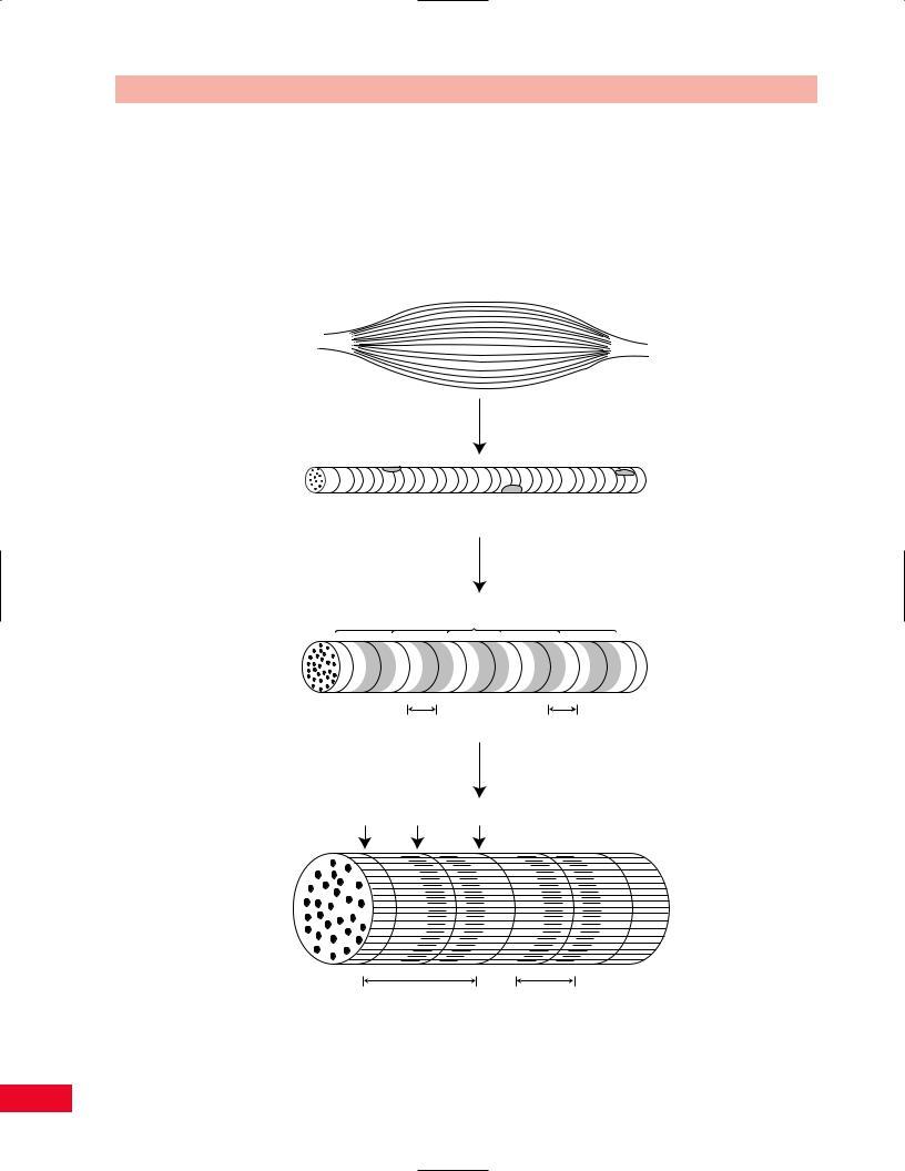
Skeletal muscle is a contractile tissue made up of fibers that contain specialized proteins (Figures 1.10 and 1.11).
Intact muscle
Muscle consists of muscle fibers
Stripes or striations
Muscle cell (fiber)
Muscle fiber is a bundle of myofibrils
Z lines
Myofibril |
A band |
I band |
|
|
Enlarged view |
Z line |
M line |
Z line |
Sarcomere |
A band |
(from Z line to Z line) |
|
Two sarcomeres
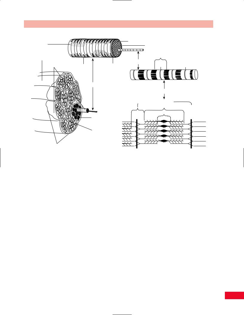
Figure 1.10 A microscopic view of muscle shows the repeated patterns of the sarcomeres and the fibrils.
10
Chapter 1 Introduction
Nuclei |
|
|
Sarcolemma |
|
|
|
Sarcoplasm |
|
|
|
|
|
|
|
Muscle |
|
|
I |
|
|
|
|
|
|
Muscle fascicles |
|
Basement |
Myofibril |
|
Satellite cell |
Z |
Z |
||
|
|
|
|
Muscle Fiber
Myofilaments
 Sarcomere
A
H
Endomysium |
Z |
M |
Z |
Perimysium
Epimysium
Figure 1.11 The organization of skeletal muscle tissue.
A loose connective tissue known as endomysium fills the space between these fibers. This tissue attaches to a stronger connective tissue that surrounds the muscle vesiculae, known as perimysium. Perimysium is in turn connected to the epimysium, which encases the entire muscle. This in turn is anchored to the fascial tissues of the nearby structures. Muscles therefore are composed of two elements: contractile tissues and inert, noncontractile tissues. The forces generated by the muscles are extrinsically applied to the muscle and will affect both types of tissue.
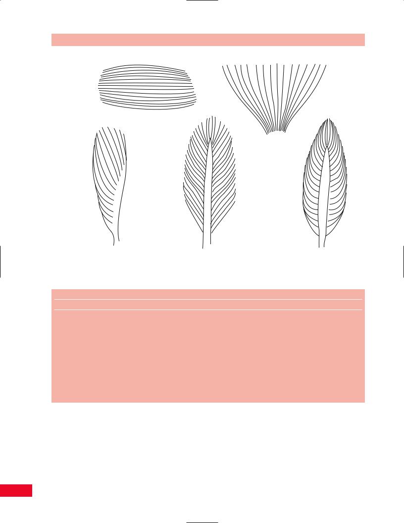
Muscles exist in many shapes and sizes. Some of these are shown in Figure 1.12.
Muscles contain three different fiber types: I, IIa, and IIb. They are defined by the chemical machinery used to generate adenosine triphosphate (ATP). Genetic makeup, training, and neuromuscular disease can affect the composition of a given muscle with respect to fiber type. Characteristics of these various fiber types are shown in Table 1.2.
Muscles act to move body parts or to stabilize a joint. As dynamic stabilizers of joints, muscles serve to duplicate the static stabilizing action of ligaments. Muscle fibers are capable of shortening to about 50% of their original length. The tension developed by a contracted muscle can be either active or passive. Active tension
is due to the contractile components, namely, actin and myosin. Passive tension results from elastic properties of the contractile tissues within the muscle.
The strength of the muscle is proportional to its cross-sectional area and mass. The force of contraction of a muscle is related to many factors, including the length of the fibers, the velocity of contraction, and the direction in which the fiber is moving at the time of its contraction. Types of muscle contraction include concentric or shortening, eccentric or lengthening, and isometric, in which the muscle does not change length. Muscles are characterized by their function; agonists are prime movers, antagonists resist the action of prime movers, and synergists support the function of the agonists. For example, in ankle dorsiflexion, the anterior tibialis is the agonist. The extensor hallucis longus and extensor digitorum longus muscles assist the tibialis anterior muscle and therefore are synergists. The gastrocnemius and soleus and plantar flexors of the toes are antagonists of the tibialis anterior.
Muscles are described in anatomy texts as having origins and insertions. It is very important to recognize that this is an arbitrary distinction. A muscle that is referred to as a hip flexor because it brings the thigh toward the torso can function just as well to bring the
11
Introduction Chapter 1
Parallel muscle fibers
Fan-shaped fibers
Unipennate |
Bipennate |
Fusiform |
muscle |
muscle |
muscle |
|
Figure 1.12 Different types of muscle-fascicle arrangements. |
|
Table 1.2 Characteristics of skeletal muscle fibers based on their physical and metabolic properties.
Muscle fiber type
Property |
Slow-twitch |
Intermediate |
Fast-twitch |
Speed of contraction |
Slow |
Intermediate |
Fast |
Rate of fatigue |
Slow |
Intermediate |
Fast |
Other names used |
Type I |
Type II B |
Type II A |
|
SO |
FOG |
FG |
Muscle fiber diameter |
Small |
Intermediate |
Large |
Color |
Red |
Red |
White |
Myoglobin content |
High |
High |
Low |
Mitochondria |
Numerous |
Numerous |
Few |
Oxidative enzymes |
High |
Intermediate |
Low |
Glycolytic enzymes |
Low |
Intermediate |
High |
Glycogen content |
Low |
Intermediate |
High |
Myosin ATPase activity |
Low |
High |
High |
Major source of ATP |
Oxidative phosphorylation |
Oxidative phosphorylation |
Glycolysis |
torso over the thigh. In order for muscles to function normally, they must be both strong and flexible.
With respect to innervation of muscles, except for the deepest layers of the vertebral muscles, the exact innervation of the limb and trunk muscles is similar between individuals, with some variability. Tables listing segmental innervation differ from text to text.
One such table appears in the Appendix of this book and should be used as a guide only. Injuries to muscles are termed strains. Analogous to ligament injuries, they are classified by severity into three grades: grade I indicates minimal damage; grade II represents an intermediate amount of damage to the muscle structure; and grade III, complete disruption.
12
Chapter 1 Introduction
Muscle
Tendon
Ligament
Figure 1.13 A tendon.
Tendons
Tendons connect muscles to other structures (see Figure 1.13). Like ligaments, tendons are also comprised of collagen, ground substance, and cells. The collagen of tendons is aligned in a very strict linear fashion and is always oriented in the line of the pull of the muscle. Tendons have been designed to transmit the force of the muscular contractile tissues to bone and other connective tissues, such as skin and ligaments, to which they are attached. Tendons are said to be able to withstand at least twice the maximum force that muscles can exert on them. The zone where the muscle blends into the tendinous tissues is called the musculotendinous junction. Muscle-tendon units represent tensile structures. As such, they may fail in the muscle, at the muscle–tendon junction, within the tendon, or at the tendon–bone insertion. Most commonly, however, failure occurs at the point of transition between two different materials (i.e., the musculotendinous junction). Some tendons are surrounded by a double-walled tubular covering, referred to as a tendon sheath or a peritendon (i.e., Achilles tendon or flexor tendons of the hand). This is lined with a synovial membrane. The sheath is used both to lubricate the tendon and to guide it toward the bony attachment. Tendon sheaths provide a pathway for the gliding movement of the tendon within the sheath. An inflamed tendon sheath can cause a locking or restricted movement, as in a trigger finger. Inflammation of the tendon structure is termed tendinitis.
Synovium and Bursae
Synovial tissue lies in the inner aspect of synovial joints and bursal sacs. It has two functions: to produce lubricating fluids and to phagocytize (remove) foreign
debris. Synovium is highly vascularized and innervated. As such, when traumatized or inflamed, synovial tissue will rapidly enlarge and produce significant pain.
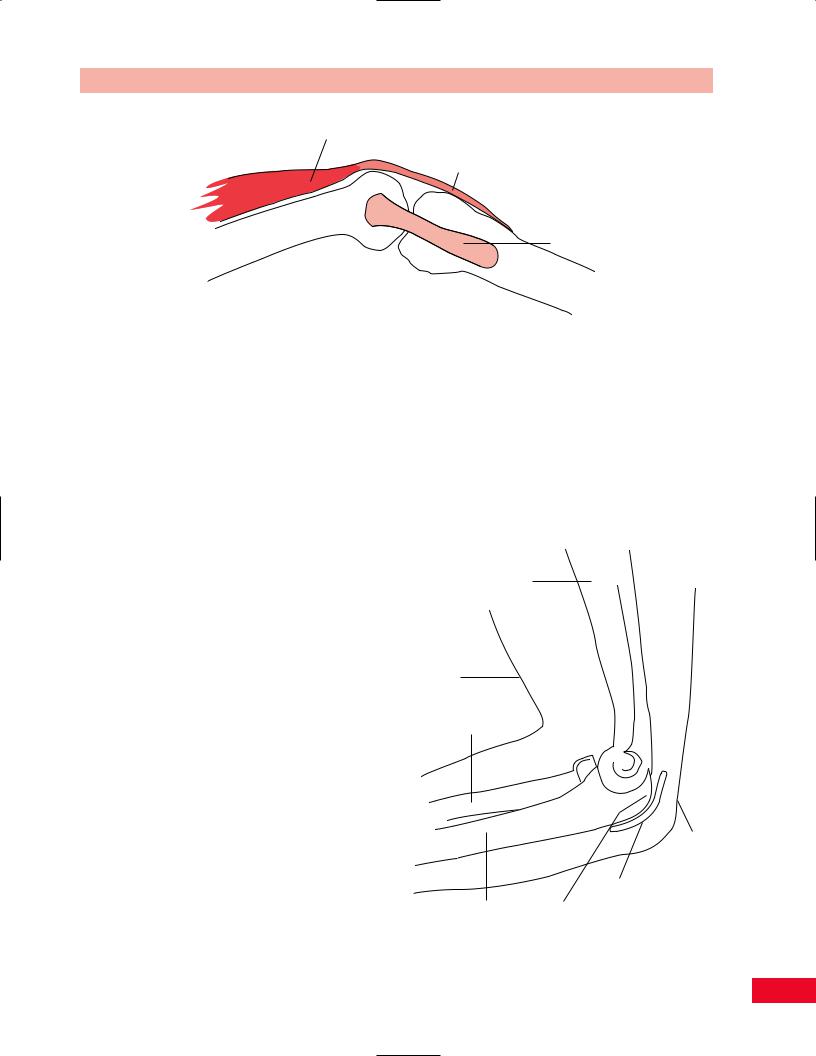
Bursal sacs serve to reduce friction. Therefore, they are located wherever there is need for movement between structures in close proximity. For example, the olecranon bursa lies between the olecranon process of the ulna and the skin overlying the posterior part of the elbow (see Figure 1.14). The subacromial
Humerus
Skin
Radius
Skin
Olecranon bursa
Ulna |
Olecranon process |
Figure 1.14 The olecranon bursa is between the skin and the olecranon process at the elbow.
13

Introduction Chapter 1
bursa lies between the acromioclavicular arch above and the rotator cuff tendons below. Inflammation of synovial or bursal tissues due to trauma, inflammatory processes, or foreign materials is termed synovitis or bursitis.
Fascia
There are three kinds of fascial tissues: superficial, deep, and subserous. The fascia is composed of loose to dense connective tissue. Superficial fascia is under the skin; deep fascia is beneath the superficial and also envelops the head, trunk, and limbs. Subserous fascia surrounds organs in the thorax, abdomen, and pelvis.
Superficial fascia contains fat, blood vessels, and nerves. It is loose in consistency and very thin. It is attached to the undersurface of the skin.
Deep fascia is dense and tough and has two layers. It wraps around regions of the body and splits to
envelop superficial muscles such as the sartorius and tensor fasciae latae. Periosteum, perimysium, and perichondrium are all elements of the deepest layer of the deep fascia. The deep fascia serves to interconnect the different muscle groups. By being continuous, it can provide tension at a distant site when pulled by a contracting muscle. Some muscles take their origin from the deep fascia. The fascia also separates groups of muscles with similar function, for example, the flexor and extensor groups of the leg. Because of the relative inelasticity of fascia, abnormally high pressure within a fascial compartment (i.e., due to injury or inflammation) can compromise the function of the nerves and blood vessels that course through that compartment. This may result in serious compromise of the tissues supplied by these nerves and vessels. Fascia may, as other tissues, experience an inflammatory reaction, fasciitis. This condition can be accompanied by moderate or even severe discomfort and scarring (fibrosis). Fibrosis can lead to stiffness and restricted movement.
14